Address for correspondence Dr. Kikkeri Narayanasetty Naveen
Professor, Department of Dermatology, No 10 , Skin OPD,
Sri Dharmasthala Manjunatheshwara College of Medical Sciences & Hospital (SDMCMS&H) Sattur, Dharwad – 580009
E-mail: [email protected]
Original Article
Cutaneous lesions in neonatal intensive care unit in a
tertiary care center
Introduction
The skin of the neonate differs from that of the
adult, in that it is thinner, delicate, has weaker
intercellular attachments and produces fewer
sweat and sebaceous gland secretions and is
more susceptible to several infections.
1Neonatal
skin disorders are classified into 4 main types:
physiologic skin disorders, acquired skin
disorders, developmental skin disorders and
iatrogenic complications.
2Kikkeri Narayanshetty Naveen, Praveen S Bagalkote*, Sachin Manohar Shetty, Umesh Dixit**
Department of Dermatology, SDM College of Medical Sciences and Hospital, Sattur, Dharwad, Karnataka, India
*Department of Pediatrics, SDM College of Medical Sciences and Hospital, Sattur, Dharwad, Karnataka, India
**Department of Community Medicine, SDM College of Medical Sciences and Hospital, Sattur, Dharwad, Karnataka, India
Abstract
Objective To document the pattern of skin lesions among the neonates admitted in intensive care unit and evaluate the association between age, gender, maturity and birth weight.Methods A total of 200 neonates, who were hospitalized in the NICU of SDMCMS&H were included in this single contact cross-sectional study. All diagnoses were made on clinical basis. The relationships between the occurrence of the lesions with the various maternal and neonatal factors were analyzed. The statistical analysis of the associations was done using percentage and student t-test.
Results The following cutaneous changes were noted. Lanugo hair was found in 121 neonates. 93 children showed Mongolian spots. The signs of miniature puberty were found in 82 children.105 children had sebaceous hyperplasia. 77 children had physiological desquamation. Erythema neonatorum was found in 59 patients and in only 3 of them it persisted beyond 7 days. 40 children had miliaria. Cutis marmorata was noted in 20 children. Other transient skin conditions seen were milia (5), vernix caseosa (5), acrocyanosis (3), neonatal acne (3), erythema toixcum neonatorum (1) and neonatal alopecia (1).
Conclusion Skin lesions are very common in the neonatal period. In the present study, most of the skin lesions were physiological and transitory. Pediatrician and dermatologists should be aware of these skin lesion in neonatal ICU and should be able to differentiate it from serious skin conditions in order to avoid unnecessary investigations and treatment in neonates.
Key words
The present study was undertaken to know the
pattern of skin lesions among the neonates
admitted in intensive care unit and evaluate the
association between age, gender, maturity and
birth weight. No such study has been conducted
in this part of Karnataka.
Methods
A total of 200 neonates, who were hospitalized
in the NICU of SDMCMS&H were included in
this single contact cross-sectional study from
November 2015 to February 2016. The neonates
receiving phototherapy were excluded from the
study. The oral cavities were not examined. All
diagnoses were made on clinical basis and no
skin biopsy was done. The maturity of the
neonates was determined by the prenatal and
obstetric medical records. Preterm was defined
as less than 37 weeks of gestational age, while
full-term was between 37 to 42 weeks and
post-term more than 42 weeks. Early neonatal period
was defined as age less than 7 days and late
neonatal was between 8 and 28 days. The data
were collected and documented using a
Microsoft Excel worksheet. The relationships
between the occurrence of the lesions with the
various maternal and neonatal factors were
analyzed. The statistical analysis of the
associations was done using percentage and
student t-test. Institutional ethical committee
clearance was obtained
Results
The total number of male children was 107 and
female children were 93 with ratio 1.15:1. 87
children were preterm, 104 were term and 9
were delivered postterm. 127 babies were in
early neonatal period while 73 in late neonatal
period. 96 neonates were delivered by lower
segment caesarian section and 104 neonates
were delivered vaginally.
The neonates were admitted for the varied
reasons. Respiratory distress was diagnosed in
34 neonates. 33 neonates were admitted for low
birth weight, 11 for very low birth weight and 3
for extremely low birth weight. 25 neonates
were born with meconium stained liquor. 17
neonates were admitted for birth asphyxia, 16
for small for gestational age, 11 for intrauterine
growth retardation, 6 neonates for twin
deliveries, 8 for preterm care and 3 for post term
care. The other reasons are as follows: anorectal
anomalies (1), congenital heart disease (4),
bilateral hydronephrosis (1), type III intestinal
atresia (1), cleft lip and cleft palate (1),
convulsions (2), dysmorphic features (1),
gastrointestinal bleed (1), hypoglycemia (3),
hyaline membrane disease (4), infant of diabetic
mother (1), neonatal seizures (1), not feeding
well (4), premature rupture of membrane (3),
renal anomalies (1), trachea-esophageal fistula
(1), tachypnea (1), weak cry (1). 2 neonates were
admitted
for
congenital
ichthyosis
with
collodion baby and 1 infant for the
Kasabach-Merritt syndrome.
In 34 neonates of respiratory distress, 7 were
low birth weight. Cellulitis, congenital heart
disease and dysmorphic features were found in 1
infant. In 25 neonates born with meconium
stained liquor, 10 of them had respiratory
distress and 6 had birth asphyxia.
persisted beyond 7 days. 40 children had
miliaria. Cutis marmorata was noted in 20
children. Other transient skin conditions seen are
milia (5), vernix caseosa (5), acrocyanosis (3),
neonatal
acne
(3),
erythema
toixcum
neonatorum (1) and neonatal alopecia (1).
In the developmental lesions found were
congenital melanocytic nevus (2), faun tail
nevus (2), hemangioma (1) and preauricular skin
tag (1).
In acquired skin lesions, intertrigo was seen in 8
neonates, icterus was seen in 4 neonates and oral
thrush was noted in 1 infant.
In iatrogenic skin diseases, 15 neonates had
bruises, 14 had ecchymoses, contact dermatitis,
plaster dermatitis and erosions were seen in 1
infant. Other skin lesions found were petechiae
(1), purpura (1), sclerema (1), loss of
subcutaneous
fat
(1),
hypospadiasis
(1),
generalized edema (1), ophthalmia neonatorum
(1), micrognathia (2), lentigines (1), simian
crease (2), umbilical hernia (1), prominent veins
(1), hairy pinna (1), hydrocele (1), phimosis (1),
insect bite (1), cleft lip and cleft palate (1) ,
cellulitis (1), scrotal dermatitis (1), triangular
face (1).
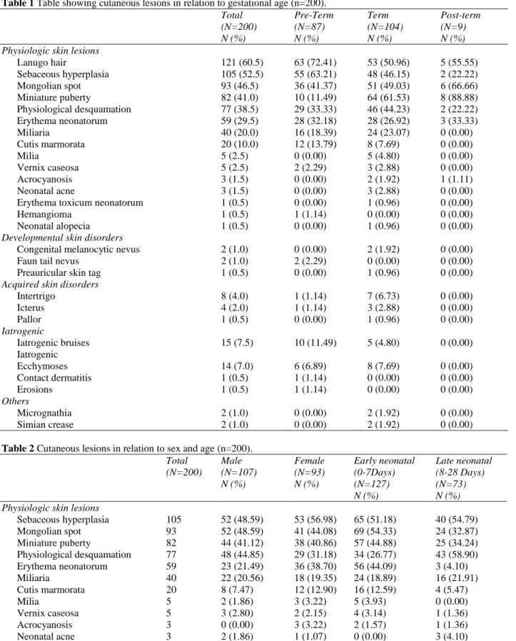
Relation between gestational age and cutaneous
lesions is shown in
(Table 1)
. Lanugo hair,
sebaceous hyperplasia, erythema neonatorum,
cutis marmorata and iatrogenic bruises were
frequently seen in preterm neonates, whereas
miniature puberty, physiological desquamation,
milia, neonatal acne, Mongolian spot, miliaria
and intertrigo were more frequently seen in term
neonates. Lanugo hairs (z=2.874,
p
=0.004) and
sebaceous hyperplasia (z= 2.21,
p
=0.027) in
preterm neonates was statistically significant,
whereas miniature puberty (z=6.921,
p
<0.001)
in post-term was statistically significant as
calculated by student t-test.
Relation between sex of children and cutaneous
lesions is shown in
Table 2
. Physiological
desquamation,
iatrogenic
bruises
and
ecchymosis were more common in male
neonates,
whereas
sebaceous
hyperplasia,
erythema neonatorum and cutis marmorata were
more common in female neonates. Erythema
neonatorum
(z=2.507,
p
=0.012)
showed
statistically significant correlation with female
preponderance.
Relation between age and cutaneous lesions is
also depicted in
Table 2
. In early neonatal
period, predominant cutaneous lesions were
miniature puberty, erythema neonatorum, cutis
marmorata, milia, vernix caseosa, Mongolian
spots and intertrigo. In late neonatal period
common cutaneous lesions were physiological
desquamation, neonatal acne, iatrogenic bruises
and ecchymosis. Erythema neonatorum (z=5.81,
p
<0.001) was statistically significant in early
neonatal
period,
whereas
physiological
desquamation
(z=4.345,
p
<0.001)
was
statistically significant during late neonatal
period.
Table 1 Table showing cutaneous lesions in relation to gestational age (n=200). Total
(N=200) N (%)
Pre-Term (N=87) N (%)
Term (N=104) N (%)
Post-term (N=9) N (%) Physiologic skin lesions
Lanugo hair 121 (60.5) 63 (72.41) 53 (50.96) 5 (55.55)
Sebaceous hyperplasia 105 (52.5) 55 (63.21) 48 (46.15) 2 (22.22)
Mongolian spot 93 (46.5) 36 (41.37) 51 (49.03) 6 (66.66)
Miniature puberty 82 (41.0) 10 (11.49) 64 (61.53) 8 (88.88)
Physiological desquamation 77 (38.5) 29 (33.33) 46 (44.23) 2 (22.22) Erythema neonatorum 59 (29.5) 28 (32.18) 28 (26.92) 3 (33.33)
Miliaria 40 (20.0) 16 (18.39) 24 (23.07) 0 (0.00)
Cutis marmorata 20 (10.0) 12 (13.79) 8 (7.69) 0 (0.00)
Milia 5 (2.5) 0 (0.00) 5 (4.80) 0 (0.00)
Vernix caseosa 5 (2.5) 2 (2.29) 3 (2.88) 0 (0.00)
Acrocyanosis 3 (1.5) 0 (0.00) 2 (1.92) 1 (1.11)
Neonatal acne 3 (1.5) 0 (0.00) 3 (2.88) 0 (0.00)
Erythema toxicum neonatorum 1 (0.5) 0 (0.00) 1 (0.96) 0 (0.00)
Hemangioma 1 (0.5) 1 (1.14) 0 (0.00) 0 (0.00)
Neonatal alopecia 1 (0.5) 0 (0.00) 1 (0.96) 0 (0.00)
Developmental skin disorders
Congenital melanocytic nevus 2 (1.0) 0 (0.00) 2 (1.92) 0 (0.00)
Faun tail nevus 2 (1.0) 2 (2.29) 0 (0.00) 0 (0.00)
Preauricular skin tag 1 (0.5) 0 (0.00) 1 (0.96) 0 (0.00)
Acquired skin disorders
Intertrigo 8 (4.0) 1 (1.14) 7 (6.73) 0 (0.00)
Icterus 4 (2.0) 1 (1.14) 3 (2.88) 0 (0.00)
Pallor 1 (0.5) 0 (0.00) 1 (0.96) 0 (0.00)
Iatrogenic
Iatrogenic bruises 15 (7.5) 10 (11.49) 5 (4.80) 0 (0.00)
Iatrogenic
Ecchymoses 14 (7.0) 6 (6.89) 8 (7.69) 0 (0.00)
Contact dermatitis 1 (0.5) 1 (1.14) 0 (0.00) 0 (0.00)
Erosions 1 (0.5) 1 (1.14) 0 (0.00) 0 (0.00)
Others
Micrognathia 2 (1.0) 0 (0.00) 2 (1.92) 0 (0.00)
Simian crease 2 (1.0) 0 (0.00) 2 (1.92) 0 (0.00)
Table 2 Cutaneous lesions in relation to sex and age (n=200). Total
(N=200)
Male (N=107) N (%)
Female (N=93) N (%)
Early neonatal (0-7Days) (N=127) N (%)
Late neonatal (8-28 Days) (N=73) N (%) Physiologic skin lesions
Sebaceous hyperplasia 105 52 (48.59) 53 (56.98) 65 (51.18) 40 (54.79) Mongolian spot 93 52 (48.59) 41 (44.08) 69 (54.33) 24 (32.87) Miniature puberty 82 44 (41.12) 38 (40.86) 57 (44.88) 25 (34.24) Physiological desquamation 77 48 (44.85) 29 (31.18) 34 (26.77) 43 (58.90) Erythema neonatorum 59 23 (21.49) 36 (38.70) 56 (44.09) 3 (4.10)
Miliaria 40 22 (20.56) 18 (19.35) 24 (18.89) 16 (21.91)
Cutis marmorata 20 8 (7.47) 12 (12.90) 16 (12.59) 4 (5.47)
Milia 5 2 (1.86) 3 (3.22) 5 (3.93) 0 (0.00)
Vernix caseosa 5 3 (2.80) 2 (2.15) 4 (3.14) 1 (1.36)
Acrocyanosis 3 0 (0.00) 3 (3.22) 2 (1.57) 1 (1.36)
Neonatal acne 3 2 (1.86) 1 (1.07) 0 (0.00) 3 (4.10)
Hemangioma 1 1 (0.93) 0 (0.00) 0 (0.00) 1 (1.36)
Neonatal alopecia 1 0 (0.00) 1 (1.07) 0 (0.00) 1 (1.36)
Developmental skin disorders
Congenital melanocytic nevus 2 1 (0.93) 1 (1.07) 2 (1.57) 0 (0.00)
Faun tail nevus 2 2 (2.80) 0 (0.00) 2 (1.57) 0 (0.00)
Preauricular skin tag 1 1 (0.93) 0 (0.00) 0 (0.00) 1 (1.36) Acquired skin disorders
Intertrigo 8 4 (3.73) 4 (4.30) 7 (5.51) 1 (1.36)
Icterus 4 3 (2.80) 1 (1.07) 1 (0.78) 3 (4.10)
Pallor 1 0 (0.00) 1 (1.07) 1 (0.78) 0 (0.00)
Iatrogenic
Iatrogenic bruises 15 11 (10.28) 4 (4.30) 6 (4.72) 9 (12.33)
Ecchymoses 14 11 (10.28) 3 (3.22) 8 (6.29) 6 (8.22)
Contact dermatitis 1 0 (0.00) 1 (1.07) 0 (0.00) 1 (1.36)
Erosions 1 0 (0.00) 1 (1.07) 1 (0.78) 0 (0.00)
Micrognathia 2 0 (0.00) 2 (2.15) 1 (0.78) 1 (1.36)
Others
Simian crease 2 1 (0.93) 1 (1.07) 1 (0.78) 1 (1.36)
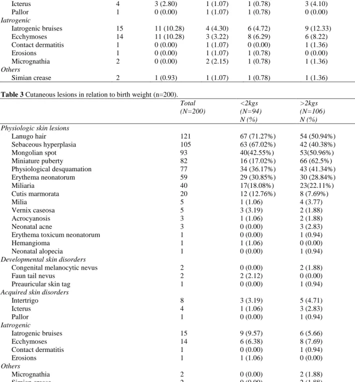
Table 3 Cutaneous lesions in relation to birth weight (n=200). Total (N=200)
<2kgs (N=94) N (%)
>2kgs (N=106) N (%) Physiologic skin lesions
Lanugo hair 121 67 (71.27%) 54 (50.94%)
Sebaceous hyperplasia 105 63 (67.02%) 42 (40.38%)
Mongolian spot 93 40(42.55%) 53(50.96%)
Miniature puberty 82 16 (17.02%) 66 (62.5%)
Physiological desquamation 77 34 (36.17%) 43 (41.34%)
Erythema neonatorum 59 29 (30.85%) 30 (28.84%)
Miliaria 40 17(18.08%) 23(22.11%)
Cutis marmorata 20 12 (12.76%) 8 (7.69%)
Milia 5 1 (1.06) 4 (3.77)
Vernix caseosa 5 3 (3.19) 2 (1.88)
Acrocyanosis 3 1 (1.06) 2 (1.88)
Neonatal acne 3 0 (0.00) 3 (2.83)
Erythema toxicum neonatorum 1 0 (0.00) 1 (0.94)
Hemangioma 1 1 (1.06) 0 (0.00)
Neonatal alopecia 1 0 (0.00) 1 (0.94)
Developmental skin disorders
Congenital melanocytic nevus 2 0 (0.00) 2 (1.88)
Faun tail nevus 2 2 (2.12) 0 (0.00)
Preauricular skin tag 1 0 (0.00) 1 (0.94)
Acquired skin disorders
Intertrigo 8 3 (3.19) 5 (4.71)
Icterus 4 1 (1.06) 3 (2.83)
Pallor 1 0 (0.00) 1 (0.94)
Iatrogenic
Iatrogenic bruises 15 9 (9.57) 6 (5.66)
Ecchymoses 14 6 (6.38) 8 (7.69)
Contact dermatitis 1 0 (0.00) 1 (0.94)
Erosions 1 1 (1.06) 0 (0.00)
Others
Micrognathia 2 0 (0.00) 2 (1.88)
Discussion
The prevalence of neonatal dermatoses varies
from 40.0% to 100% in different studies.
3,4In the
present study cutaneous lesions were present in
all the 200 neonates.
Lanugo hair (60.5%) was the most frequently
found cutaneous lesion in the present study
which was similar to other study.
5Predictably, it
was seen commonly in preterm and neonates
weighing less than 2 kg.
Sebaceous gland hyperplasia (52.5%) was the
next common dermatosis in the present study.
The prevalence was similar to other studies
which showed the prevalence varying from 3%
to 89.4%.
1,5,6,7It was commonly seen in female,
preterm and in neonates weighing less than 2kg.
Mongolian spots were found in 93 (46.5%)
patients. Majority of them (97.84%) had spots
on the sacrum. This finding is in agreement with
most of the Indian studies.
4,5,7,8,9They were
frequently seen in term neonates and early
neonatal period.
Genital or axillary hyperpigmentation, breast
enlargement, pigmented linea alba or vaginal
discharge in females were taken as the signs of
miniature puberty. It was observed in 41% of the
neonates which was similar to the study by Jain
et al
.
5A Turkish study
10noted a much lower
prevalence of scrotal hyperpigmentation and
labial hypertrophy. Boccardi
et al.
11suggested
that hyperpigmentation of the genital area was
more common in newborns of non-European
origin. They speculated that the variation in
genital hyperpigmentation might be related to
the differential activation of melanocytes.
Therefore, racial factors and skin type may be
important factors. The frequency of miniature
puberty in term neonates and in neonates
weighing more than 2 kg was high and
statistically significant. It was seen more
commonly in early neonatal period. Linea nigra
has been postulated to be a response to the
maternal and placental hormones that enter the
total circulation. Among these hormones,
estrogen and progesterone have been reported to
exert a melanocyte stimulating effect which also
causes darkening of linea alba in pregnant
women.
6,12Physiological desquamation was found in 38.5%
of the neonates comparable to a study by Jain
et
al.
5, where the frequency of occurrence was
48.33%. 25.97% of them had desquamation only
around the ankle, indicating that it could be the
initial site of desquamation as pointed out in
another study.
13It was found to be more frequent
in term, male neonates and in late neonatal
period. It did not show statistically significant
difference in terms of birth weight, though
commonly seen in neonates with birth weight
more than 2 kg. Vernix caseosa was seen in
2.5% of the neonates.
Many babies develop a striking generalized
hyperemia few hours after birth, known as
erythema neonatorum that fades spontaneously
within 24-48 h.
14It was seen in 59 (29.5%)
neonates, which was not reported in other
studies. In only 3 of them, it persisted beyond 7
days. It was seen more frequently in preterm and
female neonates and was statistically significant
(z=5.81,
p
<0.001) in early neonatal period.
Miliaria was found in 20% of the neonates
which was comparable to other Indian
studies.
5,7,8It was more common in term
neonates. Cutis marmorata was seen in 10% of
the neonates, Jain
et al.
5noted a figure of 20% in
their study, while study on Egyptian neonates
reported 3%.
1It was seen frequently in preterm,
female and early neonatal period.
were seen in 4% of the neonates which is lower
as compared to Egyptian (15.2%)
1and Pakistan
study.
15The lower prevalence may be due to
changing the napkins as frequently as required.
It was observed frequently in term neonates and
in early neonatal period.
Iatrogenic complications like bruise (7.5%) and
ecchymosis (7.0%) were noted mainly in the
ankles and dorsum of the hand, the site of
insertion of intravenous cannula. The results
were lower compared to Indian
5and Brazilian
study.
16It was observed frequently in male,
preterm, low birth weight neonates and in late
neonatal period.
We noted 2 cases of collodion baby, 2 cases of
body dysmorphic syndrome and a case of
Kasabach-Merritt syndrome as an incidental
finding.
Conclusion
Skin lesions are very common in the neonatal
period. In the present study, most of the skin
lesions were physiological and transitory.
Pediatricians and dermatologists should be
aware of these skin lesions in neonatal ICU and
should be able to differentiate them from serious
skin conditions in order to avoid unnecessary
investigations and treatment in neonates.
References
1. Shehab MM, Youssef DM, Khalil MM. Prevalence of cutaneous skin lesions in neonatal intensive care unit: A single center study. J Clin Neonatol. 2015;4:169-72. 2. Dhar S, Raychaudhury T, Banerjee R, Malakar
R. Neonatal skin disorders. In: Sacchidanand S, Oberai C, Inamadar AC, editors. IADVL Textbook of Dermatology. Mumbai: Bhalani Publishing House; 2015. p. 266-85.
3. El-Moneim AA, El-Dawela RE. Survey of skin disorders in newborns: clinical observation in
an Egyptian medical centre nursery. East Mediterr Health J. 2012;18:49-55.
4. Baruah CM, Bhat V, Bhargava R, Garg RB, Ku. Prevalence of dermatoses in the neonates in Pondicherry. Indian J Dermatol Venereal Leprol. 1991;57:25-8.
5. Jain N, Rathore BS, Agarwal AK, Bhardwaj A. Cutaneous lesions in neonates admitted in a tertiary setup neonatal intensive care unit. Indian J Paediatr Dermatol. 2013;14:62-6. 6. Haveri FTTS, Inamadar AC. A
Cross-Sectional prospective study of cutaneous lesions in newborn. ISRN Dermatology. 2014; (2014):1-8.
7. Sachdeva M, Kaur S, Nagpal M, Dewan SP. Cutaneous lesions in new born. Indian J Dermatol Venereol Leprol. 2002;68:334-7. 8. Dash K, Grover S, Rashakrishnan S, Vani M.
Clinco-epidemiologicol study of cutaneous manifestations in the neonate. Indian J Dermatol Venereal Leprol. 2000;66:26-8. 9. Nobbay B, Chakrabarty N. Cutaneous
manifestations in the newborn. Indian J Dermatol Venereal Leprol. 1992;58:69-72. 10. Gokdemir G, Erdogan HK, Koslu A, Baksu B.
Cutaneous lesions in Turkish neonates born in a teaching hospital. Indian J Dermatol Venereol Leprol. 2009;75:638.
11. Boccardi D, Menni S, Ferraroni M, Stival G, Bernardo L, La Vecchia C et al. Birthmarks and transient skin lesions in newborns and their relationship to maternal factors: a preliminary report from northern Italy. Dermatology. 2007;215:53-8.
12. Pruksachatkunakom C, Duarte AM, Schachner LA. Skin lesions in newborns. International Pediatrics. 1999;14:28-31.
13. Rivers JK, Frederiksen PC, Dibdin C. A prevalence survey of dermatoses in the Australian neonate. J Am Acad Dermatol. 1990;23:77-81.
14. Paige DG, Gennery AR, Cant AJ. The Neonate. In: Burns T, Breathnach S, Cox N, Griffiths C, editors. Rook’s Textbook of Dermatology. Oxford: Wiley-Blackwell; 2010. p.17.4.
15. Javad M. Clinical spectrum of neonatal skin disorders at Hamdard University Hospital Karachi, Pakistan. Our Dermatol Online. 2012;3:178-80.