USP6 oncogene promotes Wnt signaling by
deubiquitylating Frizzleds
Babita Madana, Matthew P. Walkerb, Robert Youngc, Laura Quickc, Kelly A. Orgelb, Meagan Ryanb, Priti Guptaa, Ian C. Henrichc, Marc Ferrerd,e, Shane Marinef, Brian S. Robertsg,h, William T. Arthurg,i, Jason D. Berndtj,k,l, Andre M. Oliveiram, Randall T. Moonj,k,l,1, David M. Virshupa,1, Margaret M. Chouc,1, and Michael B. Majorb,1
aProgram in Cancer and Stem Cell Biology, Duke-NUS Medical School, Singapore, Singapore 169857;bDepartment of Cell Biology and Physiology, Lineberger Comprehensive Cancer Center, University of North Carolina at Chapel Hill, Chapel Hill, NC 27599-7295;cDepartment of Pathology and Laboratory Medicine, Children’s Hospital of Philadelphia, University of Pennsylvania School of Medicine, Philadelphia, PA 19104;dDepartment of Automated Biotechnology, Merck Research Laboratories, North Wales, PA 19454;eNational Center for Advancing Translation Sciences/NIH, Rockville, MD 20850;fDepartment of Screening and Protein Sciences, Merck Research Laboratories, North Wales, PA 19454;gRosetta Inpharmatics, LLC, Merck & Co., Inc., Seattle, WA 98109; hHudsonAlpha Institute for Biotechnology, Huntsville, AL 35806;iSeattle Genetics, Bothell, WA 98021;jHoward Hughes Medical Institute, University of Washington School of Medicine, Seattle, WA 98195;kDepartment of Pharmacology, University of Washington School of Medicine, Seattle, WA 98195; lInstitute for Stem Cell and Regenerative Medicine at the University of Washington, Seattle, WA 98195; andmDepartment of Laboratory Medicine and Pathology, Mayo Clinic, Rochester, MN 55905
Contributed by Randall T. Moon, April 11, 2016 (sent for review January 5, 2016; reviewed by Kevin B. Jones, Peter Klein, and Masayuki Komada)
The Wnt signaling pathways play pivotal roles in carcinogenesis. Modulation of the cell-surface abundance of Wnt receptors is emerging as an important mechanism for regulating sensitivity to Wnt ligands. Endocytosis and degradation of the Wnt receptors Frizzled (Fzd) and lipoprotein-related protein 6 (LRP6) are regulated by the E3 ubiquitin ligases zinc and ring finger 3 (ZNRF3) and ring finger protein 43 (RNF43), which are disrupted in cancer. In a genome-wide small interfering RNA screen, we identified the deubiquitylase ubiquitin-specific protease 6 (USP6) as a potent activator of Wnt signaling. USP6 enhances Wnt signaling by deubiquitylating Fzds, thereby increasing their cell-sur-face abundance. Chromosomal translocations in nodular fasciitis re-sult in USP6 overexpression, leading to transcriptional activation of the Wnt/β-catenin pathway. Inhibition of Wnt signaling using Dickkopf-1 (DKK1) or a Porcupine (PORCN) inhibitor significantly de-creased the growth of USP6-driven xenograft tumors, indicating that Wnt signaling is a key target of USP6 during tumorigenesis. Our study defines an additional route to ectopic Wnt pathway ac-tivation in human disease, and identifies a potential approach to modulate Wnt signaling for therapeutic benefit.
Wnt signaling
|
ubiquitin-specific protease|
USP6|
Frizzled|
ubiquitinW
nts are a family of secreted proteins that regulate key developmental processes during embryogenesis as well as the homeostasis of adult tissues, including bone, skin, and in-testine (1). Wnt signaling is a tightly regulated process, and its deregulation is implicated in various diseases, including cancer and inflammatory and vascular diseases (2). Wnt ligands interact with multiple receptors and coreceptors, and the specific com-bination of ligand and receptor determines which downstream pathways are regulated. Wnts can signal both throughβ-catenin– dependent andβ-catenin–independent pathways (3). In the β-catenin–dependent pathway, Wnt binding to its receptors, Frizzleds (Fzds) and low-density lipoprotein-related protein 5/6 (LRP5/6), leads to the inhibition of ubiquitylation and proteaso-mal degradation of cytosolicβ-catenin (4).β-catenin is thus sta-bilized and enters the nucleus, where it binds to the TCF/LEF family of transcription factors to activate transcription of a mul-titude of target genes (5).In addition to controlling β-catenin protein stability, ubiq-uitylation regulates the subcellular localization and/or stability of multiple proteins within the Wnt signaling network, most notably membrane-proximal signaling events. For example, the cytoplasmic effector protein Dishevelled (DVL) is ubiquitylated by the KLHL12-CUL3, ITCH, and PDZRN3 ligases, resulting in its degradation or the endocytosis of Dvl/Fzd complex (6, 7). The turnover of Wnt re-ceptors Fzd and LRP6 is regulated by the transmembrane E3 ubiq-uitin ligases ring finger protein 43 (RNF43) and zinc and ring finger 3
(ZNRF3) that promote their ubiquitylation, leading to endocytosis and lysosomal degradation. RNF43 and ZNRF3 are also Wnt/ β-catenin target genes, and thereby function as negative feedback regulators of Wnt signaling (8, 9). The activity of RNF43 and ZNRF3 is regulated, in turn, by the extracellular signaling pro-teins, R-spondins (RSPO1–4), which form a ternary complex with RNF43/ZNRF3 and the coreceptors LGR4–6 (8, 10). Either loss-of-function RNF43 mutations (11, 12) or overexpression of R-spondin, arising from its translocation, leads to increased surface levels of Wnt receptors,β-catenin activation, and tumorigenesis (13, 14). Thus, diverse and tightly regulated ubiquitin ligases play a pivotal role in controlling Wnt activity.
Like ubiquitylation, regulated deubiquitylation plays a crucial role in governing information flow within the cell. Ubiquitin-specific proteases (USPs), one of the most abundant groups of deubiqui-tylation enzymes (DUBs), have fundamental roles in the ubiquitin system by specifically deconjugating ubiquitin from targeted pro-teins. Several DUBs have been shown to regulate components of the Wnt signaling pathway directly. CYLD functions as a negative regulator of Wnt signaling by deubiquitylating the cytoplasmic ef-fector DVL (15), and USP4 modulates Wnt signaling through
Significance
Ubiquitin-specific protease 6 (USP6) is a deubiquitylase that is overexpressed by chromosome translocation in two human neoplasms, aneurysmal bone cyst and nodular fasciitis. The rel-evant substrates of this ubiquitin-specific protease are not clear. Here, we identify the Wnt receptor Frizzled (Fzd) as a key target of the USP6 oncogene. Increased expression of USP6 increases the membrane abundance of Fzd, and hence increases cellular sensitivity to Wnts. USP6 opposes the activity of the ubiquitin ligase and tumor suppressor ring finger protein 43 (RNF43). This study identifies a new mechanism for pathological Wnt pathway activation in human disease and suggests a new approach to regulate Wnt activity therapeutically.
Author contributions: B.M., M.P.W., M.F., S.M., B.S.R., W.T.A., R.T.M., D.M.V., M.M.C., and M.B.M. designed research; B.M., M.P.W., R.Y., L.Q., K.A.O., M.R., P.G., M.F., S.M., B.S.R., W.T.A., J.D.B., M.M.C., and M.B.M. performed research; B.M., M.P.W., I.C.H., B.S.R., W.T.A., J.D.B., A.M.O., R.T.M., D.M.V., M.M.C., and M.B.M. analyzed data; and B.M., M.P.W., R.T.M., D.M.V., M.M.C., and M.B.M. wrote the paper.
Reviewers: K.B.J., University of Utah; P.K., University of Pennsylvania; and M.K., Tokyo Institute of Technology.
The authors declare no conflict of interest.
1To whom correspondence may be addressed. Email: [email protected],
[email protected], [email protected], or [email protected].
This article contains supporting information online atwww.pnas.org/lookup/suppl/doi:10. 1073/pnas.1605691113/-/DCSupplemental.
CELL
BIOLOGY
PNAS
like 2 (TCF7L2) (16). USP34 regulates AXIN1/2 stability and op-poses its tankyrase (TNKS)-dependent ubiquitylation, hence func-tioning as a positive regulator of Wnt signaling (17). TRABID also positively regulates Wnt signaling by deubiquitylating adenomatous polyposis coli (APC) and regulating the stability of theβ-catenin/ TCF transcription complex (18). USP47 and USP4 can directly regulate β-catenin deubiquitylation (19, 20). USP8 deubiquitylates endosome-tethered Fzd, resulting in the recycling of Fzd to the plasma membrane (21). Although genetic alterations in the ubiq-uitylation machinery have been identified in cancers (i.e., RNF43/ ZNRF3, RSPO3), to date, no mutations in DUBs that activate Wnt receptors have been documented in human tumors.
Using genome-wide small interfering RNA (siRNA) screens coupled with focused gain-of-function validation, we identified USP6 as a potent activator of the Wnt pathway. USP6, which is overexpressed upon translocation in aneurysmal bone cyst (ABC) and nodular fasciitis, potentiates Wnt signaling by increasing the abundance of Fzds and LRP6 at the plasma membrane. We show that USP6 functions by deubiquitylating Fzds and, as such, an-tagonizes the activity of the E3 ubiquitin ligases RNF43 and ZNRF3. Wnt signaling is a functionally important target of USP6, because pharmacological inactivation of Wnt signaling using a
of xenografts driven by high USP6 expression. Finally, activation of Wnt signaling is observed in human tumors harboringUSP6 translocation, supporting its clinical relevance in regulating Wnt activity. Our study uncovers a mechanism that controls Wnt receptor abundance on the cell membrane, and thus provides new targets for modulating Wnt signaling.
Results
Functional Genomic Screen ofβ-Catenin–Dependent WNT Signaling.
To identify novel regulators of Wnt signaling, an extensively validated and near-saturation genome-wide siRNA screen in HEK293T human embryonic kidney cells and HT1080 human sarcoma cells was performed. A similar strategy was previously used in DLD1 cells, a colorectal cancer cell line that expresses a mutant form of APC that disrupts theβ-catenin destruction com-plex (22). In the current screen, exogenous Wnt ligand was used to activate receptor-mediated signaling. HEK293T and HT1080 cell lines with an integrated Wnt/β-catenin–activated firefly luciferase reporter and cytomegalovirus-driven Renilla luciferase reporter were screened in the presence of WNT3A-conditioned medium in 1,536-well plates with three nonoverlapping gene-specific siRNAs in each pool. Of 28,124 siRNA pools targeting 20,042 messenger
A
B
1 10 100 1000 10000
0.00001 0.0001 0.001 0.01 0.1 1 10 100 1000
1 10 100 1000 10000
0.001 0.01 0.1 1 10 100 1000
D
USP6 siRNA
CNT CTNNB1 A B C
HEK293T
C
E
F
HEK293T HT1080
Two fold change (pooled siRNA) Pooled siRNA
DUB
USP6
APC
0 20 40 60 80 500 1000 1500
0 20 40 60 80 200 300 400 500
USP21
USP6
USP4 USP1L
USP37 USP49 USP26
HEK293T Normalized Firefly/Renilla (log2)
Relative Luciferase
Activity
Relative Luciferase
Activity
HEK293T, % of control
HT1080, % of control
HT1080 Normalized Firefly/
Renilla
(log2)
Normalized (Percent of Control) BAR reporter activity Normalized (Percent of Control)
BAR reporter activity
-log(p-value)
-log(p-value)
WNT3A CM Control CM WNT3A CM
Control CM
Control
CTNNB1USP1L USP4 USP6USP2
1
USP26USP 37
USP49
0 50 100 200 300 400 500 DUB
non-DUB
DUB non-DUB
HEK293T
**
***
***
****
*** *** **
** **
**
0.0 0.5 1.0 1.5
Fig. 1. Genome-wide siRNA screen of WNT/β-catenin signaling. (AandB) Volcano plot of the primary siRNA screen. HEK293T (A) and HT1080 (B) cells are shown; red dots represent deubiquitylases. Full cross-referenced primary screen data are provided inDataset S1. The HT1080 screen was published previously (43). (C) Scatter plot of the secondary siRNA screen. Data points represent genes targeted by a pool and at least two single siRNAs that increased or decreased the reporter by twofold or greater. Full secondary screen data are provided inDataset S1. (D) Scatter plot of the secondary siRNA screen. Selected deubi-quitylases and variance across replicates are illustrated. Normalized luciferase activity (firefly/Renilla) of DUBs whose loss of function regulated the Wnt/
RNAs, 1,877 increased or decreased Wnt/β-catenin reporter ex-pression threefold or greater in both cell lines, with aPvalue less than 0.01 (Fig. 1AandBandDataset S1). A secondary validation screen of 1,172 hits from the primary screen was performed by independently evaluating three to nine nonoverlapping single siRNAs. Hit-calling criteria for the secondary screen included an increase or decrease in the Wnt/β-catenin reporter activity of at least twofold with a Student’sttestPvalue<0.01. Additionally, at least two independent siRNAs and the repeat test of the pool had to meet a statistically significant twofold change. We identified 186 gene products that have an impact on Wnt signaling in both
HEK293T and HT1080 cells (Fig. 1CandDataset S1). Compiled and cross-listed genome-wide primary screens from DLD1, HEK293T, and HT1080 and secondary screen data from HEK293T and HT1080 are provided inDataset S1. The DLD1 primary and secondary screen data are reprinted with permission from AAAS (from ref. 22).
Of the 86 DUBs in the human genome, the genome-wide pooled siRNA screens identified 28 DUBs as putative regulators of Wnt signaling in both HT1080 and HEK293T cells. Secondary screens revealed seven DUBs that have an impact on the Wnt pathway activity (Fig. 1D). Silencing USP21 and USP6 significantly down-regulated Wnt/β-catenin reporter activity in both HEK293T and
USP6 USP6(short) USP6(CS) USP6 USP6(CS) USP6(short)
Control
ANKRD6 CTNNB1 Control
Control TGF-β USP6
USP6( CS)
USP6 (short)
USP6 BSA
USP6 (CS)
USP6 KEAP1 NFE2L2 Control
Control
USP6(CS)
USP6 (short) 4
3
2
1
0
8
6
4
2
0
6
5
3
1 4
2
0 USP6
(short)
A
B
C
D
F
H
I
G
E
HEK293T HEK293T
CTNNB1 USP6 siRNA LRP6 siRNA Control siRNA
41 153
A B 911 42 76 41
153 911 42 76
CTNNB1 USP6 siRNA LRP6 siRNA Control
siRNA AXIN1/2
A B
Nkd1
Expression
Axin2
Expression
S
B
E
R
e
p
o
rt
e
r
Activity
HQR41 Reporter
Activity
0 5 10 15 20 25 30
WNT3A CM Control CM
WNT3A CM Control CM
HEK293T HeLa
HT1080
Relative Luciferase
Activity
Relative Luciferase
Activity
Relative Luciferase
Activity
WNT3A CM Control CM
WNT3A CM Control CM WNT3A CM
Control CM
10 20 80 90 110
100
0
EtOH tBHQ
0 1 2 3 4
ANKRD6 CTNNB1 USP6
USP6(CS)
USP6(short)
Cys 541 Ser
USP Domain
220
76 105 105
76
-AXIN1/2 ****
****
** ****
***
* *
****
**** ****
* ****
**
0 10 20 30 40
****
** * *
* * *
*** *** *** *** *** *** ****
****
CTNNB1
p65 USP6/ USP6(CS) +
- - + - + - + - + - +
EV USP6
USP6 (CS) USP6 (short)
WNT3A CM
Fig. 2. USP6 activates WNT signaling. (A) Model depicting USP6 alleles. Cysteine (Cys) and histidine (His) subdomains of the USP domain are shown. Ser, serine; TBC, TBC domain. USP6(CS) harbors a point mutation in the cysteine subdomain. (B) Dose-dependent activation of Wnt signaling by USP6. HEK293T cells with an integrated Wnt/β-catenin reporter were transfected with increasing amounts of plasmids encoding USP6 or its variants. Cells were incubated with WNT3A- or control-conditioned media for 16 h before analysis. Data represent mean±SD (n=3). (C) USP6 activates Wnt signaling in HeLa cells. HeLa cells were transfected with plasmids expressing USP6 or its variants and Wnt/β-catenin reporter. The cells were incubated with WNT3A- or control-conditioned medium for 24 h before analysis. Data represent mean±SD (n=7). (D) USP6 activates Wnt signaling in HT1080 cells. HT1080 cells were transfected with Wnt/
β-catenin–firefly luciferase reporter,Renillaluciferase reporter, and the indicated plasmids. Cells were incubated with WNT3A- or control-conditioned media for 12 h before analysis. Data represent mean±SD (n=3). (EandF) USP6 does not activate TGF-βor NRF2 signaling. HEK293T cells expressing SBE (TGF-β) reporter or antioxidant response element (hQR41) reporter were transfected with USP6 or its mutants. Tert-butylhydroquinone (tBHQ; 50μM) and recombinant human TGF-β1 (10 pM) are agonists for the SBE and hQR41 reporters, respectively.Renilla-normalized relative luciferase activity was plotted. Error bars represent mean±SD (n=3). (G) USP6 expression stabilizes cytosolicβ-catenin. HeLa cells were transfected with plasmids expressing USP6-HA or its mutant isoforms and then incubated overnight with WNT3A- or control-conditioned medium. Cell lysates were analyzed for levels of endogenousβ-catenin by immunoblotting. NF-κB p65 subunit was used as a loading control. (HandI) Loss of function of USP6 down-regulates expression of WNT-responsive genes. HEK293T cells were transfected with three independent siRNAs targeting USP6 transcript. Forty-eight hours after siRNA transfection, the cells were treated with control- or WNT3A-conditioned medium. Total RNA was isolated, and expression of Wnt target genesAXIN2(H) andNKD1(I) was analyzed by quantitative RT-PCR. Data were normalized to transcript abundance forGAPDH, and represent mean±SD for biological triplicates.
CELL
BIOLOGY
PNAS
increased pathway activity (Fig. 1D). These findings partially overlap with previously reported DUBs regulating Wnt signaling, although several, including CYLD and USP8, were not identified in our screen.
To confirm and extend these findings further, we performed the converse experiment, testing whether overexpression of selected DUBs affects Wnt/β-catenin reporter activity. HEK293T cells were transfected with the indicated expression constructs, along with the Wnt/β-catenin firefly luciferase reporter andRenillaluciferase re-porter. USP6 overexpression strongly potentiated WNT3A-induced reporter activity, comparable toβ-catenin overexpression (Fig. 1E). In agreement with the siRNA screen data, USP21 and USP26 also potentiated Wnt signaling, albeit significantly less than USP6. In
susceptible to artifact and false-positive discovery, HEK293T cells were transfected with three nonoverlapping USP6-specific siRNAs that were distinct from the siRNAs used in the primary and secondary siRNA screens; all siRNAs tested suppressed Wnt signaling (Fig. 1F).
USP6 Activates Wnt/β-Catenin Signaling in Diverse Cell Types in a
USP-Dependent Manner.The full-length USP6 isoform contains the
USP domain at its C terminus and a TBC (Tre2/Bub2/Cdc16) domain at its N terminus (Fig. 2A). A naturally occurring C-terminally truncated splice variant of USP6, USP6(short), lacks an essential portion of the USP domain and is therefore catalytically inactive (23). To determine whether USP function is
required for Wnt activation by USP6, we examined Wnt/β-catenin reporter activation by USP6(short), as well as USP6 with a point mutation in a key catalytic residue, Cys541→Ser, USP6(CS). As shown in Fig. 2B, expression of USP6 in HEK293T cells enhanced Wnt signaling in a dose-dependent manner. In contrast, USP6(short) and USP6(CS) did not have a strong impact on the the Wnt/β-catenin reporter activity. Similar results were observed in HeLa and HT1080 cells (Fig. 2CandD). Furthermore, using various dilutions of WNT3A-conditioned medium, USP6 was found to activate Wnt responsiveness at all doses of treatment (Fig. S1A). In sum, these results demonstrate that the ubiquitin protease ac-tivity of USP6 is essential for activation of β-catenin–dependent Wnt signaling, and that USP6 overexpression activates Wnt sig-naling in multiple cell types of diverse tissue origin.
To test if USP6 is a general activator of transcription, we amined whether USP6 overexpression had an impact on the ex-pression of a TGF-β–responsive reporter [SMAD-binding element (SBE)] or an antioxidant responsive reporter (hQR41). USP6 over-expression weakly activated the SBE reporter (threefold), compared with 90-fold activation by TGF-β1 ligand (Fig. 2E). Moreover, USP6 did not increase transcription from the NFE2L2 transcription factor-driven hQR41 reporter (Fig. 2F), indicating that USP6 shows spec-ificity for the Wnt pathway and is not a general transcriptional activator.
We next evaluated whether USP6 activates endogenous Wnt signal transduction. Binding of Wnt ligands to Fzd and LRP5/6
receptor complexes transiently inhibits theβ-catenin destruction complex, leading to increased steady-state levels ofβ-catenin in the cytoplasm and nucleus. As shown in Fig. 2G, basal as well as WNT3A-induced total β-catenin protein levels were elevated following USP6 overexpression. This increase in endogenous β-catenin by USP6 was dependent on its catalytic activity because no increase was observed with the catalytically inactive USP6 variants (Fig. 2G). The effect of USP6 is not cell type-specific, because USP6 increased the expression of the endogenous Wnt-responsive genesNKD1andAXIN2in three cell lines of diverse origins: HEK293, HeLa, and HT1080 (Fig. S1B–G). Conversely, silencing of USP6 using three nonoverlapping siRNAs down-regulated WNT-induced expression ofAXIN2andNKD1(Fig. 2 H andI). Although these experiments required ectopic expres-sion of USP6, taken together with the USP6 siRNA data in multiple cell lines, they support the identification of USP6 as a novel potentiator of endogenous Wnt/β-catenin signaling.
USP6 Functions Upstream of the Destruction Complex and Requires
Wnt Ligand–Receptor Interactions.To elucidate the mechanism of
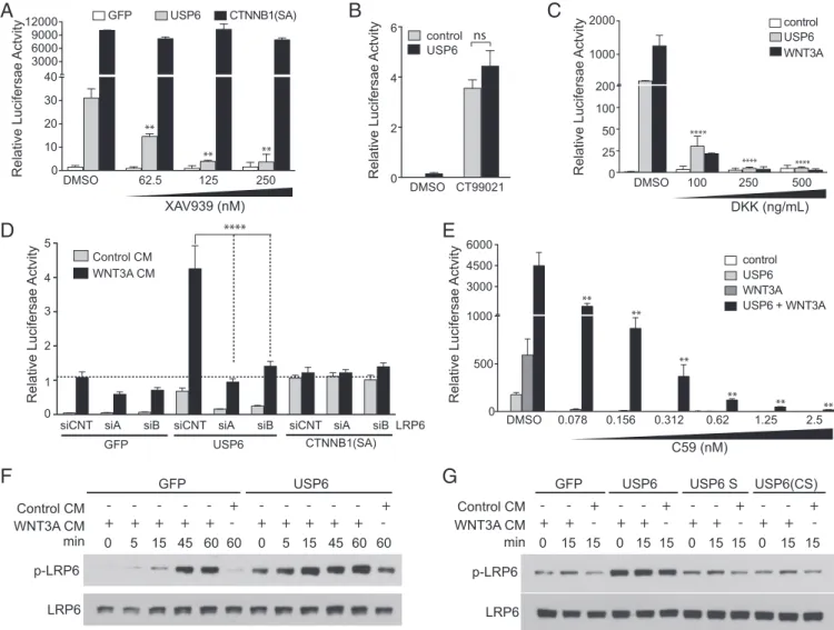
Wnt/β-catenin activation by USP6 further, we conducted a series of epistasis experiments. We first sought to determine whether USP6 functions upstream of theβ-catenin destruction complex. The β-catenin destruction complex catalyzes β-catenin phos-phorylation by AXIN-bound glycogen synthase kinase 3 (GSK3) and casein kinase 1α(CSNK1A), leading to its ubiquitylation and degradation. AXIN protein levels are negatively regulated by the poly(ADP)-ribosylation activity of the TNKS1/2 enzyme. Inhibiting TNKS1/2 with a small-molecule inhibitor, XAV939, results in increased AXIN protein levels and, consequently, decreased β-catenin activity. USP6-induced Wnt signaling was dramatically inhibited by XAV939 (Fig. 3A) indicating that USP6 activates Wnt signaling upstream of theβ-catenin destruction complex. In con-trast, XAV939 had no effect on Wnt signaling driven by a stabi-lized, constitutively activeβ-catenin mutant (Fig. 3A), confirming the epistatic specificity of the drug. Inhibition of GSK3 activity increasesβ-catenin abundance by preventingβ-catenin phosphor-ylation and its subsequent degradation. With near-complete re-pression of GSK3 activity by the GSK3 inhibitor CT99021, USP6 failed to enhance Wnt signaling further (Fig. 3B). Together, these data support a model wherein USP6 potentiates Wnt signaling upstream of theβ-catenin destruction complex.
We next tested whether the interaction of Wnts with their receptors is required for activation of the pathway by USP6. Dickkopf-1 (DKK1) is a Wnt antagonist that prevents the inter-action of Wnts with their coreceptors, LRP5/6. USP6-induced Wnt signaling was blocked by recombinant DKK1 treatment (Fig. 3C), suggesting that Wnt-LRP5/6 interaction is required for USP6 function. Further supporting a requirement for a Wnt ligand engaged receptor complex, knockdown of LRP6 with two independent siRNAs inhibited USP6-induced Wnt/β-catenin reporter activity in HEK293 cells (Fig. 3D). Importantly, sub-nanomolar amounts of the PORCN inhibitor C59, which effec-tively blocks Wnt secretion (24), inhibited basal USP6-stimulated β-catenin signaling, as well as the synergistic activation obtained upon cotransfection of WNT3A and USP6 expression plasmids (Fig. 3E).
Binding of Wnts to their cell surface receptors leads to phos-phorylation of LRP6, which triggers a cascade of events leading to stabilization of β-catenin. We observed that compared with the control, LRP6 was more rapidly phosphorylated in USP6-expressing cells upon treatment with WNT3A CM (Fig. 3F). Consistent with the observed effect of USP6 in enhancing Wnt signaling, basal as well as Wnt-induced LRP6 phosphorylation was elevated in USP6-expressing cells. This activity was dependent on the protease activity of USP6 because the USP6(short) and USP6(CS) variants were not active (Fig. 3G). These data suggest that USP6 regulates Wnt
A
B
Fluorescence Intensity (APC)
Isotype control
USP6 GFP alone
Isotype control
USP6 GFP alone
USP6 short
USP6(CS) USP6 short 10 3 10 4 10 5
400
200
0
0 0
Fluorescence Intensity (PE)
10 1 10 2 10 3
0 600
400
100 600
Counts
Frizzled
LRP6
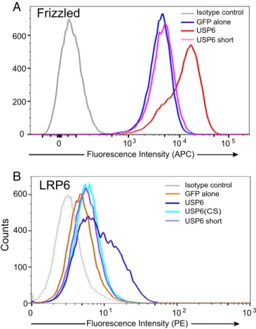
Fig. 4. USP6 increases abundance of Wnt receptors on the cell surface. (A) USP6 overexpression regulates Fzd abundance. Flow cytometric analysis of endogenous Fzd levels in HEK293 cells expressing pEGFP (GFP alone) or USP6-GFP and its variants. Fzd levels in GFP-positive cells are shown. Data are representative of three independent experiments. (B) USP6 overexpression regulates LRP6 abundance. A flow cytometric analysis of LRP6 levels in HEK293T cells transfected with USP6 and USP6 variants is shown. Data rep-resent three biological replicate experiments.
CELL
BIOLOGY
PNAS
signaling at the membrane, and that USP6 functions only in the presence of Wnt ligand secretion and Wnt receptor engagement.
USP6 Regulates Cell Surface Abundance of LRP6 and Fzds.Because
ubiquitylation is a key mechanism regulating Fzds and LRP5/6 membrane abundance, we tested if USP6, as a deubiquitylase, increased the abundance of the Wnt receptors on the cell sur-face. A USP6-GFP fusion protein was expressed in HEK293 cells, and plasma membrane levels of endogenous Fzds were measured by flow cytometry using the anti-Fzd monoclonal antibody OMP18R5 (25). Wild-type, but not inactive, USP6 markedly in-creased the cell surface abundance of Fzds, because 69.5% cells expressing USP6-GFP had high Fzd levels compared with 4.5% of GFP control cells (Fig. 4A). Wild-type USP6 similarly increased the cell surface abundance of endogenous LRP6, with 37.8% of cells showing high LRP6 levels compared with 3% of the controls (Fig. 4B). Expression of the catalytically inactive USP6(CS) and USP6(short) variants had no significant effect on surface LRP6 abundance.
USP6 Does Not Potentiate Wnt Signaling in RNF43 Mutant Cells.E3
ubiquitin ligases RNF43 and ZNRF3 negatively regulate Wnt signaling by promoting ubiquitylation and degradation of LRP6 and Fzds (8). AsPC-1 cells derived from pancreatic ductal ade-nocarcinoma (PDAC) have an inactivating mutation inRNF43, which presumably abrogates Fzd ubiquitylation, leading to Wnt-driven proliferation in vivo (14). We compared AsPC-1 with Panc 08.13 cells, a PDAC cell line with wild-type RNF43. Consistent with the loss of the RNF43 ubiquitylation activity, expressing the
ubiquitin protease USP6 in the ubiquitin ligase-deficient AsPC-1 cells caused only a twofold increase in Wnt signaling (Fig. 5A); this residual effect may be due to reversal of some ZNRF3 activity. Unlike the synergy observed inRNF43wild-type cells, in AsPC-1 cells, there was no significant enhancement of signaling in the presence of WNT3A (Fig. 5A). Consistent with the twofold acti-vation of Wnt signaling, USP6 expression drove only a modest increase in Fzd membrane abundance (Fig. 5B). In contrast, in Panc 08.13 cells, both Wnt signaling (Fig. 5C) and Fzd cell surface abundance (Fig. 5D) were markedly enhanced by USP6 to a degree comparable to the degree measured in HEK293, HT1080, and HeLa cells.
These results suggest that USP6 enhances Wnt signaling by counteracting the effects of the ubiquitin ligases RNF43 and ZNRF3. We therefore tested if titrated restoration of RNF43 activity in AsPC-1 (RNF43mutant) cells rescued the synergistic activation of Wnt signaling by USP6. As indicated by the ratio of Wnt-stimulatedβ-catenin reporter activity in the absence or presence of USP6, restoring the expression of RNF43 modestly decreased overall signaling but markedly increased the synergistic activation by USP6 (Fig. 5E). These data are consistent with a model in which deubiquitylation of Fzds by USP6 increases their cell surface abundance by reversing their RNF43/ZNRF3-mediated ubiquitylation.
Because RNF43 increases Fzd endocytosis, we tested if USP6 decreased the clearance of Fzd from the cell surface. HEK293 cells have abundant cell surface Fzd, and when protein synthesis was blocked with cycloheximide, the Fzd abundance decreased, presumably due to endocytosis (Fig. 5F, purple lines). However,
D
Fluorescence Intensity (APC) 10 -2 10 2 10 3 10 4 10 5 200
100
0 0 300 400
Counts
Counts
AsPC-1 AsPC-1
Relative Luciferase
Activity
Relative Luciferase
Activity
0 1 2 3
Control WNT3A
Panc08.13
Panc 08.13
Control WNT1
Control USP6 0 Control USP6
10 20 30 Isotype control
USP6 GFP alone
USP6 short
Isotype control
USP6 GFP alone
USP6 short
**
600
400
0 200
Fluorescence Intensity (APC)10 3
10 2 10 4 10 5
0
F
AsPC-1
RNF43 plasmid (ng)
Relative Luciferase
Activity
0 0.01 0.1 1 10
0 300 600 900 1200 1500
0 2 4 6 8 WNT3A
USP6 +WNT3A Ra
tio
U
SP6
+
W
N
T3A:
W
N
T3A
Ratio USP6+WNT3A : WNT3A
E
Counts
Fluorescence Intensity (APC) 10 3
10 2 10 4 10 5
0 2K
1K
HEK293
Isotype control
GFP + CHX GFP alone
USP6 USP6 + CHX
when USP6 was overexpressed, cell surface Fzd markedly in-creased, and its rate of clearance from the membrane decreased (Fig. 5F, brown lines). These data are consistent with USP6 working to oppose Fzd endocytosis.
USP6 Regulates Fzd Deubiquitylation.We next tested if USP6
di-rectly deubiquitylates Fzds. HEK293T cells were cotransfected with Fzd-V5, FLAG-ubiquitin (Ub), and HA-USP6 expression plasmids, and treated with control- or WNT3A-conditioned media. Fzd-V5 was immunoprecipitated using anti-V5 antibody, and levels of ubiquitylation were monitored by FLAG immuno-blotting. In immunoprecipitates from control cells, a smear of high-molecular-weight species corresponding to ubiquitylated Fzd was detected (Fig. 6A). Overexpression of USP6, but not catalyt-ically inactive USP6(CS) or USP6(short), strongly reduced Fzd ubiquitylation.
To determine if USP6 can directly deubiquitylate Fzd, in vitro deubiquitylation assays were performed. Fzd5-V5 was immuno-precipitated from HeLa cells cotransfected with Fzd-V5 and FLAG-Ub expression plasmids. The immunoprecipitated Fzd-V5 was incubated with recombinant GST-tagged USP6 (wild type or a catalytically inactive point mutant). As shown in Fig. 6B, ubiq-uitylation of Fzd-V5 was dramatically diminished by USP6, but not its catalytically inactive variant USP6(CS). USP2, a highly active nonspecific deubiquitylase, was used as a positive control. The ef-fect of USP6 on ubiquitylation of endogenous Fzd5 could not be monitored due to lack of antibodies that efficiently immunopre-cipitate Fzd5. Nevertheless, these results suggest that Fzd is a direct substrate of USP6, consistent with the model in which USP6 ac-tivates Wnt/β-catenin signaling by deubiquitylating Fzd receptors.
Human Tumors Harboring USP6 Translocation Have a Wnt/β-Catenin
Transcriptional Signature. USP6 is implicated in human
non-malignant neoplastic disorders, with chromosomal translocation-driven overexpression of USP6 occurring in two independent tumor types, ABC and nodular fasciitis, in ∼70% and 90% of cases, respectively (26, 27). To determine if a Wnt/β-catenin gene signature is activated in these tumors, gene expression profiling
by microarray analysis was performed on nodular fasciitis tumors with confirmedUSP6translocation/overexpression. The cell of or-igin in nodular fasciitis has yet to be defined, but is of mesenchymal origin. We compared the nodular fasciitis transcriptome with an averaged expression profile generated from 27 predominantly mesenchymal tumors lackingUSP6translocation. This strategy was used to exclude genes that are general mesenchymal markers or common indicators of the transformed state, and instead identify those genes selectively induced by USP6 in nodular fasciitis. Gene set enrichment analysis demonstrated strong positive correlation with multiple independent Wnt/β-catenin signatures (28–32), fur-ther supporting the model that overexpression of USP6 drives Wnt/ β-catenin signaling in human tumors (Fig. 7 andDataset S2).
Inhibition of Wnt Signaling Prevents Growth of Tumors Overexpressing
USP6.Because USP6 regulates multiple cellular signaling pathways
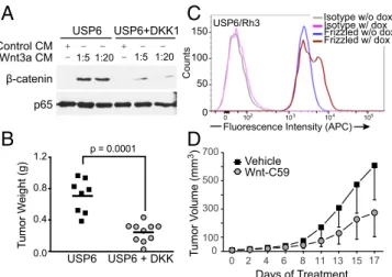
(23, 33, 34), we assessed the functional importance of Wnt/ β-catenin pathway activation in USP6-driven tumor formation. Because no immortalized ABC or nodular fasciitis cell lines exist, it was necessary to express USP6 alleles ectopically in heterolo-gous cells. Previous studies have shown that mesenchymal NIH 3T3 fibroblasts stably overexpressing USP6 (USP6/NIH 3T3) serve as a useful cellular model for ABC and nodular fasciitis. When xenografted into immunodeficient mice, USP6/NIH 3T3 cells, but not control NIH 3T3 cells, formed highly vascularized, hemorrhagic tumors, recapitulating features of the human neo-plasms, particularly ABC (34). To test whether USP6-mediated tumor formation was dependent on Wnt signaling, we introduced into USP6/NIH 3T3 cells DKK1, a secreted protein that blocks Fzd receptor activation. As shown in Fig. 8A, USP6 induction of β-catenin was significantly attenuated by DKK1, compared with cells expressing USP6 alone. The s.c. xenografting of these cells into NOG-SCID mice revealed that USP6-dependent tumor growth was significantly inhibited by DKK1 expression (Fig. 8B).
The functional role of Wnt activation by USP6 was assessed further in a second tumorigenesis model. A high percentage of primary alveolar rhabdomyosarcoma (ARMS) patient samples express high levels of USP6. Interestingly, however, in all im-mortalized ARMS cell lines examined, USP6 expression was down-regulated. Thus, to determine the effects of USP6 in the context of this cancer, it was expressed under a doxycycline-inducible promoter in the ARMS cell line, Rh3 (USP6/Rh3) at levels comparable to the levels in primary ARMS samples. In-duction ofUSP6expression significantly increased the abundance of endogenous Fzds on the cell surface (Fig. 8C), indicating ac-tivation of Wnt signaling. The s.c. injection of USP6/Rh3 cells into NOD-SCID mice led to the rapid growth of tumors. Treatment of tumor-bearing mice with the PORCN inhibitor C59 inhibited tu-mor growth by 45% when dosed at 20 mg/kg once daily by oral gavage (Fig. 8D). Taken together, these data indicate that USP6 overexpression works, in large part, through activation of the Wnt/ β-catenin pathway to drive tumorigenesis. Pharmacological agents targeting the Wnt/β-catenin pathway may therefore have thera-peutic efficacy in USP6-overexpressing cancers.
Discussion
Aberrant activation of Wnt signaling promotes the initiation and progression of diverse tumor types. Accumulating evidence re-veals that dynamic ubiquitylation/deubiquitylation of Fzd and LRP6 is a pivotal regulatory mechanism regulating Wnt/β -cate-nin signaling in a subset of human cancers. Our study further highlights the importance of maintaining appropriate surface levels of Wnt receptors for cellular homeostasis. We describe a major role for the poorly understood human oncogene,USP6, as a novel potentiator of Wnt signaling in multiple cell types. USP6 functions by deubiquitylating Fzds and perhaps LRP6 receptors, thereby increasing the abundance of both Fzds and LRP6 at the cell surface and rendering cells exquisitely sensitive to Wnt Flag-Ub
Fzd5-V5
USP6-HA short
220 150
105
76
52
_ _ _ _
_
220
150
105 _
_
_
220 150
105 76 _ _
_
_
WCL IP V5
A
B
+ + + +
(CS)
+
+ + + + + +
+ + + + +
+ +
-Flag-Ub Fzd5-V5
GST -USP6
+ +
- +
GST
+
USP6-CS USP2
+ + +
+ ++ ++
-USP6 Fzd-V5
Ub
Wnt3A + + +
-+
-+ - -
-Ub
Fig. 6. USP6 promotes Fzd deubiquitylation. (A) USP6 promotes Fzd5 deu-biquitylation in vivo. HEK293T cells were transfected with plasmids encoding Fzd5-V5, Flag-Ub, and the indicated USP6 constructs, and then treated with control or WNT3A CM. (Top) Fzd5 was immunoprecipitated using V5, and its ubiquitylation was detected with anti-Flag antibody. Whole-cell extracts were blotted with anti-V5 (Middle) or anti-USP6 (Bottom) antibodies. (B) In vitro deubiquitylation of Fzds by USP6. HeLa cells were transfected with plasmids encoding Fzd5-V5 and Flag-Ub. Fzd5 was purified using anti-V5 and then incubated with GST-tagged recombinant USP6 or USP6(CS). Fzd5 ubiquitylation was detected by immunoblotting with anti-FLAG. GST and USP2 were used as negative and positive controls, respectively. Data are representative of two independent experiments. IP, immunoprecipitation; WCL, whole cell lysate.
CELL
BIOLOGY
PNAS
ligands. USP6 activity appears to reverse the ubiquitylation of Fzds and LRP6 by RNF43/ZNRF3. Our data show that USP6 stabilizes the membrane pool of Fzd. Ongoing and future ex-periments will test the hypothesis that increase Fzd at the membrane occurs through altered ubiquitin-dependent endocy-tosis of Fzd. In human tumors harboring USP6translocations, the consequence is activation of Wnt/β-catenin signaling. Con-firming the functional importance of USP6-mediated Wnt pathway activation, inhibition of Wnt signaling either by expres-sion of DKK1 or treatment with the PORCN inhibitor C59 sup-presses USP6-mediated tumor formation in murine xenograft models. Prior studies have shown that loss-of-function mutations
inRNF43/ZNRF3and translocation-mediated overexpression of
its inhibitor, R-spondin, also drive pathological activation of Wnt signaling in human tumors. Together, these findings reinforce the critical role of this regulatory node of the Wnt pathway in human tumorigenesis.
We have previously reported that USP6 promotes deubiquity-lation of cargo proteins to prevent their lysosomal targeting and increase their surface levels through enhanced recycling (35). USP8/UBPY is also shown to deubiquitylate Fzd, rescuing it from lysosomal degradation and promoting its recycling to the plasma membrane (21).USP8was not identified in our siRNA screen for Wnt pathway activators, perhaps due to subphenotypic silencing by the siRNAs. Thus, USP6 and USP8 may possess both re-dundant and unique functions in regulating trafficking and Wnt signaling, with cell type and subcellular localization of both the USPs and cargoes contributing to their specificity. In this context, it is important to note that theUSP6gene only exists in hominoids, and thus cannot readily be studied in model organisms (36).
The current analysis demonstrates the importance of USP6’s deubiquitylating activity in the regulation of Wnt/β-catenin sig-naling. However, USP6 also contains a TBC domain, which binds to the ADP ribosylation 6 (ARF6) GTPase and promotes its activation in vivo independent of its USP domain (37). Recent studies have shown that ARF6 can activate Wnt signaling by stimulating MAPK1/ERK activation and phosphatidylinositide production to promote LRP6 phosphorylation (38, 39). In ad-dition, ARF6 is reported to regulate endocytosis of Fzd
recep-tors in the context of noncanonical Wnt signaling (40). Hence, both the deubiquitylase activity and the TBC/ARF6 interaction may contribute to the USP6-mediated activation of Wnt. This role of TBC/Arf6 interaction in regulating Frizzled endocytosis is consistent with our observation that in certain cell types, USP6(CS), an inactive point mutant of USP6 with an intact TBC domain, retains residual activity onβ-catenin reporters (Fig. 2). Future studies will examine the role of USP6’s ability to regulate Arf6 in Wnt receptor trafficking andβ-catenin activation.
USP6translocations are the key etiological agent in two human tumors, ABC and nodular fasciitis (26, 27). In both, theUSP6 rearrangement leads to overexpression of wild-type USP6 protein in a mesenchymal cell of origin. Although these tumors share some histological features, they exhibit distinct clinical behaviors (41): ABC is a locally aggressive tumor that most commonly arises in bone, whereas nodular fasciitis is a more benign s.c. lesion. Our recent studies in cellular and mouse models for these tumors revealed that NF-κB (23, 34) and signal transducer and activator of transcription 3 (STAT3) are two critical effectors of USP6 during tumorigenesis. Here, we identify Wnt signaling as an ad-ditional key pathogenic mediator of USP6, supported by the finding that inhibition of Wnt function markedly slows the growth of USP6-high tumors. Furthermore, microarray analysis of nodu-lar fasciitis samples compared with other mesenchymal tumors supports the model in which USP6 activates a Wnt/β-catenin re-sponse in vivo. Consistent with the regulation of multiple pathways by USP6, nodular fasciitis tumors showed activation of gene sig-natures associated with Wnt/β-catenin and TGF-βsignaling (Fig. 7 and Dataset S2), as well as NF-κB and STAT3 activation. The dysregulated Wnt/β-catenin pathway is also associated with a subset of cranial fasciitis, further supporting a key role for Wnt signaling in fibroproliferative lesions (42). Together, these results implicate the Wnt/β-catenin pathway as a potential therapeutic target for the treatment of human neoplasms with translocation-driven overexpression ofUSP6. Given the role of USP6 in reg-ulating NF-κB, STAT3, and Wnt signaling pathways, inhibition of these pathways or inhibition of USP6 by small molecules may benefit patients with proliferative disorders, such as ABC or nodular fasciitis, that are driven by overexpression of USP6.
0.0 0.1 0.2 0.3 0.4
LABBE_WNT3A_TARGETS_UP LABBE_TARGETS_OF_TGFB1_WNT3A_DN
LABBE_TARGETS_OF_TGFB1_WNT3A_UP 0.0
0.1 0.2 0.3 0.4 0.5
0.0 0.1 0.2 0.3 0.4 0.5
0.0 0.1 0.2 0.3 0.4 0.5 0.6
0.0 0.1 0.2 0.3 0.4
Nodular-Fasciitis (positively correlated) Nodular-Fasciitis (positively correlated) Nodular-Fasciitis (positively correlated)
Nodular-Fasciitis (positively correlated) Nodular-Fasciitis (positively correlated) Nodular-Fasciitis (positively correlated)
NES = 1.77 NES = 1.53 NES = 1.55
Enrichment Score Enrichment Score
Enrichment Score
Enrichment Score
Enrichment Score 0.0
0.1 0.2 0.3 0.4
Enrichment Score
n = 109
q-value = 0.014 n = 111q-value = 0.061
NES = 2.29 n = 81 q-value < 0.0001 NES = 2.53
n = 232 q-value < 0.0001
NES = 2.15 n = 166 q-value < 0.0001
n = 106 q-value = 0.056
Materials and Methods
Tissue Culture.HEK293T, HT1080, L-, HeLa, and Panc 08.13 cells were obtained from the American Type Culture Collection (ATCC) and grown in DMEM supplemented with 10% (vol/vol) FBS and 2 mM GlutaMAX (Life Technolo-gies) in a 37 °C humidified incubator with 5% CO2. AsPC-1 cells from the ATCC were grown in RPMI supplemented with 10% FBS in a 37 °C humidified incubator with 5% CO2. Selection and passage of HEK293T and HT1080 stable cell lines harboring the Wnt/β-catenin reporter were performed with 1μg/mL puromycin.
Primary and Secondary siRNA Screens.Near-saturation genome-wide primary siRNA screens and secondary siRNA validation screens were performed as previously described (22, 43, 44). For both screens, cells were transfected with siRNAs for 96 h; 16 h before cell lysis, cells were treated with WNT3A-conditioned media. The HT1080 primary screen data presented in Fig. 1 have been previously reported (43).
Luciferase Reporter Assays.For reporter assays in AsPC-1 and Panc 08.13 cells, cells were plated in 24-well plates and transiently transfected with aβ-catenin reporter plasmid, Super 8xTOPFlash reporter, and 50 ng of control orWNT3A
expression plasmid. Twenty-four hours after transfection, the cells were washed with PBS and lysed in 0.6% Nonidet P-40 in PBS containing protease inhibitors;
β-catenin reporter activity was measured using firefly luciferase substrate (Promega). Cell viability was determined using Lactate Dehydrogenase assay (LDH), which was then used for normalizing theβ-catenin reporter activity. For HEK293T and HT1080 cells, cells were transiently transfected in 48-well plates with 10 ng of Wnt/β-catenin reporter construct and 5 ng of control plasmid with
Renillaluciferase driven by a constitutive cytomegalovirus promoter, using Lipofectamine 2000 following the manufacturer’s protocol (Life Technologies). Twenty-four to 36 h posttransfection, activation of the reporter was measured as the ratio of firefly toRenillaluciferase activity using the Dual-Luciferase Reporter Assay System (Promega). The hQR41 reporter was a kind gift from Jeffery Johnson (University of Wisconsin, Madison, WI). For siRNA experiments, cells with integrated Wnt/β-catenin firefly luciferase reporter andRenilla
control reporter were transiently transfected with siRNA using RNAiMax or Dharmafect. Wnt/β-catenin reporter activity was measured 48–60 h
posttransfection. GCmed1 and GCmed2 control stealth siRNAs were obtained from Life Technologies. siRNA sequences are listed inTable S1.
Immunoblot.For Western blot analysis of LRP6, cells were transfected 48 h before lysis. Lysis buffer contained 0.1% Nonidet P-40, 0.1% SDS, 10% glycerol, 25 mM Tris·HCl, 0.25% sodium deoxycholate, 150 mM NaCl, 2 mM EDTA (pH 8.0), and protease inhibitor mixture (Roche). Equal amounts of proteins were resolved on a 10% SDS polyacrylamide gel and transferred to PVDF membranes. Western blots were performed according to standard methods. The pLRP6 (catalog no. 2568S), LRP6 (catalog no. 3395S), anti-rabbit IgG-HRP (catalog no. P0448), and anti-mouse IgG-HRP (catalog no. P0447) antibodies were obtained from Cell Signaling Technology, and the GSK3βand
β-catenin antibodies were obtained from BD Transduction Labs (catalog no. 610201). Anti-p65 was obtained from Santa Cruz Biotechnology (sc-372). USP6 antibody was previously described (23). The blots were developed using SuperSignal West Dura substrate (Thermo Scientific) or enhanced chemiluminescence (Life Sciences). The images were captured digitally using the LAS-3000 Life Science Imager (Fujifilm).
RNA Isolation and Quantitative RT-PCR.Total RNA was isolated from the cell lines using an RNeasy kit (Qiagen). RNA was reverse-transcribed with iScript reverse transcriptase (BioRAD) or RevertAid (Thermo Fisher Scientific). Real-time quantitative PCR was performed with SsoFast EvaGreen assay Supermix from BioRad. HPRTand GAPDH were used as housekeeping genes. The primers used are listed inTable S1.
Flow Cytometry.For flow cytometric analysis of Fzds and LRP6, cells were seeded in six-well plates and transfected with pEGFP plasmid or plasmids expressing USP6 or its catalytically inactive variants. Forty-eight hours after transfection, cells were harvested and washed with PBS containing 2–5% FBS. Nonpermeabilized cells were then stained with anti-Fzd antibody (OMP18R5) (25) or 5μg/mL LRP6 antibody (MAB1505; R&D Systems) for 45 min. Cells were washed with PBS, followed by incubation with goat anti-human IgG-APC (catalog no. 109-135-098; The Jackson Laboratory) or anti-mouse phyco-erythrin (PE) (catalog no. 115-116-146; The Jackson Laboratory) secondary anti-bodies for Fzds and LRP6, respectively. The following antianti-bodies were used as isotype controls: MAB003 (R&D Systems) for LRP6 and human IgG2 (AbD264; AbD Serotec) for Fzds. Samples were acquired on a BD Fortessa (BD Biosci-ences), and the data were analyzed using FlowJo software, version 10.0.7.
Ubiquitylation Assays. For in vitro deubiquitylase assays, HeLa cells were cotransfected with Fzd5-V5 and FLAG-Ub. After overnight incubation with anti-V5 antibody (catalog no. A7345; Sigma), immunoprecipitates were washed three times in the lysis buffer [50 mM Tris (pH 7.5), 100 mM NaCl, 2 mM MgCl2, 0.1% SDS, 0.5% sodium deoxycholate, 1% Triton X-100, 10% glycerol, 0.7μg/mL pepstatin, 1μg/mL leupeptin, 2μg/mL aprotinin, 10 mM NEM, and 10μM MG132] and with DUB assay buffer [20 mM Tris (pH 7.5), 100 mM NaCl, 0.05% Tween-20, 0.5 mg/mL BSA, and 5 mMβ-mercaptoethanol]. The sample was divided into equal portions. One aliquot was immediately boiled in sample buffer, and the other aliquots were incubated with GST or GST-USP6 or its catalytically inactive mutant (catalog no. 64-0045-050; Ubiquigent) for 1 h at 37 °C. Samples were washed once in lysis buffer, resolved on a polyacrylamide gel, and blotted with anti-FLAG antibody (catalog no. F1804; Sigma).
To monitor Fzd5 ubiquitylation in vivo, 293T cells were cotransfected with Fzd5-V5 and FLAG-Ub, with or without USP6-HA plasmids. After lysis, Fzd was immunoprecipitated with anti-V5 beads overnight at 4 °C. Immunoprecipi-tated Fzd-V5 was resolved by SDS/PAGE, and the immunoblots were probed for Ub with anti-Flag antibody.
Animal Care. BALB/c nude mice and NOD-SCID-gamma (NSG) mice were purchased from InVivos or The Jackson Laboratory. Animals were housed in standard cages and were allowed access ad libitum to food and water. The Duke-NUS Institutional Animal Care and Use Committee (IACUC) and Children’s Hospital of Philadelphia IACUC approved all of the animal studies.
Tumor Implantation and Treatment of Mice.NIH 3T3 (ATCC) and Rh3 (a gift from Peter Houghton, University of Texas Health Science Center, San Antonio) cells were stably transfected with doxycycline-inducible USP6 expression plas-mids as previously described (23). For the NIH 3T3 lines, 2×10⁶cells were s.c. injected into NOD-SCID mice; for the Rh3, 2×10⁶cells were suspended in 50% Matrigel and injected s.c. into flanks of NSG mice. Following development of palpable tumors, mice were treated with Wnt-C59 formulated in 50% PEG400 (vol/vol) in water administered by oral gavage at a dosing volume of 10μL/g of
B
A
D
C
Fluorescence Intensity (APC) Isotype w/o dox Isotype w/ dox Frizzled w/o dox Frizzled w/ dox
10 3
10 2 10 4 10 5
50
0 0 100 150
Counts
USP6/Rh3
USP6 USP6 + DKK 0.0
0.4 0.8 1.2
Tu
m
o
r W
e
ig
h
t (
g
)
p = 0.0001 USP6 + −
1:20 −
USP6+DKK1 −
1:5 1:5 1:20
− +− −
Wnt3a CM Control CM
β-catenin
p65
Tu
m
o
r V
olu
m
e (mm
3)
Days of Treatment Vehicle
Wnt-C59
0 2 4 6 8 11 13 15 17
0 100 300 500 700
Fig. 8. Inhibition of Wnt signaling prevents growth of tumors with USP6 activation. (A) Coexpression of DKK-1 reducesβ-catenin activation in USP6-expressing NIH 3T3 cells. NIH 3T3 cells transiently USP6-expressing USP6 alone or with DKK-1 were treated with various dilutions of WNT3A CM. Cell lysates were immunoblotted for endogenousβ-catenin and p65 as a control. (B) Expression of DKK-1 inhibits tumorigenesis of USP6-expressing NIH 3T3 cells. NIH 3T3 cells stably expressing doxycycline-inducible USP6 or USP6 plus DKK1 were injected s.c. into NOD-SCID mice. Tumors were harvested after 14 d, and tumor weights were recorded. (C) USP6 expression in ARMS cells increases surface abundance of Fzds. Flow cytometric analysis of cell surface Fzds in Rh3 cells upon induction of USP6 expression by doxycycline (dox). (D) PORCN inhibitor prevents growth of ARMS with inducible USP6 expression. NSG mice were injected s.c. with Rh3 cells expressing doxycycline-inducible USP6. Following establishment of tumors, the mice were gavaged with vehicle or 20 mg/kg C59, and tumor weights were recorded on study termination. Data are represented as mean±SD (n=14).P=0.0003 as determined by two-way ANOVA.
CELL
BIOLOGY
PNAS
doxycycline-containing water during the study duration.
Microarray and Gene Set Enrichment Analysis. Microarray analysis was per-formed on nine nodular fasciitis tumors with USP6 translocation and 27 addi-tional tumors, including three each of ARMS, dermatofibroma or benign fibrous histiocytoma, dermatofibrosarcoma protuberans, synovial sarcoma, embryonal rhabdomyosarcoma, melanoma, neurofibroma, and malignant peripheral nerve sheath tumor; one gastrointestinal stromal tumor; and two schwannomas. Total RNA was extracted using a miRNeasy FFPE kit (Qiagen), and microarray analysis was performed using a Human WG-DASL assay with Human HT12 v4.0 Bead-Chips (Illumina), containing 29,377 probes. Data filtering, normalization, and analysis were performed using Illumina GenomeStudio and Partek Genomics Suite software. A total of 20,818 probes remained after filtering. Differentially expressed genes in nodular fasciitis compared with other tumor types were analyzed using ANOVA.Pvalues were corrected for multiple comparison. According to source of variance analyses, the microarray batch (array slide of 12 samples) was also included as a variable in the ANOVA. Gene set
Broad Institute.
Data Analysis.Data were analyzed using Prism, version 5.0 (GraphPad). Sig-nificance for all tests was set atP≤0.05 unless otherwise stated (*P≤0.05, **P≤
0.01, ***P≤0.001, and ****P≤0.0001 in all instances).
ACKNOWLEDGMENTS.This research is supported, in part, by the National Research Foundation Singapore and administered by the Singapore Ministry of Health’s National Medical Research Council under the Singapore Trans-lational Research Investigator (STAR) Award Program (to D.M.V.). The Uni-versity of North Carolina (UNC) Flow Cytometry Core Facility is supported, in part, by a National Cancer Institute Center Core Support Grant (P30CA016086) to the UNC Lineberger Comprehensive Cancer Center. M.B.M. is supported, in part, by the NIH (New Innovator Award 1-DP2-OD007149-01). M.P.W. received support from the NIH (Grant T32-CA009156-35). M.M.C. is sup-ported, in part, by NIH Grants RO1 CA168452 and R21 CA178601. R.T.M. is an investigator of the Howard Hughes Medical Institute. A.M.O. is supported, in part, by Department of Laboratory Medicine and Pathology Mayo Clinic Award 2014.
1. Clevers H, Nusse R (2012) Wnt/β-catenin signaling and disease.Cell149(6):1192–1205. 2. Anastas JN, Moon RT (2013) WNT signalling pathways as therapeutic targets in cancer.
Nat Rev Cancer13(1):11–26.
3. Niehrs C (2012) The complex world of WNT receptor signalling.Nat Rev Mol Cell Biol
13(12):767–779.
4. MacDonald BT, He X (2012) Frizzled and LRP5/6 receptors for Wnt/β-catenin signaling.
Cold Spring Harb Perspect Biol4(12):1–23.
5. Cadigan KM, Waterman ML (2012) TCF/LEFs and Wnt signaling in the nucleus.Cold Spring Harb Perspect Biol4(11):1–22.
6. Sewduth RN, et al. (2014) The ubiquitin ligase PDZRN3 is required for vascular mor-phogenesis through Wnt/planar cell polarity signalling.Nat Commun5:4832. 7. Wei W, Li M, Wang J, Nie F, Li L (2012) The E3 ubiquitin ligase ITCH negatively
reg-ulates canonical Wnt signaling by targeting dishevelled protein.Mol Cell Biol32(19): 3903–3912.
8. Hao H-X, et al. (2012) ZNRF3 promotes Wnt receptor turnover in an R-spondin-sensitive manner.Nature485(7397):195–200.
9. Koo B-K, et al. (2012) Tumour suppressor RNF43 is a stem-cell E3 ligase that induces endocytosis of Wnt receptors.Nature488(7413):665–669.
10. Carmon KS, Gong X, Lin Q, Thomas A, Liu Q (2011) R-spondins function as ligands of the orphan receptors LGR4 and LGR5 to regulate Wnt/beta-catenin signaling.Proc Natl Acad Sci USA108(28):11452–11457.
11. Ong CK, et al. (2012) Exome sequencing of liver fluke-associated cholangiocarcinoma.
Nat Genet44(6):690–693.
12. Jiang X, et al. (2013) Inactivating mutations of RNF43 confer Wnt dependency in pancreatic ductal adenocarcinoma.Proc Natl Acad Sci USA110(31):12649–12654. 13. Seshagiri S, et al. (2012) Recurrent R-spondin fusions in colon cancer. Nature
488(7413):660–664.
14. Madan B, et al. (August 10, 2015) Wnt addiction of genetically defined cancers re-versed by PORCN inhibition.Oncogene, 10.1038/onc.2015.280.
15. Tauriello DVF, et al. (2010) Loss of the tumor suppressor CYLD enhances Wnt/beta-catenin signaling through K63-linked ubiquitination of Dvl.Mol Cell37(5):607–619. 16. Zhao B, Schlesiger C, Masucci MG, Lindsten K (2009) The ubiquitin specific protease 4
(USP4) is a new player in the Wnt signalling pathway.J Cell Mol Med13(8B): 1886–1895.
17. Lui TTH, et al. (2011) The ubiquitin-specific protease USP34 regulates axin stability and Wnt/β-catenin signaling.Mol Cell Biol31(10):2053–2065.
18. Tran H, Hamada F, Schwarz-Romond T, Bienz M (2008) Trabid, a new positive regulator of Wnt-induced transcription with preference for binding and cleaving K63-linked ubiquitin chains.Genes Dev22(4):528–542.
19. Yun S-I, et al. (2015) Ubiquitin specific protease 4 positively regulates the WNT/
β-catenin signaling in colorectal cancer.Mol Oncol9(9):1834–1851.
20. Shi J, et al. (2015) Deubiquitinase USP47/UBP64E Regulatesβ-Catenin Ubiquitination and Degradation and Plays a Positive Role in Wnt Signaling.Mol Cell Biol35(19): 3301–3311.
21. Mukai A, et al. (2010) Balanced ubiquitylation and deubiquitylation of Frizzled reg-ulate cellular responsiveness to Wg/Wnt.EMBO J29(13):2114–2125.
22. Major MB, et al. (2008) New regulators of Wnt/beta-catenin signaling revealed by integrative molecular screening.Sci Signal1(45):ra12.
23. Ye Y, et al. (2010) TRE17/USP6 oncogene translocated in aneurysmal bone cyst induces matrix metalloproteinase production via activation of NF-kappaB.Oncogene29(25): 3619–3629.
24. Proffitt KD, et al. (2013) Pharmacological inhibition of the Wnt acyltransferase PORCN prevents growth of WNT-driven mammary cancer.Cancer Res73(2):502–507. 25. Gurney A, et al. (2012) Wnt pathway inhibition via the targeting of Frizzled receptors
results in decreased growth and tumorigenicity of human tumors.Proc Natl Acad Sci USA109(29):11717–11722.
26. Erickson-Johnson MR, et al. (2011) Nodular fasciitis: A novel model of transient neoplasia induced by MYH9-USP6 gene fusion.Lab Invest91(10):1427–1433.
27. Oliveira AM, et al. (2004) USP6 (Tre2) fusion oncogenes in aneurysmal bone cyst.
Cancer Res64(6):1920–1923.
28. Kenny PA, Enver T, Ashworth A (2005) Receptor and secreted targets of Wnt-1/beta-catenin signalling in mouse mammary epithelial cells.BMC Cancer5:3.
29. Labbé E, et al. (2007) Transcriptional cooperation between the transforming growth factor-beta and Wnt pathways in mammary and intestinal tumorigenesis.Cancer Res
67(1):75–84.
30. Hoshida Y, et al. (2009) Integrative transcriptome analysis reveals common molecular subclasses of human hepatocellular carcinoma.Cancer Res69(18):7385–7392. 31. Chiang DY, et al. (2008) Focal gains of VEGFA and molecular classification of
hepa-tocellular carcinoma.Cancer Res68(16):6779–6788.
32. Onder TT, et al. (2008) Loss of E-cadherin promotes metastasis via multiple down-stream transcriptional pathways.Cancer Res68(10):3645–3654.
33. Lau AW, et al. (2010) TRE17/ubiquitin-specific protease 6 (USP6) oncogene trans-located in aneurysmal bone cyst blocks osteoblastic maturation via an autocrine mechanism involving bone morphogenetic protein dysregulation. J Biol Chem
285(47):37111–37120.
34. Pringle LM, et al. (2012) Atypical mechanism of NF-κB activation by TRE17/ubiquitin-specific protease 6 (USP6) oncogene and its requirement in tumorigenesis.Oncogene
31(30):3525–3535.
35. Funakoshi Y, Chou MM, Kanaho Y, Donaldson JG (2014) TRE17/USP6 regulates ubiq-uitylation and trafficking of cargo proteins that enter cells by clathrin-independent endocytosis.J Cell Sci127(Pt 21):4750–4761.
36. Paulding CA, Ruvolo M, Haber DA (2003) The Tre2 (USP6) oncogene is a hominoid-specific gene.Proc Natl Acad Sci USA100(5):2507–2511.
37. Martinu L, et al. (2004) The TBC (Tre-2/Bub2/Cdc16) domain protein TRE17 regulates plasma membrane-endosomal trafficking through activation of Arf6.Mol Cell Biol
24(22):9752–9762.
38. Pellon-Cardenas O, Clancy J, Uwimpuhwe H, D’Souza-Schorey C (2013) ARF6-regu-lated endocytosis of growth factor receptors links cadherin-based adhesion to ca-nonical Wnt signaling in epithelia.Mol Cell Biol33(15):2963–2975.
39. Kim W, et al. (2013) ADP-ribosylation factors 1 and 6 regulate Wnt/β-catenin signaling via control of LRP6 phosphorylation.Oncogene32(28):3390–3396.
40. Onishi K, et al. (2013) Antagonistic functions of Dishevelleds regulate Frizzled3 en-docytosis via filopodia tips in Wnt-mediated growth cone guidance.J Neurosci33(49): 19071–19085.
41. Oliveira AM, Chou MM (2014) USP6-induced neoplasms: The biologic spectrum of aneurysmal bone cyst and nodular fasciitis.Hum Pathol45(1):1–11.
42. Rakheja D, et al. (2008) A subset of cranial fasciitis is associated with dysregulation of the Wnt/beta-catenin pathway.Mod Pathol21(11):1330–1336.
43. Conrad W, et al. (2013) FAM129B is a novel regulator of Wnt/β-catenin signal trans-duction in melanoma cells.F1000Res2:134.