PHARMACOSYNTHETIC MODULATION OF SEROTONIN NETWORKS
Daniel Jason Urban
A dissertation submitted to the faculty of the University of North Carolina at Chapel Hill in partial fulfillment of the requirements for the degree of Doctor of Philosophy in the
Department of Pharmacology
Chapel Hill 2013
ABSTRACT
Daniel Jason Urban: Pharmacosynthetic Modulation of Serotonin Networks (Under the direction of Bryan Roth)
Dr. Francis Crick described the brain as “an exceedingly cunning combination of precision wiring and associative nets” (Crick, 1979). While this explanation is over 30 years old, it accurately describes the complexity of the central nervous system modern
neuroscientists are challenged with unraveling. The serotonergic network is an example of this intricate design. It originates from a small number of neurons relatively clustered together, but distributes its projections extensively throughout the entire CNS such that virtually every cell in the brain is in close proximity to a serotonin fiber. Advances using small molecules that augment serotonin concentrations such as serotonin selective reuptake inhibitors (SSRIs) have contributed immensely in constructing the frame work of the
(Crick, 1999). The recently developed chemogenetic technique termed Designer receptors exclusively activated by designer drugs (DREADDs) is ideal for selectively stimulating and inhibiting subpopulations of neurons, thereby demonstrating this delicate and precise
ACKNOWLEDGEMENTS
I would like to acknowledge my mentor, Dr. Bryan Roth, who has guided me through this journey and has helped mold me into a more productive and methodical scientist.
TABLE OF CONTENTS
TABLE OF CONTENTS ... vii
LIST OF TABLES ... x
LIST OF FIGURES ... xi
LIST OF ABBREVIATIONS ... xiii
LIST OF SYMBOLS ... xxi
CHAPTER 1. INTRODUCTION ... 1
1.1. THE SEROTONIN NETWORK ... 1
1.1.1. Serotonin and Serotonin Receptors ... 1
1.1.2. The Raphe Nucleus ... 4
1.1.3. The Serotonergic Neurons of the Dorsal Raphe Nucleus ... 6
1.1.4. Serotonergic Regulation of the Dorsal Raphe... 7
1.1.5. Other Neurotransmitters and Peptides in the Dorsal Raphe ... 8
1.2. MODULATION OF ANXIETY, DEPRESSION, AND FEEDING BY SEROTONIN ... 10
1.2.1. Serotonin and Anxiety ... 10
1.2.3. Serotonin and Feeding ... 15
1.3. SELECTIVE CONTROL OF THE SEROTONIN NETWORK ... 17
1.4. PHARMACOSYNTHETICS ... 18
1.4.1. Designer Receptor Exclusively Activated by Designer Drug (DREADD) Technology ... 18
1.4.2. Modulation of neuronal activity through DREADD activation ... 19
1.4.3. Deconstruction of complex neuronal circuits using DREADDs ... 20
CHAPTER 2. A BEHAVIORAL PROGRAM AND NEURAL NETWORK ENCODED BY DORSAL RAPHE SEROTONERGIC NEURONS. ... 23
2.1. INTRODUCTION ... 23
2.2. METHODS ... 25
2.3. RESULTS ... 34
2.3.1. hM3Dq receptors selectively expressed in DRN serotonergic neurons increase membrane excitability and 5-HT release. ... 34
2.3.2. Stimulation of DRN serotonergic neurons alters feeding in a circadian-dependent manner. ... 35
2.3.3. Acute activation of DRN serotonergic neurons induces anxiogenic and anti-depressant drug-like behavioral responses and the recruitment of discrete brain circuits. ... 38
2.3.4. Chronic activation of DRN serotonergic neurons induces an antidepressant-drug like effect. ... 41
3.1. DISCUSSION ... 44
3.2. FUTURE DIRECTIONS ... 50
APPENDIX A. Tables ... 52
APPENDIX B. Figures ... 54
LIST OF TABLES
LIST OF FIGURES
Figure 1. Dorsal raphe serotonergic neuronal projections ... 55 Figure 2. Dorsal raphe serotonergic projections highlighting the cerebral
cortex, striatum, amygdala, thalamus, olfactory regions, and motor nuclei ... 57 Figure 3. hM3Dq selectively expressed in DRN serotonergic neurons
increases membrane excitability and 5-HT release upon activation ... 59 Figure 4. Activation of DRN serotonergic neurons alters feeding in a
circadian-dependent manner ... 61 Figure 5. Acute stimulation of DRN serotonergic neurons induces
anxiogenic- and antidepressant-like behavioral responses as well
as modulation of neuronal circuits ... 63 Figure 6. Chronic stimulation of the DRN serotonergic neurons produces
antidepressant-like effects, increases exploration of a novel object,
and suppresses neuronal circuits ... 65 Figure 7. Selective expression of Cre-sensitive AAV in the DRN of
Slc6a4-Cre mice ... 68 Figure 8. Extracellular dopamine and measured metabolites ... 69 Figure 9. Administration of CNO does not alter feeding behaviors in Slc6a4-EGFP mice ... 70 Figure 10. Behavioral control studies conducted following acute
administration of CNO or vehicle ... 71 Figure 11. Behavioral control studies for chronic administration of
Figure 12. Complete DREAMM responses after chronic CNO treatment ... 76 Figure 13. DREAMM responses after chronic CNO treatment
LIST OF ABBREVIATIONS
µPET – micro Positron Emission Tomography 5-HIAA – 5-hydroxyindole Acetic Acid 5-HT – 5-hydroxytryptamine
5-HT1 – Serotonin 1 Receptor Family 5-HT1A – Serotonin 1A Receptor 5-HT1B – Serotonin 1B Receptor 5-HT1C – Serotonin 1C Receptor 5-HT1D – Serotonin 1D Receptor 5-HT1E – Serotonin 1E Receptor 5-HT1F – Serotonin 1F Receptor 5-HT2 – Serotonin 2 Receptor Family 5-HT2A – Serotonin 2A Receptor 5-HT2B – Serotonin 2B Receptor 5-HT2C – Serotonin 2C Receptor 5-HT3 – Serotonin 3 Receptor 5-HT4 – Serotonin 4 Receptor
5-HT5B – Serotonin 5B Receptor 5-HT6 – Serotonin 6 Receptor 5-HT7 – Serotonin 7 Receptor
5-HTT – Serotonin Reuptake Transporter Gene A2A – Adenosine 2A Receptor
AAV – Adeno-Associated Virus AAV – Adeno-Associated Virus AC – auditory cortex
ACh – Acetylcholine
ACTH – Adrenocorticotropin Hormone AgRP – Agouti-Related Protein
AOM – anterior olfactory nucleus (medial part) AS – Active States
ASP – Active State Probability AUC – Area Under The Curve
BDNF – Brain-derived Neurotrophic factor BNST – Bed Nucleus of the Stria Terminalis
CBN – cerebellar nuclei Cg1 – cingulate cortex Cg2 – cingulate cortex 2
CGRP – Calcitonin gene related peptide CNO – Clozapine-N-Oxide
CNS – Central Nervous System Cre – Cre-recombinase enzyme
CREB – Cyclic-AMP-Response-Element-Binding Protein CRF – Corticotropin-Releasing Factor
CSF – Cerebral Spinal Fluid CT – central thalamic nucleus CT – Circadian Time
D1 – Dopamine 1 Receptor D2 – Dopamine 2 Receptor DBS – Deep Brain Stimulation DC – Dark Cycle
DG – dentate gyrus
DMSO – Dimethyl Sulfoxide
DR – Dorsal Raphe
DRC – Dorsal Raphe Caudal DRD – Dorsal Raphe Dorsal
DREADD – Designer Receptor Exclusively Activated by Designer Drug DREAMM – DREADD Assisted Metabolic Mapping
DREAMM – DREADD Assisted Metabolic Mapping DRI – Dorsal Raphe Interfascicular
DRN – Dorsal Raphe Nucleus DRV – Dorsal Raphe Ventral
DRVL – Dorsal Raphe Ventro-lateral DS – dorsal subiculum
DTg – dorsal tegmental nucleus DVC – Dorsal Vagal Complex
EGFP – Enhanced Green Fluorescent Protein Ent – entorhinal cortex
FDA – U.S. Food and Drug Administration FDG – [18F]fluorodeoxyglucose
GAD – Generalized Anxiety Disorder GAD – Glutamate Decarboxylase
GAD65 – 65-kD isoform of glutamic acid decarboxylase GIRK – G Protein-Mediated Inwardly Rectified K+ channels GPCR – G Protein Coupled Receptor
Hb – habenula
HCM – Home Cage Monitoring System
hM3Dq – Human Muscarinic 3 receptor DREADD coupling to Gq/11 proteins hM4Di – Human Muscarinic 4 receptor DREADD coupling to Gi/o proteins HPA – Hypothalamic-Pituitary-Adrenal
HVA – Homovanillic Acid i.p. – Intraperitoneal Injections IC – inferior colliculus
IS – Inactive States
L-5-HTP – L-5-hydroxytryptophan LC – Light Cycle
LDTg – lateral dorsal tegmental nucleus LO – lateral orbital cortex
MAOI – Monoamine Oxidase Inhibitors MC - primary motor cortex
mCPP – M-Chlorophenyl Piperazine MPB – medial parabrachial nucleus MR – Medial Raphe
MRGPRB4 – MAS-related GPR, member B4 MRN – Medial Raphe Nucleus
MT – medial thalamic nucleus MVe – medial vestibular nucleus
NINDS – National Institute of Neurological Disorders and Stroke NO – Novel Object
NPY – Neuropeptide Y
OCD – Obsessive Compulsive Disorder PAG – periaqueductal grey
PBN – Parabrachial Nuclei PBS – Phosphate Buffered Saline PCPA – Para-Chlorophenylalanine PFA – Paraformaldehyde
POMC – Pro-opiomelanocortin
PTSD – Post-Traumatic Stress Disorder PV – paraventricular nucleus of the thalamus RFP – Red Fluorescent Protein
RMANOVA – Repeated Measures ANOVA RSG – retrosplenial cortex
RTWANOVA – Regular Two-Way ANOVA SC – primary somatosensory cortex
SERT – Serotonin Reuptake Transporter Slc6a4 – Serotonin Reuptake Transporter Gene SNP – Single Nucleotide Polymorphism
SNRI – Serotonin and Noradrenaline Reuptake Inhibitors SPN – spinal trigeminal nuclei
SSRI – Serotonin Selective Uptake Inhibitors SYN1 – Synapsin 1
TCA – Tricyclic Antidepressants TH – thalamus
TPH2 – Tryptophan Hydroxylase 2 TTX – Tetrodotoxin
VC – visual cortex
VGLUT3 – Vesicular Glutamate Transporter Type 3 VMAT2 – Vesicular Monoamine Transporter Type 2 VMN – Ventromedial Nucleus
LIST OF SYMBOLS
Gq - Gq type G protein alpha – stimulates phospholipase C
Gi/o - Gi-type G protein alpha – inhibits adenylyl cyclase
Gs - Gs-type G protein alpha – stimulates adenylyl cyclase
CHAPTER 1. INTRODUCTION
1.1. THE SEROTONIN NETWORK
1.1.1. Serotonin and Serotonin Receptors
Serotonin (5-hydroxytryptamine; 5-HT), is a neurotransmitter widely distributed throughout the central nervous system (CNS) and periphery. While initially being characterized as the active substance in rabbit brain extracts which caused peripheral vasoconstriction (Rapport et al., 1948), it is now well known that serotonin can influence a wide range of mammalian physiological processes from cardiovascular function and bowl motility to the regulation of mood and perception (Berger et al., 2009; Lucki, 1998). Serotonin achieves this broad modulation by tight cellular control over serotonin synthesis, reuptake, and degradation in combination with an extensive serotonergic projection network and interactions with numerous serotonin receptors. Synthesis of 5-HT begins with the rate limiting hydroxylation of the amino acid L-tryptophan to L-5-hydroxytryptophan (L-5-HTP) catalyzed by tryptophan hydroxylase (TPH). Two TPH enzymes, delineating two distinct serotonin systems, have been identified, TPH1 and TPH2 (Walther et al., 2003). TPH1 generates 95% of the body’s 5-HT, which is synthesized primarily in the gut and is
HTP occurs through L-amino acid decarboxylase converting L-5-HTP to 5-HT. Serotonin is taken up into secretory vesicles and transported to the presynaptic terminals of neurons, ready for release upon depolarization. Evidence suggests that vesicular monoamine
transporter type 2 (VMAT2) plays an important role in this process (Hendricks et al., 2003; Hoffman et al., 1998).
A majority of serotonin released from neurons are thought to come from serotonergic varicosities, allowing the neurotransmitter to diffuse into extra neuronal space (Bunin and Wightman, 1998). Following release, the high-affinity serotonin reuptake transporter (SERT, 5-HTT, Slc6a4) removes serotonin from extracellular space and plays a prominent role in regulating extraneuronal serotonin concentrations (Torres and Amara, 2007). While reuptake is important for maintaining intracellular serotonin concentrations for continual release, it also allows for the degradation of serotonin through oxidative deamination by the
mitochondrial enzyme monoamine oxidase A (MAOA) to form 5-hydroxyindole
acetaldehyde. This is then oxidized by aldehyde dehydrogenase to form 5-hydroxyindole acetic acid (5HIAA), a major metabolite of serotonin.
5-HT2A, 5-HT2B, and 5-HT2C, and is the only serotonin family that primarily couples to Gq G proteins mediating calcium release through activation of phospholipase C and hydrolysis of phosphoinositides. The 5-HT4, 5-HT6, and 5HT7 receptor families contain only one member, although multiple splice variants exist for receptors 5-HT4 and 5HT7. All three of these receptors signal primarily by coupling to Gs G proteins involved in activating adenylyl cyclase. The 5-HT5 receptor family has two members, labeled 5-HT5A and 5-HT5B, both of which primarily signal through coupling to Gi/o G proteins involved in the inhibition of adenylyl cyclase. 5-HT5A is the only function member of this family expressed in humans, while both 5-HT5A and 5HT5B are expressed in rodents (Rees et al., 1994). It is important to note that these signaling pathways described here for each family are generalized primary effector pathways. A multitude of evidence implicating additional unique primary and secondary effector pathways for most members of these families have been shown, which increases the complexity of serotonin-mediated signal transduction (Roth, 2006).
Additionally, all regions of the CNS express multiple members of serotonin receptor
families, which enable the modulation by serotonin of a wide variety of neuronal circuits and thus physiology. For example, 5HT1A and 5HT2A receptors are both expressed in Layer V pyramidal neurons in the cortex and differentially modulate pyramidal neuronal activity (Araneda and Andrade, 1991), regulating higher cortical functions such as perception, and cognition. In another example, 5HT1A and 5HT2C receptors have been found in the
complexity of the serotonin system, it is not surprising that it is involved in virtually every mammalian behavior.
1.1.2. The Raphe Nucleus
The serotonin network stems from a group of nuclei termed the raphe. The
serotonergic neurons in this relatively small group of nuclei (20,000-30,000 neurons in the rat CNS (Jacobs and Azmitia, 1992)) produce and release nearly all the serotonin in the CNS. They send ascending projections that terminate in an organized fashion throughout the
complex system in order to determine how each subdivision contributes to the modulation neural circuitry and mammalian physiology.
1.1.3. The Serotonergic Neurons of the Dorsal Raphe Nucleus
The dorsal raphe nucleus contains the largest group of serotonergic neurons, accounting for about one third of all serotonergic neurons in the brain. Most serotonin neurons are located in the rostral half and can be subdivided into four main regions: the dorsal (DRD), the ventral (DRV), the interfascicular (DRI), and the ventro-lateral (DRVL) (Baker et al., 1990). Other serotonin neurons extend laterally into the ventral periaqueductal grey area and surround the medial longitudinal fasciculus. Finally, serotonin neurons are also present in the caudal subdivision (DRC) at the boarder of the hindbrain. It is important to note that not all neurons in the dorsal raphe are serotonergic. In most subdivisions of the dorsal raphe, 50-60% of neurons are serotoninergic. This number increases to 80% for the DRV and to 100% for DRI (Hornung, 2010). Serotonin-specific projections from the DR are spread widely throughout the forebrain and are organized based on their location, such that the more rostral serotonergic neurons project selectively to specific sites such as the
neocortex, caudate putamen, and substantia nigra, whereas the more caudal serotonin neurons project to areas such as the hippocampus and entorhinal cortex (Abrams et al., 2004).
2011; Descarries et al., 1982; Steinbusch et al., 1981), suggesting the possibility that further subdivisions could be made based on these properties as well as location, which may
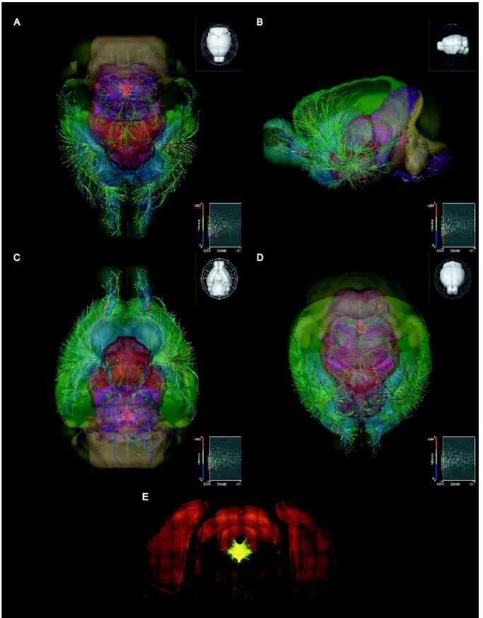
ultimately lead to the identification of discrete serotonergic groups with defined roles in neural circuitry (Ray et al., 2011). Figure 1 reveals the large diversity of serotonergic projections stemming from the dorsal raphe generated by utilizing a Cre-dependent
fluorescent neuronal tracer injected into the dorsal raphe of the Slc6a4-Cre mouse line. These regions range from the cerebral cortex, caudate putamen, amygdala, thalamus and olfactory bulb, to the cerebellar and motor nuclei in the brain stem (Fig. 2).
1.1.4. Serotonergic Regulation of the Dorsal Raphe
serotonin autoreceptor implicated in these negative feedback studies is the 5-HT1A receptor found on the soma and dendrites of serotonin neurons. Agonist-induced stimulation of the 5-HT1A receptor in the dorsal raphe hyperpolarizes and dampens neuronal activity through its activation of G protein-mediated inwardly rectified (GIRK) K+ channels mediated by interactions with G protein βγ subunits (Doupnik et al., 1997; Penington et al., 1993).
Another population of 5-HT autoreceptors is the 5-HT1B receptor, which has been located on axon terminals allowing it to directly inhibit serotonin release (Davidson and Stamford, 1995; Middlemiss and Hutson, 1990). In addition, while the contribution may be small, the 5-HT1D receptor has also been shown to modulate 5-HT release at dendrites and axons
(Pineyro and Blier, 1996; Stamford et al., 2000).
While this work focuses on the modulation of behavioral neural processes by
serotonin, it is important to mention that a large driving force of DR activity is generated by afferent projections from multiple areas in the brain. These include the lateral habenula, interpeduncular nucleus, certain hypothalamic nuclei, ventral tegmental nuclei, lateraldorsal tegmental nuclei, cingulate cortex, medial prefrontal cortex, and the central amygdala
(Behzadi et al., 1990; Hajos et al., 1998; Peyron et al., 1998). While these are primarily made of glutamatergic afferents, there are also numerous inhibitory GABAergic innervation of the DR (Wang et al., 1992).
1.1.5. Other Neurotransmitters and Peptides in the Dorsal Raphe
1977), corticotropin-releasing factor (CRF) (Commons et al., 2003; Day et al., 2004), and neurotensin (Jennes et al., 1982) have been shown in neurons within the DRN. In addition, evidence suggesting neurotransmitters such as dopamine (Ochi and Shimizu, 1978), GABA, and glutamate (Calizo et al., 2011; Fu et al., 2010) have also been shown to be present in the dorsal raphe nucleus and modulate serotonergic function in the raphe. The question of
whether any of these signaling molecules overlap with serotonin neurons has been frequently investigated. While previous studies have suggested a large co-localized population exists with substance P (Baker et al., 1991) and to a lesser degree with CRF (Commons et al., 2003; Day et al., 2004) in rats, a recent study in mice reveals the lack of co-localization of a
number of neuropeptides, including substance P, cholecystokinin, enkephalin, somatostatin, neurotensin, dynorphin, thyrotropin-releasing hormone, and CRF as well as the
neurotransmitter dopamine (Fu et al., 2010). While there is evidence of glutamate decarboxylase (GAD) in a few 5-HT neurons (Hioki et al., 2009), the atypical vesicular glutamate transporter VGLUT3 has been shown to overlap with many serotonin-containing neurons (Gras et al., 2002; Hioki et al., 2009). Incidentally, even though VGLUT3 is co-expressed with serotonin, no evidence has surfaced demonstrating that glutamate is actually released from serotonin neurons. In summary, while the dorsal raphe is made up of a
1.2. MODULATION OF ANXIETY, DEPRESSION, AND FEEDING BY SEROTONIN
While serotonin can affect virtually all mammalian behaviors, this study will focus on its modulation of three psychological disorders: anxiety, depression, and eating. Dysfunction of the serotonergic network and neurotransmission has been associated with the three
aforementioned disorders (Graeff et al., 1997; Levy and Van de Kar, 1992). The following subchapters of this work will briefly describe the role of serotonin in these disorders and serotonin-targeted medications as a treatment of these disorders.
1.2.1. Serotonin and Anxiety
Anxiety can be described as the body’s response to real or imagined aversive stimuli. Many CNS circuits and structures are involved in the response to psychologically and physically aversive stimuli. For example, stress and anxiety can stimulate the hypothalamic-pituitary-adrenal (HPA) axis, which leads to rapid release of the adrenocorticotropin
anxiogenic drugs, including the benozodiazepine inverse agonist FG-7142, the alpha2-adrenoceptor antagonist yohimbine, the non-selective 5-HT2C receptor agonist
m-chlorophenyl piperazine (mCPP), and the adenosine antagonist caffeine (Abrams et al., 2005; Singewald and Sharp, 2000). In addition, uncontrollable stress has been shown to increase the activity of serotonergic neurons in the dorsal raphe (Amat et al., 2005; Grahn et al., 1999). Increases in serotonergic activity and its implication in anxiety states can be further explained by dorsal raphe serotonergic innervation of two key anxiety centers: the amygdala and the bed nucleus of the striatum terminalis (Commons et al., 2003; Li et al., 1990). It is important to note that differential changes in serotonin concentrations in different regions of the brain after various types of stress have been observed, suggesting a more complex regulation of the stress response by serotonergic activity (Amat et al., 1998a; Amat et al., 1998b; Kirby et al., 1995; Rueter et al., 1997). Nevertheless, acute increases in serotonin through blockade of serotonin reuptake inhibitors have been shown to exacerbate anxiety in patients and in mouse models (Nierenberg et al., 2000; To et al., 1999). These results corroborate observations that anxiogenic behaviors in mice occur following the artificial elevation of serotonin levels through administration of high doses of 5-HTP (Artaiz et al., 1998). In summary, increasing serotonergic activity, whether by stressful events or
anxiety disorders, including generalized anxiety disorder (GAD), panic, social anxiety, post-traumatic stress disorder (PTSD), and obsessive compulsive disorder (OCD) (Stahl, 2008). Medications that increase serotonin levels, such as SSRIs, MAOIs, and TCAs, may seem contradictory due to the exacerbation of anxiogenic responses as mentioned above, but surprisingly, long-term or chronic treatments with these drugs are very efficacious (Den Boer and Westenberg, 1990; den Boer et al., 1987; Gorman et al., 1987). In addition, chronic treatment with SSRIs abolishes the anxiogenic effects of endocrine, social and conditioned stress in animal models (To et al., 1999; To and Bagdy, 1999; Zhang et al., 2000). Multiple theories have been constructed in an attempt to resolve this bi-phasic response of increased serotonin and its differential regulation of anxiety. Desensitization of autoreceptors such as 5-HT1A, together with the down regulation of G proteins, have been proposed as a critical step for anxiolytic efficacy with SSRIs (Hensler, 2002; Lerer et al., 1999; Li et al., 1996; Raap et al., 1999). While the available evidence supports this claim, long-term increases in serotonin, enabling the modulation of numerous neural circuits, mostly likely will result in the global remodeling of these circuits in order to adapt to this perturbation. At the moment, the changes induced by chronically increasing serotonin are still poorly understood and studies investigating how networks adapt due to this treatment are needed.
1.2.2. Serotonin and Depression
Depression refers to a heterogeneous group of psychological disorders characterized by similar symptoms that most likely originate from different abnormalities in various neural circuits. The official diagnosis of depression is still very subjective and relies on the
2002). These symptoms overlap with other disorders such as anxiety, which can have substantial co-morbidity (Hasler et al., 2004; Stahl, 2008). Early attempts to describe the biological etiology of these disorders gave rise to the hypothesis that depression is due to a deficiency of monoamine neurotransmitters such as serotonin. Currently, direct evidence for this hypothesis is lacking even though a great deal of effort has been made to identify this deficiency. Nevertheless, multiple abnormalities in serotonergic neurotransmission have been described in patients with major depression, leading to the revised hypothesis that altered serotoninergic activity is a risk factor rather than the underlying cause for this complex spectrum of disorders (Maes, 1995). In support of this hypothesis, reports of decreased 5-HT, 5-HT neurons, and dorsal raphe size in depressed suicide victims have been shown in some studies (Matthews and Harrison, ; Shaw et al., 1967). Furthermore, 5-HT depletion studies, whether by dietary restriction or by treatment with the tryptophan hydroxylase inhibitor para-chlorophenylalanine (PCPA), has led to clinical relapses of depression in patients and has decreased the mood of healthy controls (Shopsin et al., 1976; Shopsin et al., 1975; Young et al., 1985). Finally, a single nucleotide polymorphism (SNP) (G1463A) in TPH2 was found in a small subset of patients with major depression, and was demonstrated to reduce
functionality of the enzyme by 80% (Zhang et al., 2005). Analysis of this SNP in a knock-in mouse (TPH2-R441H) revealed a reduction in 5-HT and 5-HT synthesis, increased
depression- and anxiety-like behaviors, along with depression-like biomarkers such as low CSF 5-HIAA and increased expression of 5HT2A in the frontal cortex (Beaulieu et al., 2008; Jacobsen et al., 2012). In summary, dysfunction of the serotonergic system is clearly
Similar to its role in anxiety, serotonin also plays a role in the etiology and therapy of the depression spectrum. Many similar medications used to treat anxiety are also employed as first- and second-line treatments for depression (Stahl, 2008). As with anxiety, several weeks of treatment are required for improvement in depressive-like symptoms (Nurnberg et al., 1999). While it is well documented that chronic administration of these antidepressants increases serotonin neurotransmission (Blier and de Montigny, 1994; Kreiss and Lucki, 1995; Moret and Briley, 1996), it is hypothesized that this increase produces secondary neuroplastic changes that occur on a longer time and are critical for antidepressant efficiency. One example of a neuroadaptive change that is thought to be critical for increasing 5-HT concentrations, and therefore the actions of antidepressants, is the desensitization of
1.2.3. Serotonin and Feeding
Feeding behaviors are comprised of a diverse, complex set of neural circuits and peripheral hormones that regulate different aspects of ingestive behaviors such as meal initiation, meal termination, and satiety. Serotonin has been implicated in the regulation of feeding behaviors as demonstrated by early studies observing an induction of hyperphagia when 5-HT levels were decreased and hypophagia when 5-HT levels were increased (Fletcher and Paterson, 1989; Saller and Stricker, 1976). In fact, serotonergic dysregulation has been demonstrated in various eating disorders, which exhibit comorbidity with
Given that increasing serotonin concentrations in the brain has proven to be effective in attenuating food intake, manipulation of the serotonergic system has been an ongoing target of anti-obesity pharmacotherapies. Serotonin induced weight loss was initially
investigated using medications that augmented serotonin concentrations throughout the brain, such as 5-HTP, SSRIs, serotonin and noradrenaline reuptake inhibitors (SNRIs) and
fenfluramine (disrupts 5-HT vesicle storage and reverses SERT function) (Brown et al., 2001; Fletcher and Burton, 1986; Yen, 1987). All of these therapeutics have demonstrates significant weight loss in clinical trials and some have even gained FDA approval
ventromedial nucleus (VMN) of the hypothalamus causes reciprocal feeding behaviors depending on the diurnal cycle such that no overall weight loss is achieved in obese rats (Fetissov and Meguid, 2010). Further research into how these two systems interact may yield more effective serotonin-mediated treatment paradigms.
1.3. SELECTIVE CONTROL OF THE SEROTONIN NETWORK
The previous subchapters of this work have attempted to convey the magnitude and the complexity of the serotonin system and its influences on multiple physiological and psychological processes. Prior work has demonstrated significant progress towards elucidating the functionality of the serotonin network through the study of its receptors, projection areas, and sites of synthesis. Nevertheless, studies dividing this system into its parts by selectively and reversibly controlling specific regions of the serotonin network may lead to a greater understanding of how it can influence so many different processes and in addition generate new hypotheses for more targeted therapies. One way to achieve this is to dissect the raphe nuclei into its subdivisions with the goal of potentially identifying discrete groups of 5-HT neurons that are responsible for regulating different serotonergic-mediated processes. This approach has been challenging in the past and was limited to lesions or systemic and/or direct application of compounds known to induce serotonergic activity. These methods, while providing insight into the function of the raphe, lacked specificity and selectivity. Recently, new techniques have been shown to provide selective and bi-directional control over many different types of neuronal signaling and are revolutionizing neural
serotonin selectivity or failed to differentiate the specific serotonergic nuclei in the raphe (Guler et al., 2012; Ray et al., 2011; Warden et al., 2012). With the increasing number of molecular tools available for neurophysiology and neuropharmacology, serotonin research is poised to make great strides in elucidating these complex neural networks.
1.4. PHARMACOSYNTHETICS
Pharmacosynthetics, is defined as “a branch of biology which deals with the creation of pharmacological modulation using artificial components” (Farrell and Roth, 2012). This terminology refers to novel designer receptors created to probe cellular signaling and their effects on physiology. These receptors were generated to address potential confounds when investigating the physiological role of native signaling receptors, like GPCRs, that have expression profiles throughout multiple tissues and cell types. This experimental system (also referred to as chemogenetics) now allows for the activation of a particular type of signaling pathway in a specific cell type and/or tissue, thus providing a greater understating of how it may affect the physiology of the organism.
1.4.1. Designer Receptor Exclusively Activated by Designer Drug (DREADD) Technology
muscarinic orthosteric-binding pocket (Wess et al., 2013), are enough to shift the dose response curve of the highly potent endogenous ligand, acetylcholine (ACh), far enough to right that it is essentially inactive at the receptor, thus generating a native GPCR with no endogenous ligand. Simultaneously, these two mutations also shift the dose-response curve of the weak partial agonist clozapine-N-oxide (CNO), a pharmacologically inert metabolite of clozapine, to a potent full agonist, thus generating an extremely selective, highly potent exogenous ligand (Armbruster et al., 2007; Weiner et al., 2004). Using this manipulation with the human muscarinic 3 and 4 receptors, DREADDs were produced that coupled to Gq and Gi G proteins respectively. Due to the lack of a native Gs-coupling muscarinic receptor, a chimera was created by exchanging intracellular loops two and three for those of the Gs-coupled β1-adrenergic receptor (Guettier et al., 2009). Extensive validation of signal
transduction, desensitization, and internalization have demonstrated that these DREADDs are nearly identical to their non-mutant muscarinic counterparts (Alvarez-Curto et al., 2011; Armbruster et al., 2007; Conklin et al., 2008; Guettier et al., 2009; Kaufmann et al., 2013; Nawaratne et al., 2008). Combined, these orthologous ligand-GPCR pairs represent a significant advancement as tools for manipulating GPCR signaling in virtually every mammalian system.
1.4.2. Modulation of neuronal activity through DREADD activation
GPCR signaling mediates a diverse range of cellular responses including
control over neuronal signaling and insight into how this signaling translates into behavioral phenotypes. As mentioned above, DREADDs can deliver this control and have been shown to selectively, reversibly, and bi-directionally influence neuronal activity. The hM3Dq (h = human, M3 = muscarinic 3 receptor, D = DREADD, and q = coupling to Gq/11 proteins) mediates neuronal excitation through a phospholipase C-dependent manner potentially involved in the closing of potassium channels (Alexander et al., 2009), while the hM4Di (h = human, M4 = muscarinic 4 receptor, D = DREADD, and i = coupling to Gi/o proteins) mediates neuronal silencing through Gβγ activation of GIRK channels (Armbruster et al., 2007). Extensive in vivo validation of the capacities of hM3Dq and hM4Di receptors to modulation neuronal signaling has been demonstrated recently (refer to Table 1). While canonical G protein-mediated signaling cascades are initiated through the activation of these designer receptors and can lead to a wide variety of cellular effects such modulation of transcription and gene expression, most of these studies have utilized this technology to either activate or silence neurons in a cell-type and tissue selective fashion.
1.4.3. Deconstruction of complex neuronal circuits using DREADDs
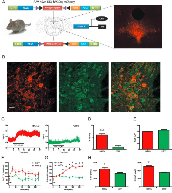
driver lines with Cre-sensitive viruses. Cre mouse driver lines have distinct cell-type and tissue-specific expression patterns of Cre recombinase (Cre), a tyrosine recombinase enzyme, due to the transgenic insertion of Cre after specific promoters of a wide variety of genes. While these driver lines were initially used for the cell-type removal of various genes when crossed with cre-dependent knock-out mouse models, recently these lines been have been combined with cre-sensitive viruses in order to gain a more selective cell-type and tissue-specific expression. Cre-sensitive viruses are created by surrounding an inverted gene (i.e. DREADD) with two pairs of heterotypic, antiparallel loxP sites (designated LoxP and
Lox2722) (Fig. 3A). Without Cre expression, the DREADD cannot express due to it inverted orientation. Upon infection of Cre containing cells, the DREADD gene is restored to its proper orientation and expression occurs (Fig. 3A). Stereotaxic injection of these
CHAPTER 2. A BEHAVIORAL PROGRAM AND NEURAL NETWORK ENCODED BY DORSAL RAPHE SEROTONERGIC NEURONS.
2.1. INTRODUCTION
The central nervous system (CNS) serotonergic network is a large and complex efferent system that impacts a highly diverse range of behavioral and physiological processes, including mood, anxiety, food intake, and energy expenditure (Berger et al., 2009). Serotonergic neurons are distributed in a series of predominantly midline raphe nuclei extending from the midbrain to the caudal medulla, and projecting widely throughout the brain and spinal cord (Jacobs and Azmitia, 1992). The CNS serotonin (5-hydroxytryptamine; 5-HT) system has long been a major focus for CNS drug development, and its
pharmacological modulation has been proposed to contribute to the therapeutic actions of medications used for a wide range of mood and behavioral disorders (Berger et al., 2009). Because some of the most commonly prescribed serotonergic drugs, such as 5-HT selective reuptake inhibitors (SSRI’s), have a global impact on serotonergic neurotransmission, the serotonergic neuronal mechanism(s) responsible for both their therapeutic and adverse effects have been difficult to discern. Achieving selective control over specific serotoninergic nuclei could begin to elucidate these neural mechanisms as well as dissect 5-HT’s influence over the diverse behavioral and physiological processes with which it is associated.
serotonergic neurons project rostrally to provide the predominant serotonergic innervation of the forebrain (Jacobs and Azmitia, 1992; Vertes, 1991; Waselus et al., 2012). Accordingly, dysregulation of DRN serotonergic neurotransmission has been implicated in psychiatric illnesses, such as anxiety and depression (Graeff et al., 1996; Lowry et al., 2008; Underwood et al., 1999) and the modulation of 5-HT release from DRN projections has been proposed as being critical for antidepressant efficacy (Stamford et al., 2000). While the selective
manipulation of DRN serotonergic neurons could theoretically illuminate these processes, this has proven difficult since many (50-75%) neurons in the DRN are non-serotonergic (Halberstadt and Balaban, 2008).
The ability to selectively modulate both acute and chronic activity of distinct serotonergic neuronal subpopulations would provide a powerful approach for unraveling neural pathways and mechanisms relevant to mood and behavioral disorders. Designer receptors exclusively activated by designer drugs (DREADDs) are uniquely suited for this type of neuronal manipulation (Alexander et al., 2009; Armbruster et al., 2007). These evolved GPCRs can modulate both short- and long-term neuronal activity, mimicking drug-like actions (Rogan and Roth, 2011; Wess et al., 2013). DREADDs are thus ideal for
selectively stimulating or inhibiting subpopulations of neurons and represent a non-invasive, reliable, reversible, and inducible approach for probing serotonergic function in behaving laboratory animals.
patterns indicative of a previously unappreciated circadian influence on the serotonergic regulation of feeding behavior. These studies further revealed that acute and chronic activation of DRN serotonergic neurons are sufficient to induce behavioral effects across multiple assays that resemble both the acute and chronic responses to SSRI antidepressants. Finally, we mapped changes in whole-brain metabolic activity induced by activating DRN serotonin neurons in behaving mice using DREAMM (DREADD-assisted metabolic mapping; (Michaelides et al., 2013)). Altogether, these findings reveal a diversity of processes by which DRN serotonergic neurons modulate network activity relevant to therapeutic effects.
2.2. METHODS
Clozapine-N-Oxide (CNO). CNO was obtained from NIH as a part of a Rapid Access
to Investigative Drug Program funded by the National Institute of Neurological Disorders and Stroke (NINDS). For all in vivo experiments, CNO was first dissolved in DMSO, and then brought to a final concentration with 0.9% saline at a concentration of DMSO at 0.4%. For all in vivo experiments, the appropriate vehicle controls (0.4% DMSO in saline) were utilized. A dose of 2 mg/kg was used for acute treatment (i.p.) at a volume 100 µl/10 g body weight. Chronic treatment consisted of 3-4 weeks of 5 mg/kg/day administered in the drinking water. The water bottles were protected from light and changed every 3 to 5 days. For microdialysis, an acute dose of 3 mg/kg (i.p.) was used.
Animal Subjects. General Procedure. All procedures were conducted in accordance
at the University of North Carolina (UNC), Duke University, and the University of California at San Francisco. Adult, age-matched, male Slc6a4-Cre mice (MMRRC: Stock number – 017260UCD) were used for all in vivo and ex-vivo experiments.
Generation of Slc6a4-hM3Dq and Slc6a4-EGFP mice. Adult (22–30 g) Slc6a4-Cre
males (MMRRC : Stock number – 017260UCD) were group-housed before surgery. Unless described otherwise, all mice were maintained on a standard 12-h light/dark cycle (lights off at 7:00) with ad libitum access to food and water. Mice were anaesthetized with a ketamine (100 mg/kg body weight) and xylazine (10 mg/kg) solution and placed into a stereotaxtic frame (Kopf Instruments). For all experiments, male mice were bilaterally injected with 0.75 – 1 μl (depending on viral titer) of purified and concentrated adeno-associated virus (AAV) (~1012 infections units/ml, packaged in serotype 8 by the UNC Vector Core Facility) into the dorsal raphe using the following stereotaxtic coordinates: -4.6 mm to bregma,
±1.93 mm lateral to midline, and −3.46 mm ventral to the skull surface at an angle of 30°. Dorsal raphe neurons were transduced with virus encoding the hM3Dq-mCherry or EGFP in reverse orientation floxed by two lox sites (lox P and lox 2722) under the control of the human synapsin (SYN1) promoter. Following surgery, the mice were individually housed and monitored for signs of complications until they recovered from surgery. We performed all experiments 4-16 weeks after surgery. All viral constructs were packaged by the UNC Vector Core Facility at a final working concentration of 4 × 10¹² to 6 × 10¹² genome
copies/ml. After all experiments, every mouse was perfused and the dorsal raphe subjected to immunohistochemistry to validate injection efficiency and viral expression.
Immunohistochemistry. Mice were anaesthetized with tribromoethanol (Avertin) and
Electrophysiology. Mice were decapitated under isofluorane anesthesia. Their brains
were rapidly removed and immediately placed in a solution of ice-cold sucrose-artificial cerebrospinal fluid (aCSF) [(in mM): 194 sucrose, 20 NaCl, 4.4 KCl, 2 CaCl2, 1 MgCl2, 1.2 NaH2PO4 , 10 glucose, and 26 NaHCO3 saturated with 95% O2/5% CO2]. Brains were sectioned at 0.06 (mm/s) on a Leica 1200S vibratome to obtain 300 µm coronal slices of the dorsal raphe, which were incubated in a heated holding chamber containing normal,
oxygenated aCSF [(in mM):124 NaCl, 4.4 KCl, 2 CaCl2, 1.2 MgSO4, 1 NaH2PO4, 10.0 glucose, and 26.0 NaHCO3] maintained at 30 ± 1˚C for at least 1 hr before recording. Slices were transferred to a recording chamber (Warner Instruments) submerged in normal,
oxygenated aCSF and maintained at 28-30˚C at a flow rate of 2 ml/min. Tetrodotoxin (TTX) was included in the bath to prevent firing of action potentials and to allow accurate
determination of changes in membrane potential. Neurons of the dorsal raphe were visualized using infrared- differential interference contrast (DIC) video-enhanced microscopy
application. Only cells with a stable access resistance (less than a 20% change from baseline to the end of the experiment) were used in the data analysis.
In Vivo Microdialysis. Mice were anesthetized with ketamine-xylazine as described
The chromatograms were analyzed using the Clarity software package (DataApex, Prague, Czech Republic). The limit of detection was considered as a signal-to-noise ratio (SNR) of 3. The data collected over time were analyzed by repeated measures ANOVA (RMANOVA), while cumulative neurochemical levels were analyzed by t-tests; a p < 0.05 was considered significant.
Home Cage Monitoring (HCM) System. Data Collection. After 5 days of acclimation
to the UCSF animal facility, mice were individually housed in 45 x 24 x 17 cm Plexiglas home-cage monitoring (HCM) enclosures. At one end of each enclosure was a photobeam-based feeding monitor and a capacitance-photobeam-based lickometer. At the far end of the enclosure, across from the feeder was a 10 x 10 x 10 cm opaque black plastic housing niche with a cotton nestlet. The enclosures were mounted atop activity platforms providing animal location data at 20 msec intervals. A detailed description of HCM hardware has been reported (Goulding et al., 2008). Animals received ad libitum access to water by the lickometer and to a powdered version of the UCSF animal facility standard chow diet (PicoLab; Mouse Diet 20).
Experimental Schedule. For all animals (hM3Dq: n = 8, EGFP: n = 8), acclimation
serotonergic neuronal activation on exploratory behavior. We examined the responses of CNO-treated Slc6a4-hM3Dq and Slc6a4-EGFP mice to a novel object placed within their HCM cages. Four hrs prior to the dark cycle onset, a metal binder clip (2 inch wide, 1 inch capacity; Officemate International) was affixed to the center of the long cage wall opposite the housing niche for a period of 4 hrs.
HCM Measures. Data quality control methods for the detection and adjustment for
device errors were performed as described (Goulding et al., 2008). Standard HCM assessment of ingestive behavior and movement patterns were performed as outlined (Goulding et al., 2008). Briefly, we reported that mice are in two fundamental mutually exclusive states: active states (ASs) and inactive states (ISs). ISs are characterized by periods of low physical activity (including sleep and resting) occurring exclusively at the nest. These alternate with ASs, during which bouts of feeding, drinking, locomotion and other behaviors occur (Goulding et al., 2008). The regulation of AS properties are distinct from those of bouts and are highly sensitive to circadian time (Goulding et al., 2008). Exploratory responses to novel object presentation were assessed by analyzing locomotor paths and determining the amount of time animals spent within a 7 cm radius of the object during the first hour of exposure.
Behavior Studies. The following behavioral paradigms were initiated 45 min after
acute administration of CNO/vehicle or after 3-4 weeks of chronic administration of either CNO/vehicle in the drinking water.
Locomotor Activity and Center Zone Time. Locomotor activity was assessed under
as previously described (Abbas et al., 2009). Locomotor activity was recorded for 1 hr. Parameters measured included total distance traveled and time spent in the center zone of the chamber. Data was extracted using Fusion software (AccuScan Instruments, Columbus, OH).
Light-Dark Emergence Test. Light and dark preference was assessed under
standardized environmental conditions in 40 x 40 x 30-cm Plexiglas chambers as described above, fit with a 16" L x 8" H x 5" W dark chamber on one side (VersaMax system;
AccuScan Instruments, Columbus, OH). Light, dark, and doorway preferences were recorded for 10 min. Data was extracted using VersaMap software (AccuScan Instruments, Columbus, OH).
Forced Swim. Mice were individually placed into a Plexiglas cylinder (21 cm
diameter, 28 cm height) containing 15 cm water (25 ± 0.5 °C) and were monitored for 6 min. Mobility, high mobility, and immobility bouts were analyzed using EthoVision XT7 for the last 4 mins of the test. After the swim session, mice were dried and placed in their home-cage surrounded by a cotton cloth. The water was changed between each animal. Animals that were unable to perform the task were not included in this study.
Novelty-Suppressed Feeding. Food was removed from the home-cage 24 hr before
testing. Mice were weighed just before food deprivation and again before testing to assess body weight loss. For testing, mice were placed into a brightly lit open field
(40 cm × 40 cm × 30 cm white plastic boxes containing 2 cm of bedding) with a food pellet at the center. Latency to begin eating the food was recorded. The total amount of food
consumed during a 5 min period after returning to the home-cage was measured to test whether feeding differences in the novel environment were due to differences in
DREADD-assisted metabolic mapping (DREAMM). Slc6a4-Cre mice expressing
hM3Dq (n=8) or EGFP (n=8) in the dorsal raphe Slc6a4 positive neurons were transported to the µPET facility the day prior to each scan to ensure habituation to the transportation
procedure and environment. All mice were then fasted overnight to attain consistency in blood glucose levels as abnormal blood glucose levels interfere with FDG uptake (Wong et al.). The following morning mice received an injection (i.p.) of either vehicle or CNO (2 mg/kg). This occurred four times, twice before (acute-vehicle and acute-CNO) and twice after 3 weeks of daily (5 mg/kg, p.o.) CNO exposure (chronic-vehicle and chronic CNO). Therefore each mouse was scanned four times. For each acute and chronic phase, vehicle and CNO scans were separated by two days. For acute phase scans, vehicle preceded CNO while for chronic phase scans, CNO preceded the vehicle (for the purpose of minimizing
withdrawal effects due to disruption of daily CNO administration after chronic exposure). Twenty min after vehicle or CNO injections mice received an injection (i.p.) of ~0.2 mCi of [18F]fluorodeoxyglucose (FDG) and were immediately placed in an open-field arena (40.64 x 40.64 x 40.64 cm) coupled to an activity monitoring system (Truscan, Coulbourn
Instruments, Whitehall, PA). Thirty min later mice were anesthetized with 1.5% isoflurane, placed in a prone position on the bed of an R4 microPET rodent scanner (Siemens Medical Solutions, Malverne, PA) and scanned using a 20 min static acquisition protocol. Scanning procedures, image processing, and analysis were performed as previously described
2.3. RESULTS
2.3.1. hM3Dq receptors selectively expressed in DRN serotonergic neurons increase membrane excitability and 5-HT release.
Selective expression of the hM3Dq receptor (Armbruster et al., 2007) in DRN
serotonergic neurons was achieved through stereotaxic injection of a previously characterized Cre-recombinase-dependent adeno-associated virus (AAV; (Krashes et al., 2011)) into the dorsal raphe of Slc6a4-Cre mice (Fig. 3A). Immunofluorescence microscopy revealed that 96.7% ±1.6, (N= 3670 cells) of hM3Dq expressing neurons co-localized with 5-HT-positive neurons with a 64.2% ±5.3 (N= 3670 cells) transduction efficiency of DRN 5-HT-containing neurons (Fig. 3A,B). A similar degree of co-localization and transduction efficiencies were observed in Slc6a4-Cre mice stereotaxically injected with a control Cre-dependent, AAV-containing EGFP virus (97.7% ±1.2 co-localization, N= 2035 cells; 73.5% ±4.6 transduction efficiency, N= 2035 cells; Fig. 7A,B). In addition, 5-HT-positive axons emanating from the dorsal raphe co-localized with hM3Dq receptors, thus demonstrating the trafficking of the hM3Dq receptor to the DRN serotonergic axons (Fig. 7C). Importantly, there was negligible expression of hM3Dq in 5-HT-positive neurons of the medial raphe (hM3Dq: 2.3% ±3.0; N= 586 cells; Fig. 7D), thus further highlighting selective expression of hM3Dq in 5-HT-positive neurons in the DRN.
Slc6a4-hM3Dq mice compared to EGFP-positive DRN neurons in Slc6a4-EGFP mice (Fig. 3C,D). Importantly, hM3Dq- and EGFP-containing neurons had similar resting membrane potentials (Fig. 3E). In vivo microdialysis was utilized to measure CNO’s effects on HT and
5-hydroxyindoacetic acid (5-HIAA) concentrations in the striatum (a major DRN projection). A significant increase in both 5-HT and 5-HIAA concentrations in Slc6a4-hM3Dq mice compared to Slc6a4-EGFP mice (Fig. 3F-I) was observed after CNO administration. Importantly, the concentration of dopamine and its measured metabolites were not altered following CNO administration (Fig. 8).
2.3.2. Stimulation of DRN serotonergic neurons alters feeding in a circadian-dependent manner.
We next utilized a quantitative, robust, and automated procedure to assess how the activation of DRN serotonergic neurons alters home cage behavioral organization. We examined behavioral patterns prior to (Days 8-15) and during the second week of continuous CNO treatment (Days 22-29) in Slc6a4-EGFP and Slc6a4-hM3Dq mice. Although no
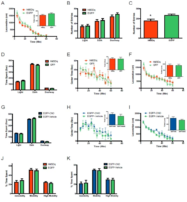
significant effects of CNO treatment on total daily food intake were observed in either group, examination of light cycle (LC) and dark cycle (DC) food intake revealed a significant effect in Slc6a4-hM3Dq mice (Fig. 4A and 9A). Whereas CNO suppressed DC food intake by 0.24 gm, CNO enhanced LC food intake by 0.26 gm (Fig. 4A). By contrast, CNO treatment produced no significant changes in DC or LC food intake in EGFP mice (Fig. 9A).
bins throughout the circadian cycle and observed significant effects at the circadian time (CT) 1 and 3 hr time points (Fig. 4B). These results indicate that DRN activation induced by CNO produced a suppression in DC food intake that was followed by an enhancement in LC food intake of similar magnitude, resulting in no net change in daily intake. Moreover, the CNO-induced increase in LC food intake occurred exclusively during the first 4 hours of the LC.
We previously demonstrated that circadian patterns of food consumption are
determined by both active state (AS) regulation and feeding-bout regulation (Goulding et al., 2008). Specifically, food consumed during a particular time of day is dependent on the probability that animals are engaged in ASs, as well as in the numbers and sizes of feeding-bouts that occur during those ASs. Moreover, AS and feeding-bout properties can be
4D). These results indicate that in Slc6a4-hM3Dq mice, CNO-induced circadian shifts in food intake are strongly mimicked by circadian shifts in ASP
We next determined the extent to which changes in feeding-bout number contributed to the shifts in the circadian pattern of feeding in CNO-treated Slc6a4-hM3Dq mice. We examined daily feeding-bout totals prior to (Days 8-15) and during the second week of CNO treatment (Days 22-29) in Slc6a4-EGFP and Slc6a4-hM3Dq mice. No significant effects of CNO treatment on daily feeding-bout totals were observed in either group. However, in accord with its effects on LC food intake and ASP, CNO treatment increased LC feeding bout numbers in Slc6a4-hM3Dq mice whereas no alterations were observed in Slc6a4-EGFP mice (Fig. 4E and 9C). For a more detailed assessment of the influence of CNO on circadian patterns of feeding-bout rates, we examined them during 2-hr bins throughout the day and revealed a significant effect at the CT1 and 3 hr time points (Fig. 4F). These results indicate that increased feeding bout rates contribute to the CNO-induced enhancement of feeding early in the light cycle in Slc6a4-hM3Dq mice.
to the CNO-induced enhancement of feeding in the first 4 hrs of the LC in Slc6a4-hM3Dq mice.
We had previously found that once ASs are initiated, a characteristic shift occurs in the probabilities of distinct behaviors as the AS progresses (Goulding et al., 2008). Notably, feeding is the predominant activity occurring during the first 2 min of ASs, after which feeding probabilities decline rapidly, in a manner presumably reflecting satiation (Goulding et al., 2008). We therefore examined the extent to which the increased food intake exhibited by Slc6a4-hM3Dq mice early in the LC is accompanied by changes in the temporal patterns of food intake within ASs. We examined ASs exhibited during the first 4 hrs of the LC on testing days prior to (Days 8-15) and during the second week of CNO administration (Days 22-29). We aligned the onsets of these ASs (Fig. 4J and K) and then determined, on a min-by-min basis from AS onset, the probabilities that animals engaged in feeding behavior (Fig. 4I). This analysis revealed significant effects at the 4, 5, 6, 7, and 10 min time-points (Fig. 4I).
2.3.3. Acute activation of DRN serotonergic neurons induces anxiogenic and anti-depressant drug-like behavioral responses and the recruitment of discrete brain circuits.
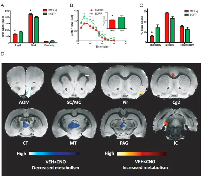
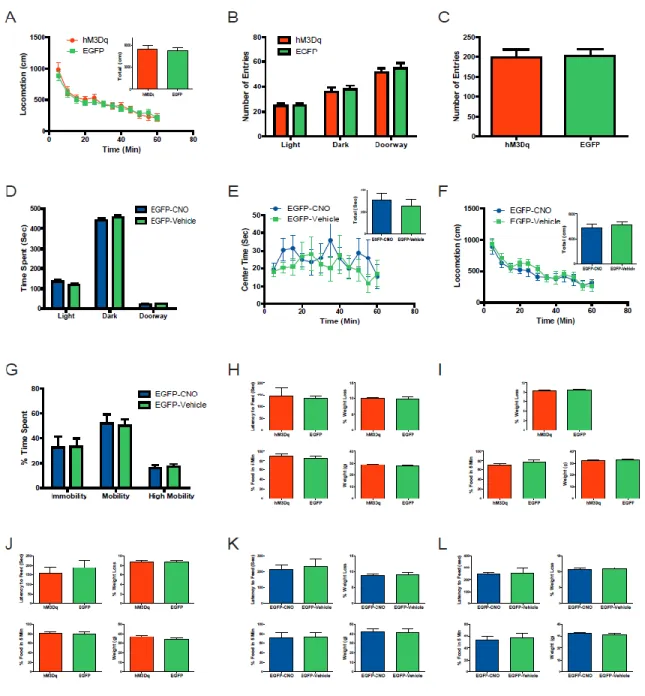
reduction of time spent in the lighted chamber and a reciprocal increase of time spent in the darkened chamber when comparing Slc6a4-hM3Dq mice to Slc6a4-EGFP control animals. Importantly, there was no difference in the number of entries into the darkened and lighted chambers when comparing Slc6a4-hM3Dq and Slc6a4-EGFP mice (Fig. 10B). Similar to the light-dark emergence test, the anxiogenic-like response after acute administration of CNO is also observed by monitoring center time in the open field when comparing Slc6a4-hM3Dq mice and Slc6a4-EGFP mice (Fig. 5B). Importantly, Slc6a4-hM3Dq and Slc6a4-EGFP mice have similar locomotion profiles after acute injection of CNO, suggesting that decreases in center time are not related to decreases in locomotion (Fig. 10A). The number of entries into the center of the open field revealed a decrease when comparing the Slc6a4-hM3Dq mice to Slc6a4-EGFP controls (Fig. 10C). An additional negative control (vehicle or absence of CNO) study was performed using these same behavioral assays, which failed to detect any differences between Slc6a4-hM3Dq and Slc6a4-EGFP mice (Fig. 10D-F). Furthermore, acute CNO treatment exerted no apparent effects in these behavioral paradigms when compared to vehicle in Slc6a4-EGFP control mice (Fig. 10G-I).
In an effort to map behavioral profiles associated with acute DRN serotonin neuron activation in the open-field to regional brain responses, we used DREADD-assisted
contrast, decreases in FDG uptake were observed in the periaqueductal grey (PAG), anterior olfactory (AOM), and medial and central thalamic areas (MT and CT) (Fig. 5D). CNO was previously shown to not have any non-specific effects on FDG uptake (Michaelides et al., 2013). The specificity of DREAMM responses to the behaviors observed in the open field after acute hM3Dq activation were validated using both within-subject (scanned after
vehicle-administered open field sessions) and between-subject controls (Slc6a4-EGFP mice), which did not show changes in time spent in the center of the open-field in response to acute DRN serotonin neuron activation.
Since acute administration of antidepressants can induce effects in some depression-related animal paradigms, we next evaluated whether acutely activating hM3Dq receptors in the DRN could induce antidepressant-like responses in the forced swim test (Porsolt et al., 1977). Following acute CNO administration, Slc6a4-hM3Dq displayed a significant
2.3.4. Chronic activation of DRN serotonergic neurons induces an antidepressant-drug like effect.
Because SSRIs require many weeks of treatment before increases in mood and/or decreases in anxiety are observed, we next investigated the consequences of chronically activating DRN 5-HT neurons by combined behavioral and functional imagining analysis. We first determined if chronic administration of CNO could alter anxiety-like behaviors. After three weeks of CNO treatment, Slc6a4-hM3Dq and Slc6a4-EGFP mice spent a similar amount of time in the both the lightened and darkened chambers (Fig. 6A) as well as a similar center time in the open field (Fig. 6B). As a control the hM3Dq and Slc6a4-EGFP mice entered a similar number of times in each of the three chambers of the light-dark exploration test and in the center zone of the open field (Fig. 11B and C). Chronic
administration of CNO when compared to vehicle did not cause any significant changes in locomotion, time spent in the lightened or darkened chambers, or time spent in the center zone of the open field in Slc6a4-EGFP control mice (Fig. 11D-F).
Although no changes in anxiety-like behaviors were observed in these two behavioral paradigms, DREAMM revealed that chronic DRN 5-HT neuron activation produced
as the medial parabrachial nucleus (MPB), dorsal and lateral dorsal tegmental (DTg and LDTg), the medial vestibular (MVe), and cerebellar nuclei (CBN) (Fig. 6G and 12). These widespread decreases in DREAMM responses were not observed in Slc6a4-EGFP control mice, which revealed no overlapping metabolic changes with CNO-exposed Slc6a4-hM3Dq mice. To investigate whether the continual activation of the hM3Dq receptor has led to the desensitization and/or down regulation of this receptor during chronic treatment, we used DREAMM again after an acute administration of CNO in chronically treated Slc6a4-hM3Dq mice. These scans revealed an intense increase in FDG uptake in the dorsal raphe area (Fig. 13) supporting the hypothesis that even following chronic activation, hM3Dq displays robust and continued activity.
To examine the effects of chronic CNO administration on the exploratory behaviors of Slc6a4-hM3Dq and Slc6a4-EGFP mice, a novel object was introduced into the home cage. Time spent by CNO-treated Slc6a4-hM3Dq and Slc6a4-EGFP mice within a 7 cm radius of the object (occupancy time) was analyzed over a 1 hr period following object placement. Time in the vicinity of the object was significantly enhanced in Slc6a4-hM3Dq, relative to Slc6a4-EGFP mice at the 20 minute time-point (Fig. 6E) and the patterns of exploratory interactions were also different (Fig 6F). Importantly, locomotor activities did not differ between the Slc6a4-hM3Dq mice and Slc6a4-EGFP control mice after chronic administration of CNO (Fig. 11A).
demonstrating robust and prolonged antidepressant-like effects from chronic treatment with CNO. Importantly, compared to vehicle, the chronic administration of CNO did not alter immobility or mobility in control mice (Fig. 11G). Further characterization of this antidepressant-like phenotype resulting from chronic treatment was evaluated using the novelty-suppressed feeding paradigm, which is reported to be one of the few behavioral tests that is differentially sensitive to the chronic versus acute effects of antidepressant treatments (David et al., 2009; Santarelli et al., 2003). Chronic administration of CNO induced a large reduction in the latency to feed in Slc6a4-hM3Dq mice compared to Slc6a4-EGFP mice, while an acute dose of CNO showed no alteration in this behavior (Fig. 6D and 11H). Importantly, weight loss, initial body weight, and percentage of food consumed in the home cage after the testing were not different between Slc6a4-hM3Dq and Slc6a4-EGFP mice (Fig. 11I). Similarly, no significant alterations between mice were observed in these same parameters after vehicle administration (Fig. 11J). In addition, acute and chronic
CHAPTER 3. DISCUSSION AND FUTURE DIRECTIONS
3.1. DISCUSSION
Serotonin has long been considered as an anorectic agent, since treatments that globally enhance serotonergic neurotransmission generally suppress feeding behaviors; however the neural mechanisms underlying these actions are incompletely understood. This limitation is in part attributable to the multitude of regions throughout the neuraxis known to both modulate feeding and to receive serotonergic innervation. Of these regions, particular emphasis has been placed upon the medial hypothalamus, which is innervated by 5-HT neurons arising from the DRN (Willoughby and Blessing, 1987).
The application of a robust and sensitive quantitative behavioral analysis revealed a level of complexity in feeding responses mediated by DRN 5-HT neurons that has not been acknowledged in current models of serotonergic satiety. Although chronically stimulating serotonergic neurons in the DRN produced no net changes in daily food intake or body weight, it did alter circadian patterns of feeding. We detected a modest suppression of DC food intake that was precisely counterbalanced by an enhancement of LC food intake. These results are in accord with a prior report indicating that chronic infusion of 5-HT into the ventromedial hypothalamus of obsess Zucker rats diminished DC food intake while enhancing LC feeding (Fetissov and Meguid, 2010).
of AS probability closely mimicked changes in the circadian pattern of food intake. These increases in both LC food intake and LC AS probability occurred predominately during the first 4 hrs of the LC. By contrast, no changes in feeding-bout size were observed at this time.
Furthermore, we have previously found in C57BL/6 mice that, within individual ASs, the probability of feeding is highest within 2 min of AS onset, and that this probability subsequently declines rapidly over time (Goulding et al., 2008). This decline in feeding probability, which was dramatically suppressed in Lepob/ob mice, was proposed to reflect the process of satiation (Goulding et al., 2008). Here, CNO-treated Slc6a4-hM3Dq mice exhibit a blunted decline in AS feeding probability during the early LC, raising the
possibility that the relative hyperphagia seen at this time is driven by a reduction in satiation. Altogether, these results reveal that a continuous selective 5-HT system manipulation can both decrease and increase food intake in a manner that is highly sensitive to circadian time. This demonstrates a level of complexity in serotonergic feeding regulation which contrasts with the common view that enhanced serotonergic neurotransmission uniformly suppresses feeding. These results indicate that a single 5-HT manipulation both can decrease and increase food intake in a manner that is highly sensitive to circadian time.
novel object was observed without any alterations in motor activity. This increase in
exploratory behavior can be interpreted as a reduction in cautious and/or inhibitory behavior, a novel serotonergic-related behavioral effect, and it has correlations to various anxiolytic-related behaviors (Crawley, 1985).
Modulation of 5-HT levels has been strongly implicated in many therapies that alleviate depression (Kroenke et al., 2001). In addition, SSRI’s have proven to be effective in various animal behavioral models that are utilized as preclinical screens (Willner, 1990). Selectively increasing DRN serotonergic neuronal activity revealed analogous
antidepressant-like effects in several of these preclinical animal models. Thus acute
stimulation of DRN serotonergic neurons produced an increase in the extracellular levels of 5-HT as well as a significant decrease in immobility and an increase in mobility in the classical antidepressant behavioral paradigm: forced swim. Chronic stimulation maintains this antidepressant-like activity, providing support for the notion that short- and long-term activation of the dorsal raphe serotonergic system plays an important role in mediating this behavioral response. A preclinical behavioral assay that better models the several week latency for clinical responsiveness to SSRIs is the novelty-suppressed feeding assay (David et al., 2009; Santarelli et al., 2003). In accord with these studies, chronic stimulation of the serotonergic neurons of the DRN in our model also reduces latencies to consume food in an aversive environment. Collectively, these data provide a strong case for implicating the involvement of this subset of serotonergic neurons in mediating antidepressant-like activity.
Mapping functional brain anatomy associated with acute and chronic
in the open field highlighted a mix of changes in FDG uptake in anxiety-related and sensory processing nuclei. In particular, significant increases in functional metabolic uptake of FDG into the left cingulate and right piriform cortices was observed. Importantly, increased cerebral blood flow and glucose uptake in the cingulate cortex region has been strongly linked to anticipatory anxiety in humans (Chua et al., 1999; Javanmard et al., 1999; Straube et al., 2009) and anxiogenic contextual conditioning in rats (Luyten et al., 2012), while increases in FDG uptake in the piriform cortex appear after acute immobilization stress in rats (Sung et al., 2009). Of additional interest were the observed decreases in FDG uptake in the periaqueductal grey area during acute activation. While anxiety-like responses, such as freezing, are usually associated with an activation of these nuclei (Vianna et al., 2001), no previous studies have examined metabolic uptake in this brain region under acute hyper-serotonergic conditions. However, previous investigations have shown that direct 5-HT administration into this area by intra-cerebral injection results in an anxiogenic-like increase in avoidance (Zanoveli et al., 2003).
In contrast to acute stimulation, chronically activating serotonergic neurons in the DRN lead to widespread decreases in FDG uptake, specifically in the left dorsal and ventral hippocampal regions (CA1, DG, and DS), CA1 region of the right hippocampus, left
2010). Evidence in support of this hypothesis has shown that hyper-activation of the habenula occurs during depressive mood symptoms induced by tryptophan depletion in humans (Roiser et al., 2009), as well as by increased excitatory synaptic activity on the lateral habenula in an animal model of learned helplessness (Li et al., 2011). Furthermore, 5-HT has been shown to suppress excitatory basal ganglia transmission onto the habenula, thereby blocking an aversive or anti-reward pathway (Shabel et al., 2012). The habenula has even been successfully targeted by deep brain stimulation (DBS) to suppress activity in a patient who failed to respond to antidepressant therapies (Sartorius et al., 2010), and there is currently an ongoing clinical trial using lateral habenula DBS for treatment-resistant
depression (NCT01798407). Another metabolic alteration of interest is the decrease in FDG uptake in the thalamic nuclei observed in both the acute and chronic activation of the DRN serotonergic neurons. Patients who were administered SSRIs have also demonstrated decreases in glucose metabolism in the thalamic regions, although some of these studies indicate this observation occurs in both responders as well as nonresponders (Brody et al., 2001; Mayberg et al., 2000). These data demonstrate the pervasive consequences of selectively activating a relatively small group of serotonergic nuclei in the dorsal raphe, further elucidating how 5-HT can induce both anxiety-like activity as well as antidepressant-like responses by differentially modulating the same neurons.
In conclusion, our findings reveal that 5-HT systems regulate feeding in a complex manner that is highly sensitive to circadian time. These results have implications for