Novel sesquiterpene lactone analogues as potent anti-breast
cancer agents
Kyoko Nakagawa-Goto
a,b,**
,1, Jo-Yu Chen
c,1, Yu-Ting Cheng
d,e,f,
Wai-Leng Lee
g, Munehisa Takeya
a, Yohei Saito
a, Kuo-Hsiung Lee
b,h,***
,
Lie-Fen Shyur
c,d,e,*
aSchool of Pharmaceutical Sciences, College of Medical, Pharmaceutical and Health Sciences, Kanazawa University,
Kanazawa 920-1192, Japan
bNatural Products Research Laboratories, UNC Eshelman School of Pharmacy, University of North Carolina, Chapel
Hill, NC 27599-7568, USA c
Graduate Institute of Pharmacognosy, Taipei Medical University 11031, Taipei, Taiwan, ROC
dMolecular and Biological Agricultural Sciences Program, Taiwan International Graduate Program, Academia Sinica,
Taipei 11529, Taiwan, ROC
eAgricultural Biotechnology Research Center, Academia Sinica, Taipei 11529, Taiwan, ROC
fGraduate Institute of Biotechnology, National Chung Hsing University, Taichung 40227, Taiwan, ROC g
School of Science, Monash University Sunway Campus, Selangor 47500, Malaysia
hChinese Medicine Research and Development Center, China Medical University and Hospital, 2 Yuh-Der Road,
Taichung 40447, Taiwan, ROC
A R T I C L E I N F O
Article history:
Received 26 January 2016 Received in revised form 15 March 2016
Accepted 16 March 2016 Available online 25 March 2016
Keywords:
Deoxyelephantopin DET derivatives
Triple-negative breast cancer
A B S T R A C T
Triple-negative breast cancer (TNBC) is associated with high grade, metastatic phenotype, younger patient age, and poor prognosis. The discovery of an effective anti-TNBC agent has been a challenge in oncology. In this study, fifty-eight ester derivatives (DETDs) with a novel sesquiterpene dilactone skeleton were organically synthesized from a bioactive nat-ural product deoxyelephantopin (DET). Among them, DETD-35 showed potent antiprolifer-ative activities against a panel of breast cancer cell lines including TNBC cell line MDA-MB-231, without inhibiting normal mammary cells M10. DETD-35 exhibited a better effect than parental DET on inhibiting migration, invasion, and motility of MDA-MB-231 cells in a concentration-dependent manner. Comparative study of DETD-35, DET and chemothera-peutic drug paclitaxel (PTX) showed that PTX mainly caused a typical time-dependent G2/M cell-cycle arrest, while DETD-35 or DET treatment induced cell apoptosis. In vivo
Abbreviations:DEAD, diethyl azodicarboxylate; DET, deoxyelephantopin; DETD, deoxyelephantopin derivative; DMAP, dimethylami-nopyridine; EDCI, 1-(3-dimethylaminopropyl)-3-ethylcarbodiimide hydrochloride; ER, estrogen receptor; HER2, human epidermal growth receptor 2; MTT, 3-(4,5-dimethylthiazol-2-yl)-2,5-diphenyl tetrazolium bromide; PTX, paclitaxel; PR, progesterone receptor; SAR, struc-ture-activity relationship; TNBC, triple-negative breast cancer.
* Corresponding author.Agricultural Biotechnology Research Center, Academia Sinica, 128, Sec. 2, Academia Road, Nankang, Taipei 11529, Taiwan, ROC. Tel.:þ886 2 26515028.
** Corresponding author.College of Medical, Pharmaceutical and Health Sciences, Kanazawa University, Kanazawa, 920-1192, Japan. Tel.:þ81 76 264 6305.
*** Corresponding author.UNC Eshelman School of Pharmacy, University of North Carolina, Chapel Hill, NC 27599-7568, USA. Tel.:þ1 919 962 0066.
E-mail addresses: [email protected] (K. Nakagawa-Goto), [email protected] (K.-H. Lee), [email protected] (L.-F. Shyur).
1Equal contribution as the co-first author.
a v a i l a b l e a t
w w w . s c i e n c e d i r e c t . c o m
ScienceDirect
www.elsevier.com/locate/molonc
http://dx.doi.org/10.1016/j.molonc.2016.03.002
Lung metastasis Paclitaxel
efficacy of DETD-35 was evaluated using a lung metastatic MDA-MB-231 xenograft mouse model. DETD-35 significantly suppressed metastatic pulmonary foci information along with the expression level of VEGF and COX-2 in SCID mice. DETD-35 also showed a syner-gistic antitumor effect with PTXin vitroandin vivo. This study suggests that the novel com-pound DETD-35 may have a potential to be further developed into a therapeutic or adjuvant agent for chemotherapy against metastatic TNBC.
ª2016 Federation of European Biochemical Societies. Published by Elsevier B.V. All rights reserved.
1.
Introduction
Breast cancer is a commonly diagnosed cancer among women. Although multiple breast cancer therapy protocols including hormone modulators and antibody preparations have been developed, the mortality rate from breast cancer has not improved over the past decade (Howlader et al., 2012). Eighty percent of breast cancers are invasive and recurrence is prob-lematic. The search for effective therapies for breast cancers is complicated by its heterogeneous pathologies and molecular profiles. It is known that many breast cancers are associated with overexpression of either one or multiple hormone recep-tors such as estrogen receprecep-tors (ER), progesterone receprecep-tors (PR), and human epidermal growth receptor 2 (HER2). Hormone therapies and/or molecular targeted therapies are applied as an effective treatment for such types of breast cancer. Howev-er, difficulties are associated with the treatment of breast can-cer with heterogeneous triple-negative type lacking ER, PR, and HER2 expression, which accounts for 15e20% of breast cancers and usually has poor prognosis, and the discovery of an effec-tive anti-breast cancer agent that targets triple-negaeffec-tive breast cancer (TNBC) has been a challenge in oncology.
Elephantopus scaber, a plant from the Asteraceae family, is widely distributed in Eastern and Southeast Asia, Africa, Australia, as well as the Indian subcontinent. It has been widely used as a traditional medicine for the treatment of various diseases and symptoms, such as hepatitis, fever, cough, asthma, arthritis, leukemia, and rheumatism (Kabiru and Por, 2013). Recent research has also revealed its varied pharmacological activities, such as antimicrobial, antidiar-rheal, antidiabetic, analgesic, anti-inflammatory, as well as antitumor activities (Hiradeve and Rangari, 2014). Deoxyele-phantopin (DET, 1), a sesquiterpene dilactone, is a major component ofE. scaber,and was first isolated in 1970 from a different Elephantopus species (Govindac et al., 1970; Kurokawa et al., 1970). Among the diverse sesquiterpene lac-tones, DET contains a germacranolide skeleton, a ten-membered ring with a trans-fused a-methylene-g-lactone, combined together with an additionalg-lactone and methac-rylate at C-8. It is believed that a,b-unsaturated ketones including an a-methylene-g-lactone group on the skeleton are associated with a variety of biological effects. This often acts as a Michael acceptor for nucleophiles such as the thiol group commonly found in the target protein and enzyme (Kupchan et al., 1971; Lee et al., 1971; Picman, 1986). Biological activities, such as hepatoprotective (Huang et al., 2013), anti-trypanosomal (Zahari et al., 2014), and anti-cancer cell
activities (Farha et al., 2014; Geetha et al., 2012; Kabeer et al., 2013; Lee et al., 2010; Lee and Shyur, 2012; Su et al., 2011; Zou et al., 2008) have been reported for plant sesquiterpene lactones. Our recent studies have also revealed that DET pos-sesses potent activity against TS/A (ERþ) mammary tumor growth and metastasis in syngeneic mice through inhibiting m-calpain activity and induction of centrosomal ubiquiti-nated protein aggregates, protein carbonylation and ER stress-mediated apoptosis that led to significant attenuation of cancer cell motility and metastasis (Huang et al., 2010; Lee et al., 2010; Lee and Shyur, 2012). In spite of the already observed anti-tumor activities of DET, whether DET or its de-rivatives is effective for inhibiting TNBC remains unknown.
In this study, we report the design and synthesis of a series of DET derivatives (DETDs), and the structure-activity relationship (SAR) of the parental DET and DETDs were addressed. The most potent DETD derivate, namely DETD-35, as well as DET, along with a reference chemotherapeutic drug paclitaxel against TNBC effectsin vitroandin vivoand the underlying mechanisms were comparatively investigated. This study provides a novel DETD that may constitute a new class of anti-TNBC agent.
2.
Material and methods
2.1. Chemicals and reagent
All chemicals and solvents were used as purchased. All melting points were measured on a Fisher-Johns melting point appa-ratus and reported without correction.1H and13C-NMR spectra were recorded on a Varian Gemini 2000 (300 MHz) or Varian Inova (400 MHz) NMR spectrometer with TMS as the internal standard. All chemical shifts are reported in ppm. NMR spectra were referenced to the residual solvent peak, chemical shifts din ppm, apparent scalar coupling constantsJin Hz. Mass spec-troscopic data were obtained on a Shimadzu LCMS-IT-TOF in-strument and JEOL JMS-700 MStation for FAB. Analytical thin-layer chromatography was carried out on Merck precoated aluminum silica gel sheets (Kieselgel 60 F-254). CombiFlash (Isco Companion systems) were used for flash chromatography. All target compounds were characterized and determined to be at least>95% pure by1H-NMR, HRMS, and analytical HPLC.
2.2. Deoxyelephantol 2
was stirred at room temperature overnight and cooled to 0C. The mixture was acidified with aqueous 2 N HCl and stirred for 30 min, and then extracted twice with AcOEt then 5% MeOH/AcOEt. The combined organic layers were washed with brine, dried over Na2SO4, and concentrated in vacuo. The residue was purified with column chromatography on SiO2 to obtain the novel dilactone alcohol 2 (0.744 g, 2.70 mmol, 91%). 1H-NMR (400 MHz, 2% CD3OD/CDCl3) 6.99 (1H, s), 6.41 (1H, dd,J¼3.1 and 1.2 Hz), 6.32 (1H, dd,J¼2.7 and 1.2 Hz), 5.47e5.43 (1H, m), 4.56 (1H, br d, J¼ 10.0 Hz), 4.27 (1H, t,J¼9.7 Hz), 4.10 (1H, ddd,J¼10.7, 6.4 and 1.6 Hz), 3.19 (1H, ddd,J¼13.1, 3.8 and 1.6 Hz), 2.89e2.82 (1H, m), 2.77 (1H, dd, J ¼ 13.5 and 4.7 Hz), 2.67 (1H, dd, J ¼ 13.1 and 10.7 Hz), 2.57 (1H, br d, J ¼ 13.5 Hz), 1.71 (3H, d, J¼1.4 Hz).13C-NMR (400 MHz, 2% CD
3OD/CDCl3) 172.9, 169.4, 155.2, 140.3, 136.3, 127.2, 126.2, 125.7, 80.9, 78.5, 67.7, 50.9, 40.5, 31.8, 19.0. [a]22
D þ42.5 (c 0.1, MeOH), HRMS-FAB: m/z (MþþNa) calcd for C15H16O5299.0890, found 299.0883. Crystal data is shown in theSupporting information.
2.3. Preparation of DET derivatives from 2
General procedure for Method A: To a solution of2(25 mg, 0.09 mmol) in methylene chloride (2.0 mL), 3-methoxycin namic acid (80 mg, 0.45 mmol), 1-(3-dimethylaminopropyl)-3-ethylcarbodiimide hydrochloride (EDCI, 92 mg, 0.48 mmol) and dimethylaminopyridine (DMAP, 12 mg, 0.1 mmol) were added. The mixture was stirred at room temperature over-night. Standard work-up was performed to obtain DETD-39 (33, 28 mg, 71%).
General procedure for Method B: 4-Bromobenzoyl chloride (55 mg, 0.25 mmol) and triethylamine (0.05 mL) at 0C were added to a solution of2(29 mg, 0.11 mmol) in methylene chlo-ride (2.0 mL). The mixture was stirred at room temperature overnight. Standard work-up was performed to obtain 46
(39 mg, 81%).
2.4. Methacrylate 7 (Method A)
1
H-NMR (400 MHz, CDCl3): 6.93 (1H, s), 6.40 (1H, d,J¼2.7 Hz), 6.10 (1H, d,J¼9.6 Hz), 5.94 (1H, d,J¼ 2.1 Hz), 5.65 (1H, s), 5.45 (1H, br s), 5.39 (1H, t, J ¼ 10.0 Hz), 4.57 (1H, d, J¼ 10.0 Hz), 4.23 (1H, dd,J¼ 10.1 and 6.5 Hz), 3.28 (1H, d, J¼13.1 Hz), 3.15 (1H, ddd,J¼10.0, 6.5 and 3.3 Hz), 2.79 (1H, dd,J¼13.6 and 4.9 Hz), 2.68 (1H, dd,J¼13.1 and 10.8 Hz), 2.54 (1H, d,J¼13.6 Hz), 1.95 (3H, s), 1.87 (3H, s). HRMS-FAB: m/z(MþþNa) calcd for C19H20O6367.1158, found 367.1171.
2.5. Cinnamate 28 (DETD-6) (Method A)
1H-NMR (400 MHz, CDCl
3): 7.71 (1H, d,J¼16.0 Hz), 7.55e7.50 (2H, m), 7.44e7.38 (3H, m), 6.94 (1H, s), 6.41 (1H, d,J¼9.6 Hz), 6.40 (1H, d,J¼16.0 Hz), 6.00 (1H, d,J¼2.5 Hz), 5.50e5.44 (2H, m), 4.51 (1H, d,J¼10.0 Hz), 4.25 (1H, ddd,J¼10.5, 6.5 and 1.6 Hz), 3.33e3.26 (1H, m), 3.19e3.11 (1H, m), 2.80 (1H, dd, J¼13.5 and 4.9 Hz), 2.69 (1H, dd,J¼13.1 and 10.7 Hz), 2.55 (1H, d,J¼ 13.5 Hz), 1.89 (3H, d,J¼ 1.4 Hz). HRMS-ESI:m/z (MþþH) calcd for C24H22O6407.1489, found 407.1475.
2.6. (E)-3-Methoxycinnamate 33 (DETD-39) (Method A)
1H-NMR (400 MHz, CDCl
3): 7.66 (1H, d,J¼15.9 Hz), 7.32 (1H, t, J¼8.0 Hz), 7.11 (1H, d,J¼7.8 Hz), 7.05e7.01 (1H, m), 6.96 (1H, dd,J¼8.0 and 2.5 Hz), 6.93 (1H, br s), 6.41 (1H, d,J¼2.9 Hz), 6.38 (1H, d,J¼15.9 Hz), 6.00 (1H, d,J¼2.9 Hz), 5.45 (1H, t, J¼10.1 Hz), 4.50 (1H, d,J¼10.1 Hz), 4.25 (1H, ddd,J¼10.5, 6.6 and 1.6 Hz), 3.84 (3H, s), 3.29 (1H, br d, J ¼ 13.0 Hz), 3.19e3.11 (1H, m), 2.80 (1H, dd,J¼13.3 and 4.9 Hz), 2.69 (1H, dd,J¼13.3 and 10.5 Hz), 2.54 (1H, d,J¼13.3 Hz), 1.89 (3H, d, J ¼ 1.2 Hz). HRMS-FAB: m/z (Mþ þ Na) calcd for C25H24O7 459.1420, found 459.1423.
2.7. 4-Methoxybenzoate 44 (DETD-32) (Method B)
1
H-NMR (400 MHz, CDCl3): 7.96 (2H, d,J¼8.0 Hz), 6.97e6.92 (3H, m), 6.35 (1H, d,J¼2.9 Hz), 5.96 (1H, d,J¼2.3 Hz), 5.58 (1H, t, J¼10.0 Hz), 5.48e5.43 (1H, m), 4.55 (1H, d,J¼10.0 Hz), 4.29 (1H, dd,J¼9.4 and 7.1 Hz), 3.31 (1H, d,J¼12.7 Hz), 3.28e3.18 (1H, m), 2.80 (1H, dd, J ¼ 13.5 and 4.9 Hz), 2.72 (1H, dd, J¼12.9 and 10.7 Hz), 2.54 (1H, d,J¼13.5 Hz), 1.91 (3H, br s). HRMS-FAB:m/z(MþþNa) calcd for C23H22O7433.1263, found 433.1250.
2.8. 2-(Naphthalen-1-yl)acetate 51 (DETD-35) (Method
A)
1H-NMR (400 MHz, CDCl
3): 7.91e7.84 (2H, m), 7.81 (1H, d, J¼8.0 Hz), 7.51 (1H, d,J¼9.6 Hz), 7.53e7.50 (1H, m), 7.44 (1H, t,J¼8.0 Hz), 7.37 (1H, d,J¼6.4 Hz), 6.85 (1H, s), 5.88 (1H, d, J¼ 3.1 Hz), 5.44e5.38 (1H, m), 5.20 (1H, t,J¼10.0 Hz), 5.06 (1H, d,J¼2.7 Hz), 4.33 (1H, d,J¼10.0 Hz), 4.13e4.00 (1H, m), 4.07 (2H, d,J¼10.8 Hz), 3.18 (1H, br d,J¼12.9 Hz), 2.93e2.86 (1H, m), 2.77 (1H, dd, J ¼ 13.4 and 4.8 Hz), 2.55 (1H, dd, J ¼12.9 and 10.8 Hz), 2.50 (1H, d, J¼13.4 Hz), 1.81 (3H, d, J ¼ 1.0 Hz). HRMS-FAB: m/z (Mþ þ Na) calcd for C27H24O6 467.1471, found 467.1475.
2.9. 2-(Naphthalen-1-yl)acetate 63 (epi-51)
2.10. Cell culture
The human normal epithelial cell line M10 and breast cancer cell lines mouse TS/A (ERþ), human MDA-MB-231 (ER, HER2, PR), and human MCF-7 (ERþ, HER2), brain cancer cell line U-87MG, colon cancer cell line HCT-116, kidney can-cer cell line A498, lung cancan-cer cell line PC6, lymphoma line U937, neuroepithelioma line SK-N-MC, stomach cancer cell line KATO III and uterus cancer cell line NES-SA obtained from the ATCC (Manassas, VA) were grown in the manufac-turers’ suggested medium supplemented with 10% heat-inactivated fetal bovine serum, 100 units/mL penicillin, and 100 mg/mL streptomycin, at 37 C in a humidified 5% CO2 incubator.
2.11. Cell viability assay
Normal or cancer cells (2103e1104cells/well) were seeded in a 96-well plate overnight and treated with different com-pounds for 24 he72 h. Cells growth were determined by MTT-based colorimetric assays as previously described (Lee et al., 2010). Viability of the test cells treated with vehicle (0.5% DMSO) only was defined as 100% viable. Survival of cells after treatment with DETDs was calculated using the following formula: viable cell number (%) ¼ OD570 (treated cell culture)/OD570(vehicle control)100.
2.12. Cell cycle analysis
To determine the effects of DET, DETD-35 and PTX on the cell cycles distribution, 5104MDA-MB-231 cells were plated in 6-well cell culture plates and filled with DMEM culture medium containing 0.5% fetal bovine serum (FBS). After 24 h, the low-serum (0.5% FBS) medium was replaced by medium contain-ing 10% FBS, and the MDA-MB-231 cells were treated with their respective half maximal inhibitory concentration (IC50) values of DET, DETD-35 and PTX for 48 h. After treatment, both adherent and floating cells were collected, washed with PBS and fixed with 1 mL of ice-cold 70% ethanol overnight at 4C. Cells were stained with 1 mL of PBS containing 20mg/ mL RNase, 1mL triton X-100 and 20mg/mL propidium iodide and incubated in the dark for 30 min at room temperature. The distribution of cells in the cell-cycle phases were analyzed from the DNA histogram using a FACS Caliber flow cytometry (Coulter Epics XL; Beckman Coulter, Miami, FL).
2.13. Boyden chamber assay
To test the anti-migratory and anti-invasive effect of DET, DETD-35 and PTX, Boyden chamber assay was carried out. MDA-MB-231 cells were placed into the upper chamber (5104cells per insert) with DMEM culture medium contain-ing 0.1% FBS. Ten percent FBS was added to the lower well of the chamber (6.5 mm diameter, 8 mm pore size; Costar, Cam-bridge, MA) as a chemoattractant and then it was filled with DMEM culture medium containing vehicle (DMSO, 0.5%), DET (2.5 and 4mM), DETD-35 (1.25 and 2.5mM), or PTX (2.5 and 4mM). After 24 h incubation at 37C in a 5% CO2 atmo-sphere, non-migrating cells were scraped from the upper sur-face of the membrane using a cotton swab, and the migrated
cells remaining in the well were fixed and stained with DAPI solution (4,6-diamidino-2-phenylindole, 1 mg/mL). The migrated cells were counted in three fields at 20 original magnifications by inverted fluorescence microscopy. To test the anti-invasive effect of the compounds, 8mm filters were pre-coated with Matrigel (30 mg per filter) and incubated at 37C for 2 h, then the protocol of migration assay described above was followed.
2.14. Compound-drug combination assay
The synergistic effects between the DETDs and PTX were determined with a range of concentrations of DETDs and PTX as indicated. Briefly, cells (1.5 103e5103cells/well) were seeded in 96-well plates and treated with compounds or drugs alone or in combination for 24 he72 h. Cell prolifera-tion was measured by MTT assay. Isobologram analysis and Chou-Talalay method were used to determine the effects of compound-drug combinations. The interaction between DETDs and PTX was determined by the combination index (CI), and the CI plot was generated using CalcuSyn software. The combined effects of two compounds can be categorized as follows: CI¼1 indicates additive interaction, CI<1 indi-cates synergism, and CI>1 indicates antagonism (Chou, 2010).
2.15. Time-lapse microscope analysis
A 12-well cell culture plate was coated with 25mg/mL fibro-nectin for 1 h and then seeded with 5104MDA-MB-231 cells in DMEM containing 10% serum. Twelve hours after seeding, time-lapse microscopy experiments were performed on an inverted confocal microscope (LSM 510 META) equipped with an environmental chamber with phase-contrast optics (im-ages taken every 30 min). Using the object-tracking applica-tion of Metamorph software (Molecular Devices), an average of 12 subsequent cell centroid displacements/30 min between two consecutive images were evaluated as cell velocities of migration. In total, cell trajectories were recorded for 24 h. These assays were made in treatments with vehicle (DMSO, 0.5%), DET (2.5, 4 and 10mM), and DETD-35 (1.25, 2.5, 4mM) added at the beginning of the time-lapse imaging.
2.16. Lung metastatic animal model of TNBC
number of tumor colonies on lungs of test mice was counted. Body weight was recorded and organ index (% organ weight/ body weight) was calculated.
2.17. Histology and immunohistochemistry
The lung organs of test mice were removed, fixed in 10% buff-ered formalin and then embedded in paraffin. Paraffin-embedded organs were sectioned (4 mm) for hematoxylin and eosin (H&E) staining. Lung tissues were subjected to immunohistochemical staining with rabbit antibodies against vascular endothelial growth factor (VEGF), Ki-67, cyclooxyge-nase-2 (COX-2), and caspase 3, respectively. The expression levels of specific immunostaining detected proteins were calculated according to the manufacturer’s protocol (Nichirei Bioscience) by AxioVision software (Carl Zeiss MicroImaging, Thornwood, NY).
2.18. Western blot analysis
Total cellular proteins were prepared from test cells as previ-ously described (Ho et al., 2012). Protein concentration was determined by the Bradford method (Bio-Rad). Protein sam-ples were resolved by 4%e20% gradient SDS-PAGE and then underwent immunoblotting. Primary antibodies against PN-1, Slug, vimentin, and VEGF were from Santa Cruz Biotech-nology (Santa Cruz, CA); E-cad, Snail, PARP, ZO-1, and ZEB-1 were from Cell Signaling Technology (Danvers, MA); and actin
was from Merk Millipore. Appropriate horseradish peroxidase-conjugated secondary antibodies were used. Pro-tein bands reacting to specific antibody were visualized by use of enhanced chemiluminescence (Amersham) with expo-sure to chemiluminescence light film (BioMax; Kodak).
3.
Results and discussion
3.1. Synthesis of DET derivatives
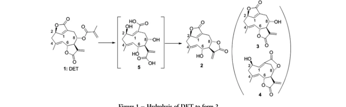
The parental compound DET was isolated and purified fromE. scaberaccording to the method published elsewhere (Huang et al., 2010), with a purity of more than 96% as judged by HPLC and NMR spectrometry analyses data. The hydrolysis of DET (1) with 1 N NaOH followed by acidic treatment pro-duced novel sesquiterpene dilactone alcohol 2 through C-7eC-8 lactonization of carboxylic acid5, which was generated by the cleavage of an ester and two lactone rings (Figure 1). This translactonization process is similar to the case of ele-phantopin, which isD4,5-epoxylated DET, to produce elephan-tol (Kupchan et al., 1969). In the case of cnicin, a sesquiterpene mono-lactone, C-8 alcohol was successfully obtained without lactone ring transformation under mild basic conditions with Na2CO3(Bruno et al., 2003). Thus, we expected that three alco-hols2e4might be produced through re-lactonization by the control of basic conditions; however, the alcohols 3 and 4
were not derived under any kind of hydrolysis conditions,
Figure 1eHydrolysis of DET to form 2.
including Na2CO3in our experiment. From the MM2 minimi-zation analysis (Chembridge software, Chem3D Pro Ver. 13.0.2.3021), alcohol 2 clearly showed low energy of 40.37 kcal/mol, while C-8 alcohol3(235.17 kcal/mol) and C-2 alcohol4(61.76 kcal/mol) indicate higher energy (Supporting information). This result suggests that the C-7eC-8 lactone ring on 2 is easier to form than the C-6eC-7 lactone on3 and the C-8eC-10 lactone on4. In addition, it became clear that the a-methylene-g-lactone ring in DET is extremely strained. The structure of C-6 alcohol2was confirmed by X-ray crystallography (Figure 2).
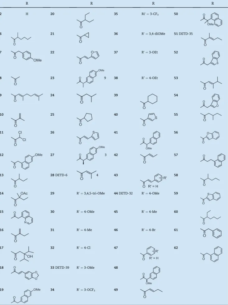
With alcohol2, various acyl groups were introduced to obtain the esters6e63(Figure 3,Table 1). An ester group often plays a crucial role not only in drug efficacy, but also as an important factor in biological activity. For instance, a lipo-philic conjugated ester group at C-6 in helenalin, a sesquiter-pene lactone, clearly contributed to its cytotoxicity against cancer cells in vitro and in vivo (Beekman et al., 1977; Lee et al., 1973). Designated ester groups were roughly classified into two groups, aliphatic group 6e27 and aromatic group
28e63. An aliphatic group might influence hydrophobicity, and its structure as well as size might affect interactions with the feature of the binding pocket of target protein/ enzyme. On the other hand, an aromatic group may contribute to planarity/hydrophobicity as well as thepep,p -anion/cation, and/orp-OH interactions with the target pro-tein/enzyme. Some of the ester groups were selected to be incorporated into2based on the structural features of other bioactive terpenoids; for example, methacrylate (DET side chain) for7, (E)-3,4-dimethylpent-2-enoate (bruceantin side chain) for16, 2-acetoxy-2-methylbutanoate (glaucarubinone side chain) for23, and cinnamate (from the SAR study of hel-enalin analogue) for28(Lee et al., 1973). Esterifications of2
were achieved by the following standard methods as shown in Figure 3: (a) Treatment of 2 with the related carboxylic acid (RCOOH), 1-(3-dimethylaminopropyl)-3-ethylcarbodi imide hydrochloride (EDCI) and dimethylaminopyridine (DMAP) to obtain the related esters7e24, 28e43and47e62; (b) Treatment of2with the related acid chloride (RCOCl) and triethylamine to obtain the esters25e27and44e46; (c) Treat-ment of2with 1-naphthylacetic acid under Mitsunobu condi-tions (Mitsunobu, 1981) to provide 63 accompanied with stereoinversion of C-6 (epi-51). Acetate6was produced as a side product during the preparation of24by removal of 2,4-dimethylpentan-3-one. The structures of all synthesized DETD were confirmed by 1H-NMR and high resolution MS spectra.
3.2. Anti-proliferative effects against breast cancer cell
lines and SAR analysis
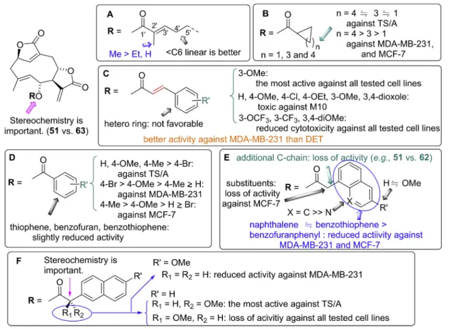
All the new DETDs2e63together with the parent DET (1) were evaluated for their anti-proliferative activities against murine mammary cancer cell line TS/A (ERþ, HER2) and human breast cancer cell lines MDA-MB-231 (ER, PR, HER2), MCF-7 (ERþ, HER2), and normal mammary M10 as shown inTable 2. The IC50values were analyzed by MTT assay in a 24-h treatment against the respective cells. The IC50of DET for TS/A and MCF-7 cells was determined at 2.3 mM, and much less effect was observed for MDA-MB-231 cells with IC50of 14.0mM. The cytotoxicity of C-6 alcohol2without an ester group is diminished in comparison with the parent DET (1). However, most of the ester analogues of 2display potent anti-proliferative activities against the tested breast cancer cell lines with less toxicity against normal mammary cells M10. Interestingly, methacrylate 7 keeping the same ester group as DET but at a different position obviously enhanced the anti-proliferative activity against human TNBC cell line MDA-MB-231, suggesting that the C-7eC-8 lactone ring contributes more anti-proliferation activity against MDA-MB231 than the C-6eC-7 lactone ring. The SAR of synthesized DETDs is summarized inFigure 4. Among all the aliphatic esters, thea,b-unsaturated ketone and/or methyl substituent ata-position have a tendency to increase the anti-proliferative activity (Figure 4A). Carbon chain length also affected the activity as indicated by the reduced activity of C1 linear 6and C7 linear 21. Of the cyclic aliphatic esters
25e27, cyclohexane 27 was more active against MDA-MB-231 and MCF-7 cells than25and26(Figure 4B). Ester substitu-ents carrying an aromatic function also displayed potent cyto-toxicity. In the case of cinnamate derivatives (Figure 4C)
28e41, 3-methoxycinnamate 33 (designated DETD-39) exhibited strong inhibitory effects against all tested breast cancer cells, which was the most potent among all synthetic analogues; however, it also showed toxicity against normal cell line M10. Decreased cytotoxicities were found in fluorine substituted and 3,4-dimethoxylated analogues 34e36. Ana-logues 42e48 with benzoate-like groups also effectively inhibited the growth of breast cancer cells. Of note, 4-bromobenzoate 46 demonstrated the most potent anti-proliferate activity against MDA-MB-231 (Figure 4D). Of the naphthalene acetate group51e61and63, the simple naph-thalene acetate 51 (designated DETD-35) displayed great anti-proliferative activity against all breast cancer cell lines, and of particular note, showed no inhibitory effect on M10
Figure 3ePreparations of ester analogues 6e63. Reagents and conditions: (A) RCOOH, EDCI, DMAP, CH2Cl2, rt; (B) RCOCl, Et3N,
Table 1eFunctional R group structures of DET analogues 2-62. Stereochemistries of all chiral centers are racemate, unless indicated.
R R R R
2 H 20 35 Ri0¼3-CF3 50
6 21 36 R0¼3,4-diOMe 51DETD-35
7 22 37 R0¼3-OEt 52
8 23 9 38 R’¼4-OEt 53
9 24 39 54
10 25 40 55
11 26 41 56
12 27 3 42 57
13 28DETD-6 4 43 58
14 29 R0¼3,4,5-tri-OMe 44DETD-32 R0¼4-OMe 59
15 30 R0¼4-OMe 45 R0¼4-Me 60
16 31 R0¼4-Me 46 R0¼4-Br 61
17 32 R0¼4-Cl 47 62
18 33DETD-39 R0¼3-OMe 48
cell growth (Figure 4E and F). Interestingly, some analogues such as56and 59can effectively inhibit the cell growth of MDA-MB-231, while less inhibition was observed against MCF-7.
Because most analogues showed similar cytotoxicity in Table 1, we therefore selected four compounds from aromatic esters28e63to see the effect of the distance between the ar-omatic ring and the terpene skeleton, the effect of the func-tional group on the aromatic ring, and the effect of p electron system. With that, 28 and 33 with the electron-withdrawing group were selected from the cinnamate group,
28e41, which has two carbons between the aromatic ring and the ketone.44was selected from 42e48, which where the aromatic ring is attached directly to the ketone.51was selected from49e61, which where one carbon exists between the aromatic ring and the ketone. Furthermore,51possesses a 10pelectron system rather than 6p. TheTable 3data show the toxicity results against different cancer cell lines by ana-logues 28 (DETD-6), 33 (DETD-39), 44 (DETD-32), and 51
(DETD-35). In addition to effectively inhibiting breast cancer cell proliferation, the four tested analogues also showed supe-rior inhibition against brain, lung, lymphoma, neuroepithe-lioma, kidney, prostate, stomach, colon and uterus cancer
cell proliferation. The most marked suppression effect among the different cancer types was observed in51(DETD-35) with no detected toxicity against M10 (Tables 2 and 3).28(DETD-6) and33(DETD-39) showed novel activity against different can-cer types, but they were also toxic to normal cells in vitro. Thus, 51(DETD-35) was chosen to be further examined for its effects on TNBC cell activity and the underlying mecha-nisms, along with the parental compound DET. The anti-cancer drug PTX was studied in parallel as a control and refer-ence drug.
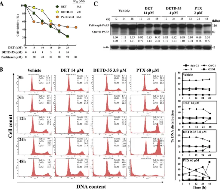
3.3. DET, DETD-35 and PTX induce cell-cycle arrest or
apoptosis
We determined the regulatory effects of DET, DETD-35 and PTX on the cell-cycle of MDA-MB-231 cells. Flow cytometric results revealed that when cells were treated for 48 h with vehicle (DMSO) or at the respective concentrations (IC50 values) of DET (14 mM), DETD-35 (3.8 mM) and PTX (60 mM) (Figure 5A) the G0/G1 phase DNA contents were decreased from 51.5% (vehicle control) to 36.2%, 39.0%, 11.2%, respec-tively, whereas the apoptotic sub-G1 DNA content was increased from 1.3% to 19.3%, 21.4%, and 66.8%, respectively
Table 2eCytotoxicity of synthesized DET analogues.
50% Inhibitory concentration (mM)a,b 50% Inhibitory concentration (mM)
TS/A MDA-MB-231 MCF-7 M10 TS/A MDA-MB-231 MCF-7 M10
1 2.3 14.0 2.3 e 34 e >15 e e
2 7.5 e e e 35 e 8.2 e e
6 3.5 e 6.7 e 36 e 9.5 e e
7 1.8 6.2 2.4 e 37 4.4 7.7 e e
8 1.9 7.5 3.7 e 38 2.1 2.7 3.3 4.0
9 1.7 4.6 3.5 e 39 2.5 3.4 3.6 4.7
10 2.1 7.0 3.9 e 40 2.2 4.9 4.1 e
11 2.7 5.9 3.4 e 41 2.7 6.0 4.5 e
12 1.9 6.7 3.4 e 42 2.4 4.3 3.7 e
13 2.3 e 3.9 e 43 2.0 4.5 5.9 e
14 4.0 e 5.8 e 44c 1.9 3.9 4.3 e
15 1.6 4.6 2.4 e 45 1.8 4.2 2.7 e
16 3.3 7.0 4.2 e 46 3.0 2.2 6.1 e
17 3.3 5.5 2.6 e 47 3.1 4.9 6.1 e
18 2.2 5.8 3.5 e 48 2.8 5.0 5.3 e
19 4.0 e 4.5 e 49 2.4 6.9 e e
20 1.8 4.3 2.5 4.7 50 2.2 7.0 e e
21 e e e e 51c 1.7 3.5 3.2
22 e e e e 52 2.0 3.1 3.1 4.7
23 2.9 e 4.2 e 53 e >15 e e
24 2.4 5.9 2.5 e 54 3.8 7.3 e e
25 2.4 8.7 7.1 e 55 4.1 3.8 4.1 4.8
26 3.0 6.7 6.2 e 56 3.1 4.9 e e
27 2.6 5.2 4.6 e 57 1.9 4.2 4.5 e
28c 1.6 4.2 1.6 4.9 58 e >15 e e
29 1.8 6.1 2.3 e 59 2.8 4.1 e e
30 1.5 4.1 3.7 4.7 60 2.1 3.8 3.8 5.8
31 2.4 3.6 3.7 e 61 3.5 5.1 e e
32 1.8 4.2 3.6 4.8 62 e >15 e e
33c 0.8 1.9 2.1 3.7 63 e 7.4 e e
a Breast cancer cell lines; TS/A (ERþ, HER2), MDA-MB-231 (ER, PR, HER2), MCF-7 (ERþ, HER2), M10 (normal mammary). b IC50was not detectable at 0e5mM for TS/A, MCF-7, and M10; 0e7.5mM for MDA-MB-231.
(Figure 5B). A typical time-dependent G2/M arrest in MDA-MB-231 cells treated with PTX was observed. In comparison, DET or DETD-35-treated cells did not show any cell-cycle arrest at specific stages, but cell apoptosis was significantly induced
at 48 h treatment. Expression of full-length and cleaved poly ADP-ribose polymerase (PARP), a protein marker for pro-grammed cell death which upon cleavage will lead to apoptosis, was evaluated by western blotting as shown in
Figure 4eSummary of structure-activity relationship of DET analogues in inhibition of proliferation of TS/A, MCF-7, MDA-MB-231 breast cancer cells or M10 normal mammary cells. (A) Aliphatic ester derivatives (6e24); (B) Cyclic aliphatic esters (25e27); (C) Cinnamate derivatives (28e41); (D) DETDs carrying benzoate-like structures (42e48); (E and F) Naphthalene acetate derivatives (49e62). Among all, naphthalene acetate 51 (DETD-35) showed significantly potent cytotoxicities against all types of tested breast cancer cell lines without growth inhibition of normal mammary M10 cells. Cinnamate 33 (DETD-39) strongly inhibited the growth of all tested breast cancer cells; however, it also inhibited normal mammary cell growth.
Table 3eSummary of the 50% inhibitory concentration (IC50,mM) of DET and the selected DETDs on inhibiting viability of various cancer cell types. The cells were treated with compound for 24 h and then subjected to MTT assays.
Cell name Tumor origin 1 DET 28 (DETD-6) 33 (DETD-39) 44 (DETD-32) 51 (DETD-35)
MCF-7 Breast 5.0 4.8 2.4 4.3 3.9
MDA-MB-231 Breast 14.0 4.0 1.9 3.9 3.5
TS/A (mouse) Breast 2.6 1.8 0.8 1.9 1.7
B16eF10 (mouse) Skin 6.00 3.90 1.70 4.20 2.70
U-87MG Brain 1.00 0.35 0.15 0.42 0.34
PC6 Lung 0.40 0.17 0.11 0.47 0.34
U937 Lymphoma 0.48 0.18 0.10 0.39 0.18
SK-N-MC Neuroepithelioma 0.51 0.32 0.11 0.51 0.26
A498 Kidney 2.82 1.11 0.48 1.48 0.79
PC3 Prostate 12.8 4.5 2.6 6.2 5.5
KATO III Stomach 1.47 0.42 0.29 0.82 0.65
HCT-116 Colon 0.28 0.11 0.10 0.36 0.14
Figure 5C. The 48-h DET treatment induced significant cleav-age in PARP, while 48-h PTX treatment reduced the expression of full-length PARP. These results indicate that different mechanisms may underlie the three treatments although they all show effects on inducing the apoptotic sub-G1 content in cell cycle analysis.
3.4. Inhibitory effects on MDA-MB-231 cell migration,
invasion, and motility
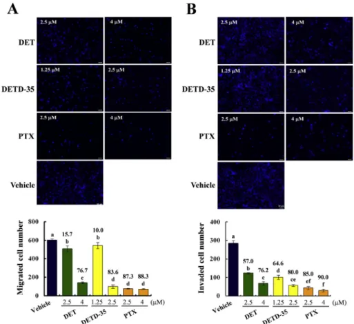
Boyden chamber assay was used to investigate the effects of DET and DETD-35 on the migration and invasion activity of MDA-MB-231 cells, and PTX was used as a reference control. Figure 6A shows that DET (2.5 and 4mM), DETD-35 (1.25 and
2.5 mM) and PTX (2.5 and 4 mM) significantly inhibited cell migration in a concentration-dependent manner under 24-h treatment. The high concentrations of DET, DETD-35 and PTX greatly reduced by 77%, 84% and 88%, respectively, cell migration, as compared to the vehicle control group, while the suppressed effect of the low concentration of DET, DETD-35 and PTX was 16%, 10% and 87%, respectively (P < 0.05). Transwell invasion assay further revealed that DET and DETD-35 inhibited invasion of MDA-MB-231 cells through a thin Matrigel matrix. It was observed that the high concentration of DET, DETD-35 and PTX significantly inhibited cell invasion by 76%, 80% and 90%, respectively, and the anti-invasion effect of the low concentrations were 57%, 65% and 85%, respectively (P<0.05,Figure 6B). These data demonstrate
that both DET and DETD-35 possess migration and anti-invasion activity against MDA-MB-231 cells, with DETD-35 exerting a better activity than DET.
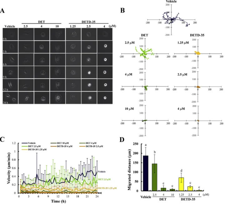
The kinetic characteristics of MDA-MB-231 cell motility treated with DET or DETD-35 for 24 h were further investigated by time-lapse confocal microscope. Trajectories of migrating MDA-MB-231 cells under different treatment conditions were monitored and analyzed over a 24-h period. Vehicle control-treated cells were observed to be moving energetically with robust membrane protrusion resulting in a continuous motion (0e24 h) (Figure 7A). As shown inFigure 7B, the most significant cell dispersion area from the origin was observed in vehicle control, and a relatively small area due to restricted movements was seen in cells treated with 2.5mM DET for 12 h. Considerably reduced cell trajectories were observed in cells treated with 4 and 10mM DET, or 1.25, 2.5 and 4mM DETD-35, respectively. The migration velocities of MDA-MB-231 cells under each treatment were further monitored and measured every 30 min. In cells treated with DETD-35 at 1.25, 2.5 and 4mM, and the higher concentrations of DET at 4 and 10mM, the suppression of movement became much more prominent with the average motility velocity decreasing significantly compared to vehicle control cells and cells treated with 2.5mM DET (Figure 7C). Furthermore, the migrated distances were significantly shorter in 2.5, 4, 10mM DET and 1.25, 2.5, 4mM DETD-35 treated cells (144 vs. 15 vs. 9 and 70 vs. 23 vs.
3mm) than that of the control (186mm) (Figure 7D). These re-sults indicate that DETD-35, a newly synthesized sesquiter-pene lactone exhibits potent and better bioactivity than DET in suppressing MDA-MB-231 cell motility.
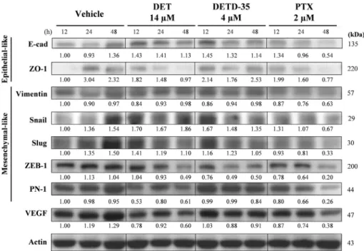
Expression profiles of several epithelial-to-mesenchymal transition protein markers in MDA-MB-231 cells were also evaluated as shown inFigure 8. Cancer cells with metastatic characteristics are known to lose adherence-related proteins, or epithelial-like proteins. DET and DETD-35 both showed a better effect on retaining E-cadherin (E-cad) expression in comparison to PTX, while DETD-35 showed the most signifi-cant activity in retaining the tight junction protein zonula occludens-1 (ZO-1) among the three treatments. DET and DETD-35 did not regulate the expression of vimentin, an inter-mediate filament protein expressed mostly in mesenchymal cells, which on the other hand was time-dependently downre-gulated by PTX. Transcription factors resulting in the repres-sion of E-cad including Snail, Slug, and ZEB-1 were expressed differently among the three treatments. DET treat-ment reduced ZEB-1 expression level to 49% at 48 h while DETD-35 exerted the same effect at an earlier stage at 24 h. Ex-pressions of VEGF and protease nexin-1 (PN-1), a serine prote-ase inhibitor, were downregulated in DET- and PTX-treatment but not in DETD-35 treatment. These protein expression data not only demonstrated the mechanistic insights of the com-pound modulation in breast cancer cell migration, invasion,
and motility, but also indicated a variation in mechanisms exerted on MDA-MB-231 cells among DET, DETD-35, and PTX.
3.5. Synergism between DETD-35 and PTX against
MDA-MB-231 cells
Combining cytotoxic drugs, such as PTX, with less toxic bio-logical agents is one way to relieve side effects, especially in adjuvant therapy (Oakman et al., 2009). The identification and validation of the potential benefits of phytocompounds with low toxicity but effective for cancer inhibition has thus become an important area of pharmaceutical science (Newman and Cragg, 2007). The pressing need for develop-ment of new therapeutic or preventive agents for cancer
diseases has spurred the search for bioactive phytocom-pounds or their derived analogues with novel modes of action or improved efficacy (Lin et al., 2012; Newman and Cragg, 2007; Pan et al., 2010). To investigate whether DETD-35 acts syner-gistically with PTX against MDA-MB-231 cell activity, anti-proliferation assay using MTT reagent was first performed to obtain IC values of each compound. IC40of PTX and DETD-35 on MDA-MB-231 cells were shown as 23.8 nM and 3.32mM, respectively (Figure 9A). Further, cells treated with a fixed con-centration of DETD-35 at 1mM were co-treated with ascending concentrations of PTX (0e25 nM) and displayed better inhibi-tory activity on cancer cell proliferation than PTX treatment alone (Figure 9A and B). Based on the IC40value of each com-pound, we used concentrations of DETD-35 ranging from
0 to 4mM and PTX from 0 to 100 nM for compound-drug com-bination study. Classic isobologram analysis and Chou-Talalay method were conducted to investigate the synergistic effect of compound and drug. Combination index (CI) was calculated with the CalcuSyn software (Version 2.0, Biosoft) and expressed as CI vs. Fa (fraction affected). CI<1 indicates synergy; CI¼1 indicates additive effect; and CI>1 indicates antagonism. We observed that DETD-35 in combination with PTX acts synergistically in inhibiting the growth of MDA-MB-231 cells (Figure 9C and D).
3.6. DETD-35 potently suppressed lung metastasis of
MDA-MB-231 cells
To examine the drug efficacyin vivo, DETD-35 and the refer-ence drug PTX were administrated before and/or after MDA-MB-231 cell injection into xenograft mice. An alternate treat-ment group was included in this study to evaluate the syner-gistic effect of DETD-35 and PTX in animals in which mice were administrated alternatively with a half of dose of either compound in the single-treatment group. This treatment was designed to seek a strategy to potentially reduce PTX-related side effects but retain the same treatment efficacy.
DETD-35 pre-treatment at a dosage of 10 mg/kg (pre-DETD-35-10) was shown to significantly reduce the number of tumor foci (83%) in the lungs of tumor-bearing mice especially when treated before tumor occurrence, suggesting its potential application as a preventive agent (Figure 10A). DETD-35 and PTX post-treatment at 10 mg/kg and 5 mg/kg showed 50% and 62%, respectively, reduction of lung tumor foci. DETD-35 at 2 mg/kg, however, showed little effect. Interestingly, the alternate treatment group PTX-5þDETD-35-2 with adminis-tration of DETD-35 at 2 mg/kg (10 doses) and PTX at 5 mg/kg
(11 doses) exerted a similar effect to that of PTX treatment alone at 5 mg/kg (21 doses), implying a possible strategy to reduce PTX cytotoxicity while retaining treatment efficacy at the same time, as none of the treatments showed a negative effect on the organ index of liver, kidney, spleen, and lung in the tested animals (Figure 10B).
3.7. Histopathological and immunohistochemistry
analysis of lung tissues
Histopathology was performed using hematoxylin and eosin (H&E) staining to depict the lung architecture within different treatment groups. As shown inFigure 11, the sham control mice showed typical lung alveoli architecture, and the tumor control group showed fully grown tumor mass, with the nuclei becoming larger and more irregular compared with the sham control group. DETD-35 inhibited histological alterations in lung tissue in a dose dependent manner. The PTX groups also expressed fewer alveolar defects compared to the tumor control group. Compared to other treatment groups, pre-treatment pre-DETD-35-10 and alternate-pre-treatment PTX-5 þ DETD-35-2 significantly inhibited tumor growth and metastasis in mouse lung tissues.
The protein markers used in IHC staining correlate with proliferation (Ki67), angiogenesis (VEGF), inflammation (COX-2) and apoptosis (caspase 3) in lung mammary tumor sites. Our results revealed that the protein expressions of these markers were significantly attenuated in DETD-35 treat-ment compared with the corresponding tumor vehicle con-trols in mice lung tissues (Figure 11). For lung samples taken after 71 days of cancer cell injection into the tail vein, the over-expression of Ki67 and VEGF were detected in the tumor con-trol group which were suppressed in the DETD-35 and PTX
groups. DETD-35 showed dose-dependent inhibition of Ki67 and VEGF expression in lung tissue. Pre-treatment with DETD-35 and alternate-treatment of DETD-35 with PTX signif-icantly inhibited the protein expression levels of VEGF and Ki67 as well as on pro-inflammatory enzyme COX-2 in lung tu-mor foci, whereas increased expression of apoptotic marker caspase 3 was found in the DETD-35, PTX and alternate-treatment of DETD-35 with PTX groups (Figure 12). Together with results fromin vitrocell-based assays, analysis of protein expression levels in metastatic MDA-MB-231 tumor xenograft
provides strong evidence to support the suppressive effect of DETD-35 on TNBC metastasis.
4.
Conclusion
In summary, a novel sesquiterpene di-lactone2was obtained from a bioactive natural product, deoxyelephantopin (DET,1) by lactone ring transformation. Fifty-eight ester analogues of
2 were designed, synthesized and evaluated for anti-breast
Figure 9eCompound-drug combination studies of DETD-35 and PTX against MDA-MB-231 cell proliferation. (A) Inhibition of cell proliferation by PTX or DETD-35. Cells were treated with indicated concentrations for 24 h. Cell viability was determined by MTT assay. (B) Bar graph showing the combination effect of PTX and DETD-35. (C) Classic isobologram analysis graph and (D) combination index (CI) plot of DETD-35 co-treatment with PTX. CI<1 indicates synergy; CI[1 indicates additive effect; and CI>1indicates antagonism.
cancer activity. Most of the synthesized analogues exhibited better anti-proliferative activity against TNBC cell line MDA-MB-231 than that of parental DET. SAR study revealed the a,b-unsaturated ketone and/or methyl substituent at thea po-sition have a tendency to increase the anti-proliferative activ-ity in the case of the aliphatic esters. Analogues with cinnamates such as 33 (DETD-39), benzoates such as 44
(DETD-32), and naphthalene acetates such as51 (DETD-35), also showed potent anti-breast cancer activity. The most potent analogue51(DETD-35) and the parent DET were further investigated for their anti-TNBC effectsin vitro and in lung metastasis xenograft mouse model using an MDA-MB-231 cell line. By employing various assays including cell viability assay, Boyden chamber analysis, time-lapse microscopy, and immunohistochemistry analysis, DETD-35 was shown to exhibit a better effect than DET on inhibiting cell migration, in-vasion, and motility of MDA-MB-231 cells in a concentration-dependent manner. Among the designed compound and drug administration strategies in the animal study, DETD-35
pre-treatment (at 10 mg/kg body weight) showed the most sig-nificant reduction in metastatic pulmonary foci (83%) of MDA-MB-231 cells in mice; the alternate treatment of DETD-35þPTX also greatly reduced metastatic pulmonary foci (71%). Our observation suggested that DETD-35 may have the potential for further development into a complementary or sensitizing agent to chemotherapeutic drugs especially for TNBC.
Grant support
This work was supported by a Grant from the National Research Program for Biopharmaceuticals (NRPB) (NSC 101-2325-b-001-007), Taiwan and institutional grant awarded to L.F.S. This study was also supported in part by a Grant-in-Aid from the Ministry of Education, Culture, Sports, Science and Technology (MEXT KAKENHI, Japan) awarded to K.N.G. (Grant Number 25293024 & 25670054), and a NIH grant
from the National Cancer Institute awarded to K.H.L. (CA-177584).
Conflict of interest statement
The authors declare no competing financial interest.
Acknowledgments
The authors thank the technical assistance of the X-ray crys-tal structure determination of DETD-2 by the X-ray Facility at Institute of Chemistry, Academia Sinica, Taiwan.
Appendix A.
Supplementary data
Supplementary data related to this article can be found at http://dx.doi.org/10.1016/j.molonc.2016.03.002.
R E F E R E N C E S
Beekman, A.C., Woerdenbag, H.J., Uden, W., Pras, N.,
Konings, A.W., Wikstr€om, H.V., Schmidt, T.J., 1977. Structure-cytotoxicity relationships of some helenanolide-type sesquiterpene lactones. J. Nat. Prod. 60, 252e257.
Bruno, M., Rosselli, S., Maggio, A., Raccuglia, R.A., Napolitano, F., Senatore, F., 2003. Antibacterial evaluation of cnicin and some
natural and semisynthetic analogues. Planta Med. 69, 277e281.
Chou, T.C., 2010. Drug combination studies and their synergy quantification using the Chou-Talalay method. Cancer Res. 70, 440e446.
Farha, A.K., Dhanya, S.R., Mangalam, S.N., Geetha, B.S., Latha, P.G., Remani, P., 2014. Deoxyelephantopin impairs growth of cervical carcinoma SiHa cells and induces apoptosis by targeting multiple molecular signaling pathways. Cell Biol. Toxicol. 30, 331e343.
Geetha, B.S., Nair, M.S., Latha, P.G., Remani, P., 2012.
Sesquiterpene lactones isolated fromElephantopus scaberL. inhibits human lymphocyte proliferation and the growth of tumour cell lines and induces apoptosis in vitro. J. Biomed. Biotechnol. 2012.
Govindac, T.R., Sidhaye, A.R., Viswanat, N., 1970.
Deoxyelephantopin, a new sesquiterpene fromElephantopus scaberLinn. Indian J. Chem. 8, 762.
Hiradeve, S.M., Rangari, V.D., 2014. A review on pharmacology and toxicology ofElephantopus scaberLinn. Nat. Prod. Res. 28, 819e830.
Ho, B.Y., Lin, C.H., Apaya, M.K., Chao, W.W., Shyur, L.F., 2012. Silibinin and paclitaxel cotreatment significantly suppress the activity and lung metastasis of triple negative 4T1 mammary tumor cell in mice. J. Tradit. Complement. Med. 2, 301e311. Howlader, N., Noone, A.M., Krapcho, M., Neyman, N., Aminou, R.,
Waldron, W., Altekruse, S.F., Kosary, C.L., Ruhl, J.,
Tatalovich, Z., Cho, H., Mariotto, A., Eisner, M.P., Lewis, D.R., Chen, H.S., Feuer, E.J., Cronin, K.A. (Eds.), 2012. SEER Cancer Statistics Review, 1975e2009 (Vintage 2009 Populations). National Cancer Institute.
Huang, C.C., Lo, C.P., Chiu, C.Y., Shyur, L.F., 2010. Deoxyelephantopin, a novel multifunctional agent, suppresses mammary tumour growth and lung metastasis and doubles survival time in mice. Br. J. Pharmacol. 159, 856e871.
Huang, C.C., Lin, K.J., Cheng, Y.W., Hsu, C.A., Yang, S.S., Shyur, L.F., 2013. Hepatoprotective effect and mechanistic insights of deoxyelephantopin, a phyto-sesquiterpene lactone, against fulminant hepatitis. J. Nutr. Biochem. 24, 516e530.
Kabeer, F.A., Sreedevi, G.B., Nair, M.S., Rajalekshmi, D.S., Gopalakrishnan, L.P., Kunjuraman, S., Prathapan, R., 2013. Antineoplastic effects of deoxyelephantopin, a sesquiterpene lactone fromElephantopus scaber, on lung adenocarcinoma (A549) cells. J. Integr. Med. 11, 269e277.
Kabiru, A., Por, L.Y., 2013. Elephantopus species: traditional uses, pharmacological actions and chemical composition. Adv. Life Sci. Technol. 15, 6e13.
Kupchan, S.M., Aynehchi, Y., Cassady, J.M., Schnoes, H.K., Burlinga, A.L., 1969. Tumor inhibitors. 40. Isolation and structural elucidation of elephantin and elephantopin, 2 novel sesquiterpenoid tumor inhibitors fromElephantopus elatus. J. Org. Chem. 34, 3867e3875.
Kupchan, S.M., Eakin, M.A., Thomas, A.M., 1971. Tumor inhibitors. 69. Structure-cytotoxicity relationships among sesquiterpene lactones. J. Med. Chem. 14, 1147e1152. Kurokawa, T., Nakanish, K., Wu, W., Hsu, H.Y., 1970.
Deoxyelephantopin and its interrelation with elephantopin. Tetrahedron Lett. 11, 2863e2866.
Lee, K.H., Huang, E.S., Piantado, C., Pagano, J.S., Geissman, T.A., 1971. Cytotoxicity of sesquiterpene lactones. Cancer Res. 31, 1649e1654.
Lee, K.H., Meck, R., Piantado, C., Huang, E.S., 1973. Antitumor agents. 4. Cytotoxicity and in vivo activity of helenalin esters and related derivatives. J. Med. Chem. 16, 299e301.
Lee, W.L., Wen, T.N., Shiau, J.Y., Shyur, L.F., 2010. Differential proteomic profiling identifies novel molecular targets of paclitaxel and phytoagent deoxyelephantopin against mammary adenocarcinoma cells. J. Proteome Res. 9, 237e253. Lee, W.L., Shyur, L.F., 2012. Deoxyelephantopin impedes
mammary adenocarcinoma cell motility by inhibiting calpain-mediated adhesion dynamics and inducing reactive oxygen species and aggresome formation. Free Radic. Bio. Med. 52, 1423e1436.
Lin, C.H., Lee, W.L., Shyur, L.F., 2012. An overview of the current development of phytoremedies for breast cancer. In: Cho, W.C.S. (Ed.), Materia Medica for Various Cancers, Evidence-based Anticancer Complementary and Alternative Medicine. Springer, pp. 47e67.
Mitsunobu, O., 1981. The use of diethyl azodicarboxylate and triphenylphosphine in synthesis and transformation of natural-products. Synthesis-Stuttgart 1, 1e28.
Newman, D.J., Cragg, G.M., 2007. Natural products as sources of new drugs over the last 25 years. J. Nat. Prod. 70, 461e477. Oakman, C., Pestrin, M., Cantisani, E., Licitra, S., DeStefanis, M.,
Biganzoli, L., Di Leo, A., 2009. Adjuvant chemotherapyethe dark side of clinical trials. Have we learnt more? Breast 18, S18eS24.
Pan, L., Chai, H.Y., Kinghorn, A.D., 2010. The continuing search for antitumor agents from higher plants. Phytochem. Lett. 3, 1e8. Picman, A.K., 1986. Biological-activities of sesquiterpene lactones.
Biochem. Syst. Ecol. 14, 255e281.
Su, M.X., Chung, H.Y., Li, Y.L., 2011. Deoxyelephantopin from
Elephantopus scaberL. induces cell-cycle arrest and apoptosis in the human nasopharyngeal cancer CNE cells. Biochem. Bioph. Res. Co. 411, 342e347.
Zahari, Z., Jani, N.A., Amanah, A., Latif, M.N.A., Majid, M.I.A., Adenan, M.I., 2014. Bioassay-guided isolation of a sesquiterpene lactone of deoxyelephantopin from
Elephantopus scaberLinn. active on Trypanosome brucei rhodesience. Phytomedicine 21, 282e285.