Dr Satyendra Mohapatra et al JMSCR Volume 08 Issue 02 February 2020 Page 496
MRI Evaluation of Compressive Myelopathy
Authors
Dr Satyendra Mohapatra
1, Dr Braja Behari Panda
2*,
Dr Savitri Bhagat
3, Dr Amulya Kumar Panda
41
PG Resident, Veer Surendra Sai Institute of Medical Sciences and Research (VIMSAR), Burla
2
Associate Professor, Department of Radiodiagnosis, Veer Surendra Sai Institute of Medical Sciences and Research, Burla
3
Professor and H.O.D., Department of Radiodiagnosis, Veer Surendra Sai Institute of Medical Sciences and Research, Burla
4
Senior Resident, Department of Radiodiagnosis, Veer Surendra Sai Institute of Medical Sciences and Research, Burla
*Corresponding Author
Dr Braja Behari Panda Abstract
Background:Compressive myelopathy is the term used to describe the spinal cord compression either from outside or within the cord. The role of MRI is to distinguish compressive from noncompressive cause of myelopathy.
Material & Method:A cross sectional study of 60 patients with clinical history of compressive myelopathy are evaluated for various causes by 1.5 Tesla MRI scanner.
Result: Out of 60 cases of compressive myelopathy various different causes are trauma (26), Infection (14), Primary Neoplasm (10), Secondary Neoplasm (10).
Conclusion: MRI is very definitive, Sensitive, Accurate, Noninvasive, Radiation free modality for evaluation of compressive myelopathy.
Keyword: MRI, Compressive myelopathy, Primary Neoplasm, Secondary Neoplasm.
Introduction
Compressive myelopathy is the term used to describe the spinal cord compression either from outside or within the cord itself. Compression may be due to trauma, displaced vertebrae, epidural abscess, epidural hemorrhage, intradural and extradural neoplasm.
Spinal cord injury is the major cause of quadriplegia and disability. Plain Radiograph has a low sensitivity for identifying traumatic spinal lesion. In patients who have negative plain film for spinal injury but high clinical suspicion of
spinal injury should undergo MR for a more definitive evaluation of spine. MRI is a definite modality for assessing soft tissue injuries.
In case of suspected cord compression due to neoplasm, MRI serves as an excellent method for imaging tumour involving spinal column, canal and cord. Of all the areas of spinal pathology, It may be in the field of spinal tumour that MRI has had most impact.
Spinal tumour are categorized as extradural, intradural extra medullary, intra medullary.
http://jmscr.igmpublication.org/home/ ISSN (e)-2347-176x ISSN (p) 2455-0450
Dr Satyendra Mohapatra et al JMSCR Volume 08 Issue 02 February 2020 Page 497 Secondary tumour may arise from lungs
carcinoma, breast carcinoma, lymphoma and renal cell carcinoma, maximum lesion are extradural in location.
Infectious etiology of spinal compression are mainly due to tubercular. Maximum infectious lesions are extra dural.
Many spinal cord diseases are reversible if recognized and treated in early stage.
Material & Method
The study was conducted in VIMSAR, Burla. We included 60 patients of both sex.
The study was conducted from September 2017 to August 2019, 2 years.
All the patients underwent MRI evaluation.
Inclusion Criteria
All age group> 10 yr, both sex, all cases of compressive myelopathy.
Exclusion Criteria
Cases of non-compressive myelopathy
Degenerative disc herniation.
MRI evaluation is performed by GE 1.5 TESLA Electromagnet machine.
Whenever possible patients were followed up for histopathological diagnosis in case of neoplasm and outcome in case of spinal trauma
Observation
All the patients with compressive myelopathy were subjected to MRI evaluation.
MRI evaluate various cause of compressive myelopathy
MRI used to classify the lesion based on location in to extra dural and intra dural
MRI also evaluate age and sex distribution of various causes of compressive myelopathy
Table 1: Causes of Compressive myelopathy
MR diagnosis Number of patients(n=60) %
Traumatic Myelopathy 26 43.3
Infection/TB 14 23.3
Primary neoplasm 10 16.7
Secondary Neoplasm/Metastases 10 16.7
Most common cause for compressive myelopathy in our study was spinal trauma (43.3%) followed by spinal infection (23.3%).
Table 2: Level of Spinal Injury
Level of lesion Number of patients(n=26) %
C: Cervical 12 46.2
T: Thoracic 14 53.8
LT: Lower Thoracic 12 46.2
UT: UpperThoracic 2 7.7
TL: Thraco – Lumbar 0 0.0
L: Lumbar 4 15.4
Thoracic is the most common site of spinal injury.
Table 3: Infectious Causes of Compressive Myelopathy
Infection No. of Patients (Total 14)
Tubercular 12
Pyogenic Epidural Abscess 1
Hypertrophic Pachymeningitis 1
Dr Satyendra Mohapatra et al JMSCR Volume 08 Issue 02 February 2020 Page 498
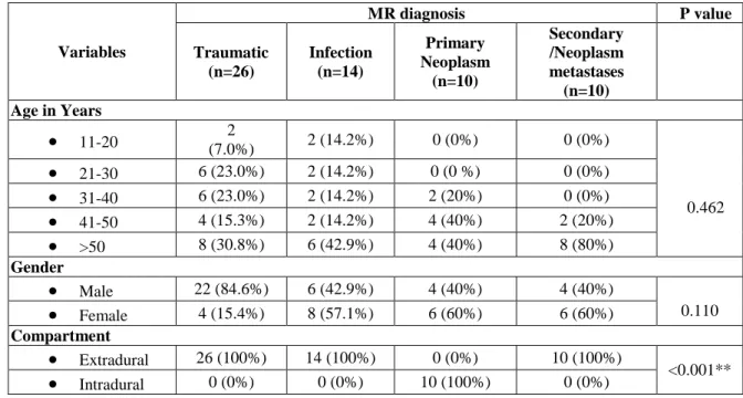
Table 4: Age, Gender & Compartmental Distribution of Various Causative factors according to the MRI Finding
Variables
MR diagnosis P value
Traumatic (n=26)
Infection (n=14)
Primary Neoplasm
(n=10)
Secondary /Neoplasm metastases (n=10) Age in Years
11-20 (7.0%) 2 2 (14.2%) 0 (0%) 0 (0%)
0.462
21-30 6 (23.0%) 2 (14.2%) 0 (0 %) 0 (0%)
31-40 6 (23.0%) 2 (14.2%) 2 (20%) 0 (0%)
41-50 4 (15.3%) 2 (14.2%) 4 (40%) 2 (20%)
>50 8 (30.8%) 6 (42.9%) 4 (40%) 8 (80%)
Gender
Male 22 (84.6%) 6 (42.9%) 4 (40%) 4 (40%)
0.110
Female 4 (15.4%) 8 (57.1%) 6 (60%) 6 (60%)
Compartment
Extradural 26 (100%) 14 (100%) 0 (0%) 10 (100%)
<0.001**
Intradural 0 (0%) 0 (0%) 10 (100%) 0 (0%)
Table 5: Characterisation of Traumatic Spinal Injuries by MRI
MRI finding No. of patients (n=26) %
1.Stable fractures 12 46.2
2.Unstable fractures 14 53.8
3.Posterior elements fracture 14 53.8
4.Ligamentous injury 14 53.8
5.Cord changes 24 92.3
6.Epidural hematoma/ soft tissue component 12 46.2
7.Pre and paravertebral collection 12 46.2
Table 6: Primary Tumour Classification
Primary neoplasms No. of patients
Neurofibroma 04
Meningioma 06
Total 10
Meningioma is the most common cause of primary tumour causing compressive myelopathy.
Table 7: Location of Primary Tumour
Diagnosis Cervical Thoracic Lumbar
Meningioma 0 4 2
Neurofibroma 2 2 0
Meningioma are more common in thoracic region where as neurofibroma are common in thoracic & cervical region.
Table 8: Site of Metastasis
Levels of lesions in
Secondary neoplasm/metastases
Number of patients (n=10)
%
Cervical 2 20.0
Thoracic 8 80.0
Upper 4 40.0
Lower 4 40.0
Thoraco lumbar (T12-L1) 0 0.0
Lumbar 2 20.0
Dr Satyendra Mohapatra et al JMSCR Volume 08 Issue 02 February 2020 Page 499
Table 9: Location of the Pathology
Compartment Number of patients %
Extradural 50 83.3
Intradural – Extramedullary 10 16.7
Total 60 100.0
Extradural compressive lesions (83.3%) are the most common cause for compressive myelopathy.
Image 1: T2Sag Intradural Extramedullary
Neurofibroma
Image 2: Post Contrast sag Meningioma
Image 3: T2Sag Tuberculosis of Spine with Cold
Abscess
Image 4: T2Sag Sclerotic Metastasis - Carcinoma
Prostate
Image 5: Fracture Dislocation Causing Cord Edema Cord Compression
Discussion
A total of 60 patients referred were studied for compressive myelopathy using MRI in department of Radiodiagnosis, Veer Surendra Sai Institute of Medical Sciences and Research, Burla.
Dr Satyendra Mohapatra et al JMSCR Volume 08 Issue 02 February 2020 Page 500 In traumatic cases level of injuries were thoracic
(53.8%), cervical (46%), lumbar (15.4%) this comparable with study conducted by Kerslake et al1.
The age of patients in our study range from 12 to 70 year ,mean 42 year and 22 were male and 4 were female (male:female-11:2). This is in comparission with study done by Yamashita et al2. Among 26 cases of traumatic compressive myelopathy RTA (70%), fall from height (30%), which is similar to study conducted by Kulkarni et al3.
Among 26 patients 24 had cord changes and 2 had no changes.24 patiets showed hypointensity on T1WI, and hyperintensity on T2WI and FLAIR suggestive of cord edema and contusion .The signal changes are in consistence with study done previously by Hackney et al4.
In our study infectious causes of compressive myelopathy were (14). 12 cases were in thoracic region and 2 in lumbar region. Epidural component compressing the cord was seen in all the 14 cases. Study done by Roos DEA et al5 showed thoracolumbar region is the most common affected site as in our study.
We had 10 cases of primary neoplasm, among which 4 were neurofibroma & 6 cases were meningioma. All the primary neoplasm were intradural extra medullary.
We had 10 cases of secondary neoplasm, out of which 6 patients (60%) showed more than 1 lesion,this is comparison to study done by Lien et al 6.Among 10 patients, we had 3 patients with primary carcinoma bronchus,2 had breast carcinoma, 2 had lymphoma,1 had carcinoma of prostate , 1 had RCC &1 with unknown primary. In our study compartmental distribution of pathology. Out of 60 patients, 50 showing extradural, 10 showing intradural extra medullary pathology.
Conclusion
MRI could successfully characterize the spinal tumour based on location into extradural/ intradural and assess the integrity of spinal cord,
intervertebral discs and ligament after acute trauma. So in the end MRI is very definitive, sensitive, accurate, though costly but very specific, noninvasive, radiation free modality for evaluation of compressive myelopathy
References
1. Kerslake RW, Jaspan T, Worthington BS. Magnetic resonance Imaging of Spinal trauma: The British Journal of Radiology. 1991;64:386-402.
2. Yamashita Y, Takahashi M, Matsuno Y, Kojima R, Sakamoto Y, Oguni T, et al. Acute Spinal cord Injury. Magnetic resonance Imaging correlated with myelopahty: The British Journal of Radiology. 1991;64:201-209.
3. Kulkarni MV, Mc Ardle CB, KapanickyD,Miner M, Cotler HB, Lee KF, etal. Acute Spinal Cord Injury. MR Imaging at 1.5 T: Radiology. 1987; 164: 837-843.
4. Hackney DB, Asata R, Sci DM, Joseph PM, Carvlin MJ, McGrath JT, Grossman RI, et al. Hemorrhage and edema in Acute spinal cord compression. Demonstration by MR Imaging: Radiology. 1986; 161: 387-390.
5. Roos DEA, Persijn V, Meerten EL, Bloem JI. MRI of Tubercular spondylitis: AJR. 1986; 146: 79-82.