TOOLS FOR PROBING 2A SEQUENCE SPACE
Helena Escuin Ordinas
A Thesis Submitted for the Degree of PhD at the
University of St. Andrews
2008
Full metadata for this item is available in the St Andrews Digital Research Repository
at:
https://research-repository.st-andrews.ac.uk/
Please use this identifier to cite or link to this item: http://hdl.handle.net/10023/692
Tools for Probing 2A Sequence Space
By
Helena Escuin Ordinas School of Biology University of St. Andrews
A thesis submitted for the Degree of Doctor of Philosophy at the University of St. Andrews
ABSTRACT
Foot-and-mouth disease virus (FMDV) 2A is an oligopeptide composed of
only 18 amino acids that can mediate a co-translational cleavage at its own
C-terminus. It has been observed that 2A sequences do not show cleavage activity
within bacterial organisms. Why 2A lacks activity in a prokaryotic organism such as
E.coli is unclear. A series of plasmids designed to provide a phenotypic screen for
2A-mediated cleavage (in prokaryotes) were developed. Even though no active 2A
sequences were found in bacteria, this system can easily be adapted to eukaryotic cells
and will also be very useful in mutagenic studies on 2A sequences. Furthermore,
2AFMDV has been used in the construction of a reporter of stress in the cell. This may
allow us to open a new approach in the use of 2A oligopeptide, which had already
been widely used to co-express genes of interest with reporter proteins, in
biotechnology and gene therapy.
Theiler’s murine encephalomyelitis cardiovirus (TMEV) 2A has the same role as
in FMDV but is 150 aa in length instead of the 18 aa in FMDV. It also presents the
same C-terminal motif but what is the function of the remaining -85% of the
cardiovirus 2A sequence remains a mystery. To this end we have produced antibodies
against TMEV-2A, to study the role of 2ATMEV within the cell.
Database searches probing for 2A’s C-terminal conserved motif
(-DxExNPGP-) has identified many 2A-like sequences, not only within picornaviruses
but also in trypanosomes, insect and cellular genes. These remarkable findings
indicate that the control of protein synthesis by 2A is not solely confined to the
Picornaviridae. Bioinformatics analyses of all the known 2A-like sequences,
comparing all the different upstream sequences, show a clear pattern on the
organization of residues in the upstream region.
The discovery of this 2A oligopeptide has led to a breakthrough in protein
expression technology. It has been used as a highly effective new tool for the
co-expression of multiple proteins from a single ORF in plant biotechnology and also
gene therapy applications. Although we have gained substantial insights into the
general features and biological significance of this process, a great deal still needs to
be uncovered about the structural and mechanistic details of this unique mechanism of
Declarations
i) I, Helena Escuin Ordinas, hereby certify that this thesis, which is approximately
40,000 words in length, has been written by me, that it is the record of work carried
out by me and that it has not been submitted in any previous application for a higher
degree.
ii) I was admitted as a research student in September 2004 as a candidate for the
degree of Doctor of Philosophy in Molecular Virology; the higher study for which
this is a record was carried out in the University of St Andrews between 2004 and
2008.
Date ………. Signature of candidate ………
iii) I hereby certify that the candidate has fulfilled the conditions of the Resolution
and Regulations appropriate for the degree of Doctor of Philosophy in the University
of St Andrews and that the candidate is qualified to submit this thesis in application
for that degree.
Date ……….. Signature of supervisor ………
Unrestricted copyright declaration
In submitting this thesis to the University of St Andrews I understand that I am
giving permission for it to be made available for use in accordance with the
regulations of the University Library for the time being in force, subject to any
copyright vested in the work not being affected thereby. I also understand that the title
and abstract will be published, and that a copy of the work may be made and supplied
to any bona fide library or research worker, that my thesis will be electronically
accessible for personal or research use, and that the library has the right to migrate my
thesis into new electronic forms as required to ensure continued access to the thesis. I
have obtained any third-party copyright permissions that may be required in order to
allow such access and migration.
Acknowledgments
I would like to thank all the people who have given me their support and the
necessary strength during the process of completing this project.
Special thanks to my supervisor Martin Ryan for his guidance, valuable advice and
encouragement. The completion of this thesis would not have been possible without
him. Thanks also to the members of Ryan’s lab, always ready to help and answer all
my infinite questions.
Special thanks also to Garry and Jane, my Scottish family—thanks for all your
support and for making me feel at home! I have to mention Jane’s carrot cake, the
best in the world!
My friends deserve a special mention too for giving me all the reassurance needed and
for always believing in me. Thanks to my three little grasshoppers, Kacie, Claire and
Mandy, for always being there and for all the wonderful moments spent together.
Xenilin ‘danke shoen’ for always listening to me and Sandrita, my amazing office
neighbour—always so supportive! Sylvie for all the ‘shakira’ moments and Neil,
thanks for your encouragement at any time of the day; thanks to Andy and Elisa for
those relaxing lunch, coffee and yoga times together; and thanks to Pitu, Joe and
Carlitos (the three other musketeers) for always making me smile no matter what.
Sara, Rakel (ewes), Almusan y Noe (minime) por vuestros buenos consejos y todo el
cariño que me dais. Gregorio por todos eso momentos de risas juntos, tan necesarios
durante la tesis. A la Topita, Markius i Trini, per fer-me saber que sempre puc contar
amb vosaltres, encara que la distancia ens separi.
Finally, I would like to give immense thanks to my family who have been an
invaluable source of strength. Moritz y Suspi, gracias sisters por estar ahi en los
buenos y, tambien, malos momentos. Mami y papi (super cluecos) gracias por no
dejar de creer en mi, por vuestro apoyo y amor incondicional. Me siento muy
Contents
1. Introduction 1
1.1Viruses……….1
1.2Positive stranded RNA viruses………..2
1.3Picornaviruses……….2
1.3.1 Picornavirus genera………...4
1.3.1.1 Enteroviruses………..4
1.3.1.2 Rhinovirus………..6
1.3.1.3 Cardiovirus……….6
1.3.1.4 Aphthovirus………7
1.3.1.5 Hepatovirus………8
1.3.1.6 Parechovirus………...8
1.3.1.7 Erbovirus………...8
1.3.1.8 Kobuvirus………...9
1.3.1.9 Teschovirus………....9
1.3.1.10 Proposed novel genera………...9
1.3.1.11 Novel species………...9
1.3.2 Genome structure and organization………...10
1.3.3 Replication cycle……….13
1.3.4. Picornavirus polyprotein………15
1.3.5.1 L protein………...15
1.3.4.2 L* protein……….15
1.3.4.3 Capsid proteins……….16
1.3.4.4 Protein 2A………17
1.3.4.5 Protein 2B………18
1.3.4.6 Protein 2C………18
1.3.4.7 Protein 3A………19
1.3.4.8 Protein 3B (VPg) ………19
1.3.4.10 Protein 3Dpol………...20
1.3.4.11 Cleavage intermediates………..20
1.3.5. Polyprotein processing ………..21
1.4. Control of protein biogenesis within positive stranded RNA viruses……….22
1.4.1. Ribosomal Frameshifting………...22
1.4.3. Leaky scanning………...24
1.4.4. Reinitiation………...26
1.4.5. Suppression of termination………27
1.4.6. Subgenomic mRNA………...29
1.4.7. Nested subgenomic mRNAs………..31
1.52A oligopeptide……….33
1.5.1 Aphthovirus 2A………...33
1.5.1.1 Characteristics………...33
1.5.1.2 Studies on FMDV 2A………...35
1.5.1.4 Translational Model and Mechanism of 2A Action………38
1.5.1.5 Ribosome-Nascent peptide interactions………..42
1.5.1.6 Length of 2A oligopeptide……….45
1.6. 2A-like sequences………46
1.6.1 Insect 2A-like sequences……….46
1.6.2. Other 2A-like sequences………50
1.6.3 Picornavirus 2A-like sequences………..53
1.6.3.1 TMEV 2A……….53
1.6.3.1.1 Studies on TMEV 2A………53
2. Materials and Methods 55
2.1 Cloning………..55
2.1.1 Polymerase Chain Reaction (PCR)……….55
2.1.3 Analytical Restriction Enzyme Digests………...55
2.1.4 Agarose-gel Preparation………...56
2.1.5 Gel Electrophoresis………...56
2.1.6 Purification of DNA fragments from Agarose gel...56
2.1.7 Ligations………...56
2.1.8 Preparation of E. coli (JM109) by Calcium Chloride Method...56
2.1.9 Transformation of Competent E. coli (JM109) cells...57
2.1.10 Mini-preparation of plasmid DNA...57
2.1.11 Midi-preparation of plasmid DNA...57
2.1.12 DNA sequencing...58
2.1.13 Alkaline Phosphatase, Calf Intestinal treatment (CIAP)...58
2.1.14 TOPO® cloning………...58
2.1.15 pGEM®-T Easy cloning...59
2.1.16 Oligonucleotide annealment...60
2.1.17 Primer sequences...61
2.2 Analysis of translation profiles………62
2.2.1 Antibodies………...62
2.2.2 Denaturing Polyacrylamide Gel Electrophoresis (SDS-PAGE)……….63
2.2.3 Preparation of samples to run on SDS-PAGE……….63
2.2.4 Coupled in vitro Transcription/Translation reactions (TNT)………..63
2.2.5 In vitro Immune precipitation (IP) ……….64
2.2.6 Visualization of Radiolabelled Translation products……….64
2.2.7 Western blot analysis………..64
2.3 Bacterial protein expression and purification………...65
2.3.1 Glutathione S-transferase (GST) gene fusion system……….65
2.3.1.1 GST-tagged protein expression………66
2.3.2 Polyhistidine gene fusion system (His-tag) ………66
2.3.2.1 His-tagged protein expression………..67
2.3.4 Purification of the His-tagged protein using Nickel column………...67
2.3.5 Purification of the GST-tagged protein using Glutathione agarose columns…..68
2.3.5.2 Pre-packed Glutathione-Agarose Column………68
2.3.6 Ultrafiltration………...69
2.3.7 Thrombin protease digestion………...69
2.3.8 Silver staining………..69
2.4 Cell culture………70
2.4.1 Cell lines………..70
2.4.1.1 Mammalian cell lines………...70
2.4.2 Splitting cells………..70
2.4.3 Mammalian protein expression………...70
2.4.3.1 Transfection reagents………...71
2.4.3.1.1 Chemical reagents………...71
2.4.3.1.1.1 GeneJuice transfection reagent………..71
2.4.3.1.2 Cationic lipids………...71
2.4.3.1.2.1 FuGENE 6 transfection reagent……….72
2.4.4 Analyses of protein expressed in mammalian cells………72
2.4.4.1 Lysis of cells and Western Blotting……….72
2.4.4.2 Fixing cells………...73
2.4.4.3 Immunofluorescence………73
2.4.4.4 Imaging………...74
2.4.4.5 In vivo Immune precipitation (IP) ………...74
3. RESULTS 75
3.1 Development of a bacterial screen for 2A activity……….75
3.1.1 Construction of the destabilized reporter pHE12………75
3.1.2 pJT184……….77
3.1.3 Testing the system………...77
3.1.4 Semi-random Mutagenesis………..84
3.1.5 Searching for active 2As in prokaryotic systems………86
3.1.6 Improvement of the bacterial screen system………...88
3.1.6.2 Western Blot anti-V5………...90
3.2 Purification of TMEV-2A………92
3.2.1 Optimization of bacterial expression systems…..………...92
3.2.2 Small scale induction, purification and thrombin digestion trials………...97
3.2.3 Large scale induction, purification and thrombin digestion………..102
3.2.4 Induction of large amounts of protein for TMEV-2A antibody production…..104
3.3. Creation of a reporter of stress in the cell using 2AFMDV protein………….107
3.3.1 Testing pHE27 in vitro………..110
3.3.2 Testing pHE27 in cells: Western Blot, Immunofluorescence and Immunoprecipitation………..110
3.4 2A-like sequences………118
3.4.1 Insect virus 2A-like sequences………..118
3.4.2 Strongylocentrotus purpuratus 2A-like sequences………...123
3.6 Bioinformatic Analyses……….127
3.6.1 Picornaviruses………..129
3.6.2 Mammalian 2A-like sequences……….143
3.6.3 Insect 2A-like sequences………..147
3.6.4 Trypanosomal 2A-like sequences……….150
3.6.5 Strongylocentrotus purpuratus 2A-like sequences………...154
3.6.5.1 CATERPILLER sequences………154
3.6.5.2 Non-LTR sequences………...157
3.6.6 Non-LTR 2A-like sequences……….160
4. DISCUSSION 163
4.1 Development of a bacterial screen for 2A activity………...163
4.1.1 Predicted model of 2A oligopeptide within the ribosome……….167
4.1.2 Involvement of the Release factors in 2A’s mechanism of action………168
4.1.4 TmRNA rescue system versus Release factors function in 2A’s mechanism of
action?...172
4.1.4.2 TmRNA rescue system………...173
4.2 Purification of TMEV-2A………..174
4.3 Creation of a reporter of stress in the cell using 2AFMDV protein…………..176
4.4 2A-like sequences………179
4.4.1 Insect virus 2A-like sequences………..179
4.4.2 Strongylocentrotus purpuratus 2A-like sequences………...182
4.4.2 2A-like sequences within Dicistroviridae……….………184
4.5 Bioinformatic Analyses……….186
4.5.1 Picornaviruses………...186
4.5.2 Trypanosomes, insects and mammalian 2A-like sequences……….189
4.6 Summary……….196
5. REFERENCES 198
1. INTRODUCTION
1.1. Viruses
Viruses are obligate intracellular parasites which consist of a genetic material (RNA or DNA) encapsidated within a protein coat which may also be surrounded by a lipid membrane.
The concept of infectious particles smaller than a bacterium, such as viruses, was developed in 1892 by Dimitri Ivanosfsky (1864-1920), who found such particles in the sap of mosaic tobacco plants (reviewed by Horzinek, 1997 and Lustig & Levine, 1992). These studies were followed by Martinus Beijerinck (1851-1931) and lead him to propound a new concept: a filterable agent too small to observe in the light microscope but able to cause disease by multiplying in living cells.
In 1898 Friedrich Loeffler and Paul Frosh isolated the first infectious filterable particle from animals, foot and mouth disease virus (FMDV). In 1901 Walter Reed described the first human virus, the causative agent of yellow fever.
Further studies not only helped in the description of new viruses and their properties but also in the successful production of vaccines to prevent specific diseases. From the 1960’s virologists began to use viruses as tools to gain an in-depth knowledge and understanding of life processes, from the replication of nucleic acid to protein synthesis and transport.
1.2. Positive stranded RNA viruses
Positive stranded RNA viruses comprise over one-third of all virus genera and include pathogens such as poliovirus, hepatitis C virus, severe acute coronavirus syndrome SARS, winter vomiting calicivirus, among others. These viruses have a relatively small genome that can, directly, be translated in the first step of infection without having to be transcribed first. Their RNA acts like cellular mRNA and can be translated by the host’s cells machinery. They need to encode their own RNA-dependent RNA-polymerase to replicate their RNA and they use different strategies to express their proteins. These expression strategies will be discussed in section 1.4.
1.3. Picornaviruses
Picornaviruses are non-enveloped positive-stranded RNA viruses, which encode a single, long, open reading frame (ORF) comprising a polyprotein of ~225 kDa. The Picornaviridae is one of the largest families of human and animal pathogens
and contains many important human and animal viruses, including: poliovirus, hepatitis A virus and foot-and-mouth disease virus.
The Picornaviridae consists of 9 genera: Enterovirus (Poliovirus, Human enterovirus A, Human enterovirus B, Human enterovirus C, Human enterovirus D,
Simian enterovirus A, Bovine enterovirus and Porcine enterovirus B.), Rhinovirus (human rhinovirus1-2), Cardiovirus (Encephalomyocarditis virus and Theilovirus),
Aphthovirus (Foot-and-mouth disease virus, Equine rhinitis A virus, Bovine
rhinovirus 2), Hepatovirus (Hepatitis A virus and Avian encephalomyelitis-like virus),
Parechovirus (Human parechovirus and Ljungan virus), Erbovirus (Equine rhinitis B
virus), Kobuvirus (Aichi virus and Bovine Kobuvirus) and Teschovirus (Porcine Teschovirus). Three new genera have been proposed and provisionally named: Sapelovirus (Porcine enterovirus A, SV2-like virus, Duck Picornavirus TW90A),
Senecavirus (Seneca valley virus) and Tremovirus (Avian encephalomyelitis virus, which now belongs to Hepatovirus). In the near future, the genera Rhinovirus will be
removed, its two members placed in the genus Enterovirus. In addition, two new species have recently been identified: duck hepatitis virus 1 and seal picornavius 1.
These will form two novel genera. The family Picornaviridae will thus consist of 13
Family Picornaviridae
Genus Species
Enterovirus
Aphthovirus
Hepatovirus
Parechovirus
Kobuvirus
Teschovirus
Senecavirus
Tremovirus
New unnamed genera:
Poliovirus, Human enterovirus A, Human enterovirus B, Human enterovirus C, Human enterovirus D, Simian enterovirus A, Bovine enterovirus and Porcine enterovirus B
Human rhinovirus 1-2
Encephalomyocarditis virus and Theilovirus
Foot-and-mouth disease virus and Equine rhinitis A virus Bovine rhinovirus
Hepatitis A virus and Avian encephalomyelitis-like virus
Human parechovirus and Ljungan virus
Equine rhinitis B virus
Cardiovirus
Aichi virus and Bovine Kobuvirus
Porcine Teschovirus
Porcine enterovirus A, SV2-like virus,Duck Picornavirus TW90A
Sapelovirus
Seneca valley virus
Avian encephalomyelitis virus, which now belongs to Hepatovirus
Erbovirus
duck hepatitis virus 1 and seal picornavirus 1
Rhinovirus
Table 1. The Picornaviridae. Table showing the classification of all the genera and species within the
1.3.1 Picornavirus genera
1.3.1.1 Enteroviruses
Enteroviruses have been implicated in chronic as well as acute diseases. These chronic diseases include dermatomyositis, polymyositis, dilated cardiomyopathy and diabetes mellitus.
Poliovirus is a well-known virus within this family, which causes poliomyelitis, an acute viral infectious disease that spreads from person to person via
the faecal-oral route.
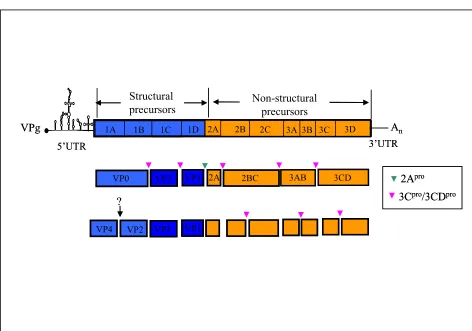
[image:15.595.212.358.439.685.2]Most of these virus infections are asymptomatic, although, in a few cases, the virus can enter the central nervous system leading to acute flaccid paralysis (figure 1). Poliovirus is a widely studied virus, whose genome (~7400 nucleotides) functions as single genome sized RNA and is representative of most positive-sense RNA viruses (shown in figure 2). Extensive studies of this virus allied to successful vaccine production, and vaccination program have led to the almost complete eradication of polio. This will be the second virus eradicated from the world, smallpox being the first.
2Apro
3Cpro/3CDpro
VP1 VP3 VP4 VP2
2A 5’UTR
3B 3C 3D 1C 1D
1A 1B 2B 2C 3A
VPg An
Structural
precursors Non-structural precursors
3’UTR 2A
VP0 VP3 VP1
?
2A 2BC 3AB 3CD 2Apro
3Cpro/3CDpro
VP1 VP3 VP4 VP2
2A 5’UTR
3B 3C 3D 1C 1D
1A 1B 2B 2C 3A
VPg An
Structural
precursors Non-structural precursors
3’UTR 2A
VP0 VP3 VP1
?
[image:16.595.90.562.166.497.2]2A 2BC 3AB 3CD
1.3.1.2 Rhinovirus
Rhinoviruses, aetiological agents of the common cold, are the most commonly isolated viruses from individuals experiencing mild upper respiratory illnesses (Figure 3). In contrast to enteroviruses, rhinoviruses do not replicate in the intestinal tract. Constant efforts towards producing effective vaccines have been unsuccessful and remain as an important goal. This genus contains 99 human rhinovirus serotypes classified into 2 species, Human rhinovirus A and B. This genus will disappear soon,
its 2 species moved into the enterovirus genera (http://www.picornaviridae.com/).
Figure 3. One of the classic symptoms caused by rhinovirus in the common cold.
1.3.1.3 Cardiovirus
This genus consists of two species: encephalomyocarditis virus (EMCV), which is represented by a single serotype of the same name, and theilovirus, which comprises 4 different serotypes: Theiler´s murine encephalomyelitis virus (TMEV), Vilyuisk human encephalomyelitis virus (VHEV), Theiler’s-like virus (TLV) isolated from rats and human pathogen Saffold virus (SAF-V). These are distinguished from other genera by special features of their genome organization (presence of a poly(C) tract - except TMEV - and the dissociability of their capsid at pH 5-7, among others).
Several strains of EMCV have been determined: encephalomyocarditis virus (strain EMC-B nondiabetogenic), Encephalomyocarditis virus (strain EMC-D
diabetogenic), Maus-Elberfeldvirus, Mengo virus and Porcine encephalomyocarditis
TMEV strains were first isolated by Max Theiler at the Rockefeller Foundation during the 1930’s, (Lipton, 1975) from the central nervous system of paralysed mice and later from the intestine of apparently uninfected mice.
These investigations demonstrated that the virus caused widespread, asymptomatic, enteric infection and chronic progressive demyelination in mice. Such symptoms were later identified as being similar to those observed in humans with multiple sclerosis (Lipton, 1975; Roos, 2002).
There are several strains classified on the basis of differences in their biological activities: i) GDVII sub-group (GDVII and FA), which are extremely
virulent and produce an acute disease that is similar to poliomyelitis; and ii) TO sub-group (DA, BeAn, TO, WW, Yale, etc), which produce a biphasic disease (reviewed
by Racaniello, 2001; Roos, 2002).
1.3.1.4 Aphthovirus
Foot-and-mouth disease virus (FMDV) is a member of this genus and is responsible for Foot-and-mouth disease (FMD). This disorder principally affects domesticated and wild cloven-hoofed animals and is known to spread by direct contact between infected and susceptible animals by the airborne route, by animal products such as meat and milk, and mechanical transfer via people, wild animals,
birds, and by vehicles. It is accepted as a significant epidemic disease that infects primarily cattle, goats, pigs, and sheep and rarely humans (reviewed by: Rossmann, 2002; Agol, 2002).
FMD is one of the most contagious animal diseases, with important economic losses. It is endemic in Asia, Africa, the Middle East and South America. On the domestic front, the 2001 outbreak cost the UK something in the region of £10 billion with the loss of eight million cattle, sheep, pigs and goats (DEFRA, March 2002)
Unfortunately, presently available inactivated vaccines are not entirely effective. Vaccination blocks disease symptoms, making detection of infection difficult, but does not always block transmission of the virus to other animals. Sheep can harbour the virus for several months; cows for up to a year or even longer. Occasional vaccine-linked disease outbreaks occur as a result.
(Pringle et al., 1999) and is the only non-FMDV member of this genus. The genome
organisation of this virus possesses two features which distinguish it from FMDV; (i) there is no poly C tract and (ii) ERAV has only one copy of 3B (VPg) (Wutz et al.,
1996).
1.3.1.5 Hepatovirus
Hepatitis A is an acute infectious disease caused by Hepatitis A virus (HAV). It is transmitted via the faecal-oral route and may be mistaken for flu. Symptoms
typically appear 2 to 6 weeks after the start of infection and may return over the following 6-9 months. The most common symptoms are: fatigue, fever, nausea, abdominal pain and diarrhoea.
In contrast with other picornaviruses, Hepatovirus 2A protein does not show any proteolytic function with regards to polyprotein processing. Cleavage between 2A/2B, leading to separation of 5’ terminal structural proteins from the non-structural proteins, is achieved by the 3C protease (Jia et al., 1993; Martin et al., 1995).
1.3.1.6 Parechovirus
Two species form this genus, Human parechovirus (HePV), which comprises five serotypes (Ito et al., 2004; Al-Sunaidi et al., 2007), and the recently described
Ljungan virus (LV), which may be comprised of two or more serotypes.
Human parechovirus causes mostly mild gastrointestinal or respiratory illness, with a few cases of myocarditis and encephalitis. It commonly infects children between 2 to 5 years old. In contrast, Ljungan virus causes diabetes, neurological disease, myocarditis and intrauterine foetal death in humans (Niklasson et al., 2007).
It is a zoonotic virus, which was isolated from bank voles (Niklasson et al., 1999). Its
genome contains two 2A proteins in tandem. The N-terminal 2A1 has the
-DxExNPGP- motif and is related to the type of 2As encoded by aphthoviruses, cardioviruses, erboviruses and teschoviruses. In contrast, the 2A2 belongs to the type
of 2As encoded by kobuviruses and hepatoviruses (Johansson et al., 2002).
1.3.1.7 Erbovirus
Equine rhinitis B virus (ERBV) is the only member of this genus and causes acute upper febrile respiratory disease in horses (Dynon et al., 2007). There are two
symptoms they cause more closely resemble those produced by rhinovirus. ERV-1 proteins are very similar to the FMDV proteins, whereas, most ERV-2 proteins are more closely related to EMCV proteins (Wutz et al., 1996).
1.3.1.8 Kobuvirus
This genus consists of 2 species, Aichi virus, which infects humans (Yamashita et al., 1998) and Bovine Kobuvirus, which infects cattle (Yamashita et al., 2003).
Aichi virus, first recognized in 1989 as the cause of oyster-associated non-bacterial gastroenteritis in humans, has been recently classified into the new Kobuvirus genus (Pham et al., 2007)
1.3.1.9 Teschovirus
Teschovirues constitute a recently defined genus within the Picornaviridae. It
is represented by a single species, Porcine Teschovirus (PTV), which causes porcine enteroviral encephalomyelitis disease. PTV-1 Talfan is the reference strain for this genus, and, as mentioned earlier, its genome contains a significantly shorter IRES compared to the other genera, and requires part of the 5´UTR sequence for its activity (Kaku et al., 2002).
1.3.1.10 Proposed novel genera
Sapelovirus consists of three species: Porcine enterovirus A, SV2-like virus, Duck Picornavirus TW90A. The name of this proposed genus derives from Simian, Avian and Porcine Entero-Like viruses.
Senecaviruscontains Seneca valley virus (SVV), whose proteins closely resemble some cardiovirus proteins (P1, 2C, 3Cpro and 3Dpol). However, the 5’UTR, leader, 2A and 3A proteins are very different to all known picornaviruses. SVV-2A protein presents the same conserved motif found in aphthoviruses (Luke et al., 2008)
Tremovirus consists of a single species, Avian encephalomyelitis virus (AEV), which currently belongs to hepatovirus.
1.3.1.11 Novel species
Duck hepatitis virus 1 (DHV-1) is one of the three viruses causing Duck virus hepatitis, an acute highly contagious disease affecting young ducklings up to the age of 28 days. Its genome features three in-tandem 2A genes. The 2A1 and 2A3 proteins
are aphthovirus-like and human parechovirus-like 2A proteins respectively, whereas 2A2 is not related to any known picornavirus protein. These 2A proteins are only 12
amino acid long (Tseng et al., 2007b) Another exclusive feature of this new species is
the length of 3’ UTR, which is composed of 314 nucleotides, the largest among the picornaviruses (Tseng et al., 2007a). On the bases of these findings it is proposed that
DHV-1 should be assigned to a new genus in the Picornaviridae.
Seal picornavius 1 (SePV-1) is a new picornavirus species that was isolated from Arctic ringed seals (Phoca hispida) in Canada. The SePV-1 genome contains
two 2A genes, whose sequence correspond to the canonical co-translational cleavage site DxExNPG↓P, found in cardioviruses, apthoviruses, teschoviruses and erboviruses (explained in sections 1.4.5 and 1.4.6). Furthermore, the absence of a predicted maturational cleavage site between 1A and 1B (VP0) was also observed in other species within the family, such as LV, HPeV and DHV.
1.3.2 Genome structure and organization
The name Picornaviridae conveys two important features of the family, the
small size (Pico-) and the type of nucleic acid that the viral genome carries
(-rnaviridae). This family has played a major role in the development of modern
virology. Foot-and-mouth disease virus was the first animal virus discovered, followed by poliovirus, which was isolated ten years later. Picornavirus virions are spherical particles with a diameter of 30 mm and are simply composed of a protein shell surrounding the naked RNA genome. Their capsid is composed of four structural proteins (VP1, VP2, VP3, and VP4), excepting parechoviruses, DHV-1 and SePV with only three (VP1, VP3 and VP0, which is equivalent to the uncleaved VP2 plus VP4 precursor of other picornaviruses).
called VPg (3B; virion protein, genome linked). FMDV is the only one in the family that has three Vpgs instead of one (Forss & Schaller, 1982). The picornavirus genome can be divided in three regions: a 5´untranslated region (5´UTR), which is
600 to 1,200 nucleotides long, the coding region, which comprises 6,500 to 7000
nucleotides and a short 3´untranslated region (3´UTR) that contains a
heteropolymeric segment and a poly(A) tail (Rueckert, 1996; Palmenberg, 1990). The 5’UTR contains a 5’terminal stem-loop, cloverleaf-like structure, and a type I or II internal ribosome entry site (IRES), which allows a cap-independent mode of translation by binding to eIF4G (Kolupaeva et al., 1998, 2003; Clark et al., 2003).In
aphthoviruses, cardioviruses (except TMEV) and erboviruses the 5’UTR also includes a poly(C) tract. The poly(C) tail is longer in cardioviruses and is associated with higher virulence in animals (Duke et al., 1990; Hahn & Palmenberg, 1995; Wutz et al., 1996). IRES’s enable the eukaryotic ribosome to bind directly to the internal site
without first having to scan from the 5’ terminus, thus allowing 5’cap-independent translation (reviewed in Jang, 2005). There are four different IRES types, one of them belatedly discovered in porcine teschovirus-1 (Kaku et al., 2002). These viral
elements are widely used in vectors for gene therapy and their ability to enhance expression of an upstream gene in dicistronic vectors, has been recently discovered (Niepman, 2007). The 3’ poly (A) tail is essential for infectivity. Its removal in poliovirus leaves the viral genome non-infectious (Spector & Baltimore, 1974).
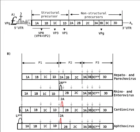
The picornavirus polyprotein is divided into three regions: P1, which encodes the structural proteins, and P2-P3, which encodes the non-structural proteins that are involved in polyprotein processing (2A or 2Apro, 3Cpro and 3CDpro) and genome replication (2B, 2C, 3AB, 3BVPg, 3CDpro, 3Dpol).
Unlike other genera, cardio-, aphtho-, erbo-, kobu-, tescho- and sapelovirus have an L protein region at the N-terminus of the polyprotein (figure 4). In aphtho- and erboviruses this acts as a protease (Lpro), cleaving at its own C-terminus, to produce the first cleavage of the polyprotein (van Pesch et al., 2001; Hinton, 2002;
van Eyll et al., 2002).
The next stage in processing is the primary even that separates the encapsidation functions (P1; capsid proteins) from the replicative functions of the polyprotein (referred to as the P2-P3 region).
the 3C and L-proteases are involved in the degradation of certain host-cell proteins, to enhance virus replication (for reviews see: Leong et al., 2002; Grubman et al., 1995;
Ryan & Flint, 1997; Piccone et al., 1995; Robert & Belsham, 1995; Gradi et al.,
2004). Each of these proteins are described in the following sections and important intra-genera variations discussed.
5’UTR
3B 3C 3D
1C 1D
1A 1B 2A 2B 2C 3A
.
VPg An
VP0 VP3 VP1 L VPg Structural precursor Non-structural precursors 3’UTR (VP4+VP2) A) 5’UTR
3B 3C 3D
1C 1D
1A 1B 1C 1D 2A 2B 2C 3A 3B 3C 3D
1A 1B 2A 2B 2C 3A
.
VPg An
VP0 VP3 VP1 L VPg VPg Structural precursor Non-structural precursors 3’UTR (VP4+VP2) A) 1B 1A 1A 1C 2B 2B Cardiovirus Aphthovirus Lpro 2A 2A 1D 1B 1C 1D 3Cpro 3B 3A 3D 3Cpro 3B 3A 3D 2C 2C L 3Cpro 3B 3A 1C 1A 2B
P1 P2 P3
Hepato- and Parechovirus Rhino- and Enterovirus 2Apro 1D 1B 1C 1D 1A 1B
2A 2B 2C 3A3B3Cpro 3D
3D 2C B) 1B 1A 1A 1C 2B 2B Cardiovirus Aphthovirus Lpro 2A 2A 1D 1B 1C 1D 3Cpro 3B 3A 3D 3Cpro 3B 3A 3D 2C 2C L 3Cpro 3B 3A 1C 1A 2B
P1 P2 P3
Hepato- and Parechovirus Rhino- and Enterovirus 2Apro 1D 1B 1C 1D 1A 1B
2A 2B 2C 3A3B3Cpro 3D
3D 2C B) 1B 1A 1A 1C 2B 2B Cardiovirus Aphthovirus Lpro 2A 2A 1D 1B 1C 1D 3Cpro 3B 3A 3D 3Cpro 3B 3A 3D 2C 2C L 3Cpro 3B 3A 1C 1A 2B
P1 P2 P3
Hepato- and Parechovirus Rhino- and Enterovirus 2Apro 1D 1B 1C 1D 1A 1B
2A 2B 2C 3A3B3Cpro 3D
3D 2C B) 1B 1A 1A 1C 2B 2B Cardiovirus Aphthovirus Lpro 2A 2A 1D 1B 1C 1D 3Cpro 3B 3A 3D 3Cpro 3B 3A 3D 2C 2C L 3Cpro 3B 3A 1C 1A 2B
P1 P2 P3
Hepato- and Parechovirus Rhino- and Enterovirus 2Apro 1D 1B 1C 1D 1A 1B
2A 2B 2C 3A3B3Cpro 3D
3D 2C B) 3Cpro 3B 3A 1C 1A 2B P1
P1 P2P2 P3P3
Hepato- and Parechovirus Rhino- and Enterovirus 2Apro 1D 1B 1C 1D 1A 1B
2A 2B 2C 3A3A3B3B3C3Cpropro 3D3D
3D 2C
[image:23.595.90.547.176.607.2]B)
Figure 4. Picornavirus genome and proteolytic processing. A) General representation of picornavirus genome. Leader protein (L) is only present in aphthoviruses and cardioviruses and is a protease in the former genus. B) Proteolytic processing in picornaviruses. Grey vertical arrow: 3Cpro
cleavages, also including those for which 3CDpro are required. Black curved arrow: 2Apro, a trypsin
1.3.3
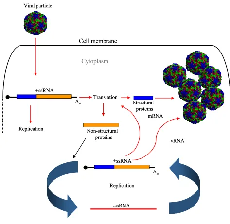
Replication cycle
The replication cycle starts with attachment of the viral particle to specific cell receptors on the membrane, followed by uncoating of the capsid to release the RNA genome inside the cell. The entire replication process takes place in the cytoplasm, where the RNA is translated to yield viral proteins necessary for replication, polyprotein processing and encapsidation of new virus particles (figure 5).
The single positive RNA strand replicates itself by producing a negative stranded RNA intermediate that is used as a template to create new single positive strands. This process occurs in small membranous vesicles that are stimulated by certain virus proteins. The synthesis rates of the positive RNA strand is ~100 fold greater than the negative strand since the daughter positive strands may fulfil one of three functions (i) as templates for the negative strand synthesis, (ii) they act as mRNAs to direct the synthesis of viral proteins or (iii) they are encapsidated into virions.
Once the pool of capsid proteins is sufficient for encapsidation, coat protein precursor P1 is cleaved and assembled into pentamers. These associate with newly synthesized positive-stranded RNAs to form fresh infectious particles that are released from the cell in a number of ways depending on the virus type. The majority of picornaviruses release their new particles by cell lysis, in others, such as Hepatitis A virus, release occurs in the absence of as cytophatic effect.
An +ssRNA
Translation
-ssRNA Replication
mRNA
Non-structural proteins
vRNA
An +ssRNA
Viral particle
Structural proteins
Replication
Cytoplasm
Cell membrane
An +ssRNA
An +ssRNA
Translation
-ssRNA Replication
mRNA
Non-structural proteins
vRNA
An +ssRNA
An +ssRNA
Viral particle
Structural proteins
Replication
Cytoplasm
[image:25.595.93.566.75.536.2]Cell membrane
Figure 5. Picornavirus replication cycle. The entire replication takes place in the cytoplasm. The viral capsid attaches to specific cell membrane receptors and releases the positive single stranded RNA (+ssRNA) genome into the cytoplasm. The genome is translated into a large polyprotein, which can process itself during and after translation leading to the production of non-structural and structural proteins, required for replication and capsid formation, respectively. The replication machinery produces a negative-stranded RNA from the input positive-stranded template. This new strand will be used as a template to produce new positive-stranded RNAs that will be encapsidated once the pool of capsid proteins is large enough. Thus, new infectious viruses will be produced.
1.3.4. Picornavirus polyprotein
1.3.5.1 L protein
In contrast to other picornaviruses, the first protein encoded in the genome of cardioviruses, aphthoviruses, erboviruses, kobuviruses, teschoviruses as well as two members of the proposed genera Sapelovirus (Human enterovirus-8 and simian virus 2), is the Leader protein (L) (Zell et al., 2005). In aphthoviruses, it is a protease (Lpro)
(Strebel & Beck, 1986), which appears in two different forms (Lb and Lab) in FMDV (Clarke et al., 1985). Lpro cleaves at its own C-terminus releasing itself from the
N-terminus of VP4. Furthermore, it also cleaves the translation initiation factors eIF4GI and eIF4GII, together with 3Cpro, leading to inhibition of cap dependent translation in infected cells (Gradi et al., 2004; Belsham et al., 2000). Like aphthovirus, erbovirus L
protease cleaves at the L/VP4 junction but does not have a role in eIF4G cleavage (Hinton et al., 2002).
In cardioviruses, the L protein does not have proteolytic activity but plays other important roles during infection. The TMEV L protein interferes with trafficking of the cytoplasmic interferon regulatory factor 3 (IRF-3), a factor critical for transcriptional activation of alpha/beta interferon genes (Delhaye et al., 2004) and
interacts with Ran-GTPase, disrupting nucleocytoplasmic transport (Porter et al.,
2006). L protein also plays an essential role in persistence by inhibiting the production of alpha/beta interferon (van Pesch et al., 2001). Moreover, Cardiovirus L protein also
has a role in alteration of nucleocytoplasmic traffic, although this function is not essential for viral reproduction (Lidsky et al., 2006).
1.3.4.2 L* protein
L* protein is a unique feature of translation of the TO subgroup strains of TMEV (DA, BeAn). It encodes an 18 kDa protein, which is translated from an alternative open reading frame starting 13 nucleotides downstream from the AUG codon of the main protein (Kong & Roos, 1991; Yamasaki et al., 1999). In vivo
studies of the L* protein have shown a direct effect in viral persistence of infections and also enhancement in infection of macrophage cell lines (reviewed by Brahic et al., 2005). This complementary AUG codon appears as an ACG in other strains, such
(van Eyll & Michiels, 2000). Saffold virus (SAF-V) is a novel human cardiovirus, which has been recently discovered. In contrast to TMEV strain DA, which has 156 amino acids, the L* open reading frame of SAF-V encodes only 57 amino acids. It is still unknown if this open reading frame encodes for a protein (Jones et al., 2007).
1.3.4.3 Capsid proteins
The structural proteins are encoded towards the 5’end of the open reading frame, which appears to fold into a structure closely related antigenically to the viral proteins VP0, VP1 and VP3 (Jackson et al., 2003). The structural capsid proteins,
VP1, VP2, VP3, and VP4 (VP1, VP3 and VP0 in parechoviruses) form an assembled icosahedral structure (shown in figure 6).
VP1 (1D)
VP2 (1B)
VP3 (1C)
VP1 (1D)
VP2 (1B)
[image:27.595.88.534.326.672.2]VP3 (1C)
Figure 6. Icosahedral symmetry of the capsid if picornaviruses. The structural capsid proteins, VP1, VP2 and VP3 are shown in different colours. VP4 is not shown because it is internal to the capsid. Figure by courtesy of Professor Martin Ryan.
1.3.4.4 Protein 2A
The 2A protein is variable between genera, in contrast to the other non-structural proteins, which have the same structure. Kobuviruses, HePV, DHV-1 (2A3)
and hepatovirus 2A proteins are not involved in polyprotein processing (Jia et al.,
1993; Schultheiss et al., 1995; Yamashita et al., 1998). In other genera, 2A is
involved in the separation of the P1 structural proteins from the P2 and P3 non-structural proteins. In enteroviruses and rhinoviruses 2A is a protease (2Apro), while in cardio-, aphtho-, erbo- and tescho-, LV (2A1), DHV-1 (2A1) and SePV (2A1-2) it is an
oligopeptide also responsible for the primary cleavage between structural and non-structural proteins (Donnelly et al., 2001a). This ‘cleavage’ is not proteolysis but a
translational effect -‘ribosome skipping’ (Ryan et al., 1991; Ryan & Drew, 1994;
Ryan et al., 1999; Donnelly et al., 2001b; Ryan et al., 2002). This novel mechanism is
explained in more detail in section 1.2.5. 2A in the aphtho-, erbo- and teschovirus is an oligopeptide of ~18 aa whereas in the cardioviruses 2A is a longer track (~150 aa). EMCV-2A protein activates the translation initiator factor 4E binding protein 1, which binds eIF4E, the cap binding subunit of the initiation factors complex. Thus, it suppresses cap-dependent translation (Gingras et al., 1996; Aminev et al., 2003 a, b;
Svitkin et al., 2005). In entero- and polioviruses 2A protease also plays a role in
translation repression by cleaving eIF4GI, both directly and indirectly, through the activation of cellular proteases (Zamora et al., 2002). In addition, 2A protease,
together with 3C protease, cleaves the poly(A)-binding protein (PABP) during infection, and thus, inhibits host cell translation (Joachims et al., 1999;
Kuyumcu-Martinez et al., 2002). Poliovirus 2A protease also increases RNA stability (Jurgens et al., 2006).
New 2A sequences with the same conserved motif ‘DxExNPGP’ (where x= any amino acid) found in aphtho- and cardioviruses have also been found within the
Picornaviridae. In addition, this motif has been identified in species outwith this
1.3.4.5 Protein 2B
Enterovirus 2B protein is a viroporin, a transmembrane pore-forming protein, which participates in different viral functions (Gonzalez & Carrasco, 2003). This kind of protein alters membrane permeability through the formation of pores, and thus, induces disassembly of the Golgi complex (Sandoval & Carrasco, 1997). Coxsakievirus 2B protein inhibits vesicular protein transport by reducing the endoplasmic reticulum (ER) and Golgi´s Ca2+ content. This disturbance on intracellular Ca2+ homeostasis leads to two different events: the enhancement of viral RNA genome replication and the suppression of apoptotic host-cell responses (Kuppeveld et al., 2006; Doedens & Kirkegaard, 1995). Little is known about the
function of the other picornavirus 2B proteins, although it is been recently shown that rhinovirus 2B is also localized in the ER and Golgi apparatus and functions similarly to enterovirus 2B. In contrast, HAV, FMDV and EMCV 2B protein is not localized in the ER and Golgi apparatus and does not cause relevant effects on Ca2+ homeostasis and intracellular protein trafficking (de Jong et al., 2008).
1.3.4.6 Protein 2C
Protein 2C is a highly conserved non-structural protein that binds to membranes and RNA, and is crucial in poliovirus replication (reviewed in Goodfellow et al., 2003). It also plays a role in encapsidation (Vance et al., 1997) and
also has ATPase/GTPase activity (Rodríguez & Carrasco, 1993). 2C and its precursor 2BC are responsible for poliovirus RNA binding to the cytoplasmic vesicles, whose formation is also induced by this precursor. Its mechanism of action has yet to be determined. FMDV 2C protein is localized in juxtanuclear structures, vesicles that could be derived from Golgi compartements. However, the origin of these vesicles is not clear yet; recent studies argue against the relation with the Golgi (Knox et al.,
2005; Moffat et al., 2005). For instance, in FMDV and EMCV infected cells treated
with Brefeldin A (BFA), which inhibits membrane transport between the ER and the Golgi by preventing the formation of COPI-dependent secretory transport vesicles (Duden et al., 1994), 2C juxtanuclear localization and replication is not inhibited
(Gazina et al., 2002). In contrast, replication is inhibited in PV and Rhinoviruses
1.3.4.7 Protein 3A
3A protein has a role in disrupting ER-to-Golgi protein trafficking, which inhibits the secretion of cytokines such as interleukins (IL-6, IL-8) and interferon-β (IFN-β) (Dodd et al., 2001). It has an anti-apoptotic effect; it provokes
the release of intracellular calcium via permeabilization of cellular membranes (Liu et al., 2003). Poliovirus 3A exists as a dimer and is a critical component of the viral
replication complex (Strauss et al., 2003). Additionally, it also inhibits TNF-induced
apoptosis by elimination of TNF receptor from the cell surface due to the inhibition of protein trafficking (Neznanov et al., 2001). FMDV 3A plays an essential role in the
determination of host-range. For instance, it has been shown that a single mutation within 3A mediated adaptation of FMDV to the guinea pig (Nuñez et al., 2001).
Furthermore, mutations within poliovirus 3A also affected host-range (Lama et al.,
1998).
1.3.4.8 Protein 3B (VPg)
Protein 3B is covalently bound to the 5’end of viral RNA and functions as a primer for the initiation of viral genome replication. FMDV 3B encodes three different types of VPg, which are uridylylated by 3Dpol, making possible the initiation of the viral RNA replication (Nayak et al., 2005). The poliovirus VPg NMR structure
has been solved and will hopefully improve our understanding of the mechanism of action of VPg, which is essential for virus replication, and also the interactions between this protein and the viral polymerase (Shein et al., 2006).
1.3.4.9 Protein 3C
Protein 3C is a chymotrypsin-like protease responsible for the primary 2C/3A cleavage of the polyprotein (Palmenberg et al., 1992; reviewed by Ryan & Flint,
1997; Ryan et al., 2004). In the case of HePV, kobuvirues, DHV-1 (2A3) and
hepatoviruses the primary 2A/2B polyprotein cleavage is mediated by the 3C protease (Stanway & Hyypiä, 1999). 3Cpro also induces the cleavage of the translation initiator factors eIF4A and eIF4GI-II (Belsham et al., 2000) and is involved in cell apoptosis
in poliovirus and enterovirus (Barco et al., 2000). Poliovirus 3Cpro, combined with
unknown host-cell activity, degrades p53 (Weidman et al., 2001). Furthermore, this
(CTD) that interacts with several translation factors. This mechanism of translation inhibition complements the effect of eIF4G cleavage by 2Apro (Kuyumcu-Martinez et al., 2004). In addition, 3Cpro contains RNA-binding domains (Blair et al., 1998).
1.3.4.10 Protein 3Dpol
3Dpol is a RNA-dependent RNA polymerase (RdRp) that does not possess proof-reading activity. Its mechanism of action is still unknown. Studies have shown specificity of 3Dpol for each virus type and specificity also between 3Dpol and the other viral replicative proteins within the same virus species. A chimeric poliovirus, its 3Dpol replaced by coxsackievirus 3Dpol, showed a lack of replication due to the inefficient recognition of the P1 protein substrate by the chimeric 3CD protease (3CDpro) (Bell et al., 1999). Atomic structures of 3Dpol are available for poliovirus
(Hansen et al., 1997) and FMDV (Ferrer-Orta et al., 2004), both showing the classical
architecture: ‘fingers’, ‘palm’ and ‘thumb’ domains.
1.3.4.11 Cleavage intermediates
There are three different intermediates, 2BC, 3AB and 3CDpro, which have different roles from their cleavage products. Accumulation of small ER- and Golgi-derived membrane vesicles in the cytosol has been observed in enterovirus-infected cells, and is where viral replication takes place. 2BC is responsible for this accumulation (Bienz et al., 1994). 3AB is thought to be an integral membrane protein
(Ciervo et al., 1998) and it also induces 3Dpol activity, most likely by recruiting 3Dpol
to the 3’termini of chain elongation sites (Richards & Ehrenfeld, 1998). On the other hand, 3CDpro binds the 5’ RNA ‘cloverleaf’ structure, an essential step in replication
(Blair et al., 1998).
Poliovirus 3CD forms a ribonucleoprotein complex (RNP), together with a 3kDa ribosome-associated cellular protein at the 5’ UTR region. This complex is essential for positive RNA synthesis but not for the negative strands (Andino et al.,
1.3.5. Polyprotein processing
In picornaviruses the first step of infection, following cell entry, is translation of the RNA genome into a polyprotein (reviewed in Palmenberg, 1990). The viral RNA, which has a poly (A) sequence at its 3’ end, like cellular mRNAs, sequesters the cell’s own translational machinery for its protein synthesis. One important peculiarity of most viral mRNAs is their uncapped 5’ end. Picornaviruses have a ~22 amino acid long protein, VPg, which is covalently attached to its 5´end that has an important role in initiation of RNA synthesis and may also have a role to play in the virulence of the virus (reviewed in Reuckert, 1996). This polyprotein precursor does not appear in infected cells because it undergoes co-translational cleavage while it is being translated. The different products appear in equimolar quantities due to their common origin from a single precursor (reviewed in Palmenberg, 1990). 3Cpro and 3CDpro are responsible for the main polyprotein processing events cleaving all the structural and non-structural protein precursors, except i) Lpro, which cleaves itself at its C-terminus, ii) 1AB precursor, whose cleavage mechanism is still unknown, and iii) 2AB, except in hepato- and parechoviruses. 2Apro, is the other enzyme involved in
polyprotein processing and is responsible for the 1D/2A cleavage in rhino- and enteroviruses. 2Apro separates the structural protein domain from the non-structural domain and it was first thought to be present in a wide range of picornaviruses. However, we now know that the majority of picornaviruses use another method (see section 1.5.1.4: ribosome skipping). This novel 2A protein, first discovered in FMDV (Ryan et al., 1991) expands the repertoire of translation strategies used by RNA
viruses.
1.4. Control of protein biogenesis within positive stranded RNA viruses
1.4.1. Ribosomal Frameshifting
The majority of RNA viruses are single-stranded. The majority of these are positive-sense, single-stranded RNA viruses, such as the family Picornaviridae, Coronaviridae, Caliciviridae and Flaviviridae. Their genome is transcribed into a
polycistronic mRNA, which is translated into a polyprotein that is subsequently cleaved into individual mature proteins.
One of the replication strategies that positive-sense, single-stranded RNA viruses RNA viruses use is programmed ribosomal frameshifting, in which ribosomes change reading frame within the mRNA, leading to the synthesis of alternative proteins. There are two essential signals needed for this process to occur; (i) a ‘slippery’ hepta-nucleotide sequence (eg. UUUAAAC), where the ribosome changes frame and (ii) an RNA pseudoknot structure situated downstream of the slippery sequence and consisting of two helical segments connected by single-stranded regions or loops. The current model proposes that the ribosome finds the pseudoknot while the slippery sequence is being translated causing the ribosome to pause on the slippery sequence, where it slips back one nucleotide and subsequently continues translation in the -1 reading frame (Somogyi et al., 1993). This recoding mechanism
.
ORF 1a (0 Frame)
ORF 1b (-1 Frame)
Cap Poly A
Slippery sequence
U U U A A A C G G G U A C G G G G U A G C A G U C G A G C C U U C C C C A U C G U C A G C U C G G
5’ 3’ UUGCUAGUGGAUGUGAUCCUGAUGUUGAUAAAG G A Stem 1 Loop 1 Stem 2 Loop 2 Translation
ORF 1a (0 Frame)
ORF 1b (-1 Frame)
Cap Poly A
Translation A)
B)
Protein 1a
Protein 1a-1b
ORF 1a (0 Frame)
ORF 1b (-1 Frame)
Cap Poly A
Slippery sequence
U U U A A A C G G G U A C G G G G U A G C A G U C G A G C C U U C C C C A U C G U C A G C U C G G
5’ 3’ UUGCUAGUGGAUGUGAUCCUGAUGUUGAUAAAG G A Stem 1 Loop 1 Stem 2 Loop 2 Translation
ORF 1a (0 Frame)
ORF 1b (-1 Frame)
Cap Poly A
Translation A)
B)
ORF 1a (0 Frame)
ORF 1b (-1 Frame)
Cap Poly A
Slippery sequence
U U U A A A C G G G U A C G G G G U A G C A G U C G A G C C U U C C C C A U C G U C A G C U C G G
5’ 3’ UUGCUAGUGGAUGUGAUCCUGAUGUUGAUAAAG G A Stem 1 Loop 1 Stem 2 Loop 2
ORF 1a (0 Frame)
ORF 1b (-1 Frame)
Cap Poly A
Slippery sequence
U U U A A A C G G G U A C G G G G U A G C A G U C G A G C C U U C C C C A U C G U C A G C U C G G
5’ 3’ UUGCUAGUGGAUGUGAUCCUGAUGUUGAUAAAG G A Stem 1 Loop 1 Stem 2 Loop 2 Translation
ORF 1a (0 Frame)
ORF 1b (-1 Frame)
Cap Poly A
Translation Translation
ORF 1a (0 Frame)
ORF 1b (-1 Frame)
Cap Poly A
Translation A)
B)
Protein 1a
Protein 1a-1b
1.4.3. Leaky scanning
The majority of eukaryotic mRNAs are monocistronic and initiate translation when the 40S ribosomal subunit and initiation factors bind to the 5’ end of the mRNA (m7G cap). This preinitiation complex moves in a 3´direction on the mRNA in a process called scanning, until it reaches (generally) the first AUG codon. Then initiation factors are released allowing the 60S subunit to associate with the small subunit. The efficiency of initiation is affected by the nucleotide sequence surrounding a certain codon. The consensus sequence accepted as the most efficient in eukaryotic cells is 5’GCC(A/G)CCAUGG 3’, where the presence of a purine at the position -3 followed by a G at position +4 is essential for high levels of translation. Nevertheless, only 5% of eukaryotic sequences contain this ideal consensus sequence, most of them have suboptimal ones (reviewed in Flint et al., 2000). Some viral
mRNAs encode two or more proteins in overlapping reading frames, initiation of translation occurring not only at the first 5’ AUG but also at downstream AUGs. This occurs when ribosome preinitiation complexes bypass the 5´AUG due to the fact that this codon is surrounded by suboptimal nucleotide sequences. An example of this can be found in Sendai virus P/C gene mRNA, a member of the family Paramyxoviridae
that contains a non-segmented negative sense single-stranded RNA genome, from which 6 mRNAs are transcribed. One of them, the P/C mRNA, is polycistronic and has two open reading frames that start near the 5´end (Girogi et al., 1983; figure 8).
The P protein starts at the 5’ proximal ATG in the first open reading frame, the last 95 amino acids being expressed as a different protein (X). The second open reading frame produces a nested set of non-structural C proteins (C’, C, Y1, and Y2). Translation of the first 3 initiation sites on P/C mRNA is arranged in such a manner that leaky scanning is enhanced. The first non-structural protein, C’, starts at an unusual non-ATG unusual codon, ACG, whereas the rest of proteins initiate on AUGs. Although ACG is in a good context, because it is an unusual start codon, this leads to an inefficient initiation, in which some ribosomes bypass ACG and initiate translation at the next initiator codon (AUG). This second initiator, which translates P protein, is an AUG but is in a poor initiation context (a pyrimidine at position -3), while the third AUG is in good context (an A at -3) and translates for C protein (Curran & Kolakofsky, 1989).
negative-sense single-stranded RNA viruses. Influenza B virus RNA segment 6 is bicistronic and encodes NB and NA proteins in overlapping reading frames. NB translation starts at the 5’ proximal AUG codon, while the NA AUG codon is four nucleotides downstream. Even if the NA initiation codon is in a good context for translation initiation compared to the NB one, both proteins accumulate in almost equal amounts in infected cells. This suggests a unique model in which ribosomes randomly choose which AUG to use. Certain cellular factors and surrounding mRNA sequences might have an effect on the selection of these codons (Williams & Lamb, 1988).
5’ P/ 104 3’
C’/ 81 C/ 114
Y 1/ 183 Y 2/ 201
X/ 1523
ACG
Termination (726)
Termination (1808)
5’ P/ 104 3’
C’/ 81 C/ 114
Y 1/ 183 Y 2/ 201
5’ 3’
5’ P/ 104 3’
C’/ 81 C/ 114
Y 1/ 183 Y 2/ 201
X/ 1523
ACG
Termination (726)
Termination (1808)
1.4.4. Reinitiation
Reinitiation is a very common strategy among prokaryotic cellular and viral RNAs. It is another mechanism by which two proteins are produced from a single mRNA. An example for this strategy is influenza virus RNA segment 7, which encodes two proteins, M1 and BM2, from a bicistronic mRNA. The ATG initiation codon for BM2 protein overlaps the termination codon for the M1 protein. Therefore, BM2 protein synthesis is dependent upon the initiation and termination of the upstream protein M1 (figure 9). A coupled translational termination-initiation mechanism is essential for the downstream protein to be produced (Horvath et al.,
1990).
In caliciviruses, which are non-segmented positive single-stranded RNA viruses, translation initiation of the 3’terminal open reading frame is also achieved via
a termination-reinitiation process. Its genome contains two open reading frames for member of the genera Lagovirus and Sapovirus, and three open reading frames in Vesivirus and Norovirus (Green et al., 2000). The minor capsid protein VP2 is
expressed via reinitiation of translation after termination of the upstream VP1 protein
synthesis. A sequence of about 80 nucleotides, called termination upstream ribosomal binding site (TURBS), is essential for this termination-reinitiation mechanism to occur. TURBS has a specific conserved motif, which is complementary to 18S rRNA, and may be important to prevent release of post-termination ribosomes, hence, increasing the chance of reinitation (Meyers, 2007).
5’ M1 ORF 3’
-UAAUG-BM2 ORF
5’ M1 ORF 3’
-UAAUG-BM2 ORF
1.4.5. Suppression of termination
Suppression of termination can lead to the generation of a second protein with an extended carboxy-terminus. This occurs when one of the three termination codons UAG (amber), UGA (opal) and UAA (ochre) are suppressed as a result of leaky termination.
In murine leukaemia virus (MuLV), which is member of the family
Retroviridae, the gag and pol coding regions are separated by an in-frame UAG
termination codon. Read-through suppression of the UAG codon results in the production of a gag-pol fusion protein, which is cleaved later to produce the Pol proteins (protease, reverse transcriptase and integrase). This event happens because the tRNA is misreading the UAG termination codon for a Gln codon. A purine-rich sequence 3’ of the termination codon, as well as a pseudoknot structure further downstream enhances suppression of termination (Yoshinaka et al., 1985; Feng et al.,
1992) (shown in figure 10).
Another example of translational suppression is observed in Sindbis virus, which belongs to the genus alphaviruses from the Togaviridae family. This
Translation
5’ 3’
Translational read-through Gag protein
Gag-Pol fusion protein UAG
gag pol
Proteolytic Processing
protease Reverse transcriptase integrase
Translation
5’ 3’
Translational read-through Gag protein
Gag protein
Gag-Pol fusion protein Gag-Pol fusion protein
UAG
gag pol
Proteolytic Processing
protease Reverse transcriptase integrase
Figure 10. Supression of termination in MuLV. Read-through suppression of the termination codon (UAG) produces the gag-pol fusion protein, which is, later, processed to produce the Pol proteins. This event happens because the tRNA is misreading the UAG termination codon for a Gln codon.
1.4.6. Subgenomic mRNA
Translation
Replication
Proteolytic Processing
5’ 3’
Proteolytic processing
nsP1 nsP2 nsP3 nsP4
Translation
Non-structural proteins
Structural proteins
3’ 5’
Subgenomic mRNA
5’ 3’
Capsid E3 E2 6K E1 Replication
Translation
Replication
Proteolytic Processing
5’ 3’
Proteolytic processing
nsP1 nsP2 nsP3 nsP4
Translation
Non-structural proteins
Structural proteins
3’ 5’
Subgenomic mRNA
5’ 3’
Capsid E3 E2 6K E1 Replication
1.4.7. Nested subgenomic mRNAs
The order Nidovirales includes the Coronaviridae (torovirus and coronavirus) Roniviridae and Arteriviridae. They have single-stranded, polycistronic RNA
genomes of positive polarity. The non-structural proteins are encoded at the 5’end, and structural proteins at the 3’end. After uncoating inside the host cell cytoplasm, the genome is translated into two replicase open reading frames (ORF1a and ORF1b) by the host ribosome, ORF1b is produced by ribosomal frameshifting. The large polyprotein precursor is autoproteolytically cleaved to produce a membrane-bound replicase/transcriptase complex that mediates the synthesis of the genome RNA and a nested set of subgenomic RNAs. Structural proteins are synthesized from these 3´coterminal set of subgenomic RNAs, which are composed of a leader and a body (illustrated in figure 12). The leader and body are transcribed from sequences in the 3’ end and 5’-terminal one third of the genomic negative-strand, respectively, by discontinuous RNA synthesis. These two segments are connected by a conserved junction site sequence, which is found both at the 3’ end of the common leader sequence and at the 5’ end of the mRNA body (Snijder & Meulenberg, 1998). During synthesis of the negative-stranded RNA, the nascent RNA strand is transferred from one site in the genomic template to another to yield subgenomic RNA molecules. This process is guided by conserved transcription-regulating sequences (TRSs) found at the genomic positive-stranded RNA (Pasternak et al., 2004).
The number of subgenomic RNAs produced varies between families, being 4 in Torovirus, 7 in Arteriviridae and 9 in Coronavirus(Gorbalenya et al., 2006; Snijder
Proteolytic processing Translation Protein 1a Protein 1a-1b Replicase/transcriptase complex Translation ORF 1a ORF 1b 5’ 3’
Ribosomal frameshift site
2 3 4 5 6 7 Discontinuous Subgenomic mRNAs synthesis 7
5’
3’
3 4 6 5 25’
5’
5’
5’
5’
3’
3’
3’
3’
3’
Translation Structural proteins Genome replication Subgenomic mRNA transcription Proteolytic processing Translation Protein 1a Protein 1a-1b Replicase/transcriptase complex Translation ORF 1a ORF 1b 5’ 3’Ribosomal frameshift site
ORF 1a
ORF 1b
5’ 3’
Ribosomal frameshift site
2 3 4 5 6 7 Discontinuous Subgenomic mRNAs synthesis 7
5’
3’
3 4 6 5 25’
5’
5’
5’
5’
3’
3’
3’
3’
3’
Translation Structural proteins 75’
3’
3 4 6 5 25’
5’
5’
5’
5’
3’
3’
3’
3’
3’
3 4 6 5 25’
5’
5’
5’
5’
3’
3’
3’
3’
3’
Translation Structural proteins Genome replication Subgenomic mRNA transcription1.5 2A oligopeptide
1.5.1 Aphthovirus 2A
1.5.1.1 Characteristics
2A is a short peptide located between the capsid protein domain and the downstream replicative domain. It is a proteolytic enzyme in some viruses, such as enteroviruses and rhinoviruses whereas in cardioviruses, aphthoviruses, erboviruses, DHV-1 (2A1), LV (2A1), SePV (2A1-2) and teschoviruses, it appears to be an
oligopeptide with a self-cleaving activity (reviewed by Glaser et al., 2003).
FMDV-2A is an oligopeptide of only 18 amino-acids in length, whereas in Cardioviruses it is ~150 aa in length. This protein is responsible for the separation of the structural protein domain from the non-structural domain during primary polyprotein processing. The ‘cleavage’ of the 2A protein occurs at its C-terminus, during translation. Thus, 2A remains attached to the upstream capsid protein precursor (P1) after processing. Subsequent secondary polyprotein processing, mediated by 3Cpro and 3CDpro, separates 2A from P1, “delineating” 2A as just 18 amino acids.