COMPARATIVE ANALYSIS OF DIMENSION OF FRONTAL SINUS WITH
DIFFERENT SKELETAL PATTERNS
1
Nishi N. Kapasiawala,
1
Bharati Vidyapeeth Deemed University Dental College
2
Department of Orthodontics, Bharati Vidyapeeth Deemed University Dental College
ARTICLE INFO ABSTRACT
Objective:
and compare the relationship of the frontal sinus with the different skeletal malocclusion and also to find a relationship between the length of the mandible and the d
Materials and Methods:
according to the criteria and grouped into 3 groups, group 1: Class I (n=20), group 2: Class II (n=20) and group 3: Class III(n=20)
i.e. the following linear measurements were recorded: maximum height, maximum width, area of frontal sinus region and the length of the mandible.
Statistical Analysis:
skeletal classes. Multiple comparison test was performed with post hoc with Bonferoni and Sidak statistical tests were performed.
Results:
showed statistically insignificant differences in Class I, Class II, and Class III (p 0.12 respectively).
Conclusion:
area of
not so reliable in depicting skeletal malocclusions.
Copyright©2016, Nishi N. Kapasiawala et al. This is an open access article distributed under the unrestricted use, distribution, and reproduction in any medium, provided the original work is properly cited.
INTRODUCTION
A lateral cephalometric radiograph is a standardized, reproducible radiograph used primarily for orthodontic diagnosis and treatment planning since the introduction of radiography by Broadbent in 1931 (Albarakati
Devereux et al., 2011; Nijkamp et al., 2008;
1991). Various anatomical landmarks of lateral cephalogram are used for assessment of different malocclusion and its treatment planning (Endo et al., 2010; Salehi
landmarks used in this study are paranasal sinus, because they can be easily accessed by radiographic methods especially by lateral cephalogram as it does not provide duplicate information (Durão et al., 2013).Paranasal sinuses
of four paired air-filled spaces that surround the nasal cavity. The four paranasal sinuses present in human body are: maxillary sinus, frontal sinus, ethmoidal sinus, sphenoidal sinuses. The frontal sinus is used in this study as it can be easily identified in lateral cephalogram of most of the patients.
*Corresponding author: Amol S. Patil
Department of Orthodontics, Bharati Vidyapeeth Deemed University Dental College and Hospital, Pune, India.
ISSN: 0975-833X
Vol.
Article History:
Received 03rd September, 2016
Received in revised form 09th October, 2016
Accepted 16th November, 2016
Published online 30th December,2016
Key words:
Frontal sinus, Skeletal types, Skeletal malocclusions.
Citation: Nishi N. Kapasiawala, Nakul R. Raval and Amol S. Patil
patterns”, International Journal of Current Research, 8, (1
RESEARCH ARTICLE
COMPARATIVE ANALYSIS OF DIMENSION OF FRONTAL SINUS WITH
DIFFERENT SKELETAL PATTERNS
Kapasiawala,
1Nakul R. Raval and
2,*Amol S. Patil
Bharati Vidyapeeth Deemed University Dental College and Hospital, Pune, India
Department of Orthodontics, Bharati Vidyapeeth Deemed University Dental College
ABSTRACT
Objective: The aim of the study was to investigate the normalcy of the dimensions of frontal sinus and compare the relationship of the frontal sinus with the different skeletal malocclusion and also to find a relationship between the length of the mandible and the dimensions of the frontal sinus.
Materials and Methods: A total of 60 pretreatment digital lateral cephalograms were selected according to the criteria and grouped into 3 groups, group 1: Class I (n=20), group 2: Class II (n=20) and group 3: Class III(n=20). Lateral cephalograms were traced and analysed on basis of frontal sinus i.e. the following linear measurements were recorded: maximum height, maximum width, area of frontal sinus region and the length of the mandible.
Statistical Analysis: One-way ANOVA test was performed to compare the difference between the skeletal classes. Multiple comparison test was performed with post hoc with Bonferoni and Sidak statistical tests were performed.
Results: The linear measurements of maximum height, maximum width, a showed statistically insignificant differences in Class I, Class II, and Class III (p 0.12 respectively).
Conclusion: We observed there is a no significant difference between maximum height, width and area of frontal sinus with respect to Class I, Class II and Class III. Hence, we can say frontal sinus is not so reliable in depicting skeletal malocclusions.
is an open access article distributed under the Creative Commons Att use, distribution, and reproduction in any medium, provided the original work is properly cited.
radiograph is a standardized, reproducible radiograph used primarily for orthodontic diagnosis and treatment planning since the introduction of Albarakati et al., 2012; 2008; Atchison et al.,
Various anatomical landmarks of lateral cephalogram are used for assessment of different malocclusion and its Salehi et al., 2012). The landmarks used in this study are paranasal sinus, because they can be easily accessed by radiographic methods especially by lateral cephalogram as it does not provide duplicate Paranasal sinuses are a group filled spaces that surround the nasal cavity. paranasal sinuses present in human body are: maxillary sinus, frontal sinus, ethmoidal sinus, sphenoidal The frontal sinus is used in this study as it can be easily identified in lateral cephalogram of most of the patients.
Department of Orthodontics, Bharati Vidyapeeth Deemed University
It is present in frontal bone and it is widely used in forensic science due to its irregular shape and individual characteristics making it unique for each individual same as fingerprints (Kullman et al., 1990). Thus, we decided to investigate the relation between frontal sinus and different skeletal malocclusions.
MATERIALS AND METHODS
60 pretreatment digital lateral cephalograms
the criteria as mentioned below. All cephalograms were of the same dimension, magnification and printed from the same machine. Criteria for selection of the Cephalograms are as follows:
Subject should be healthy with no systemic signs of trauma or a congenital disease.
Subject should not have any paranasal sinuses pathology.
Subject should show no sign of previous orthodontic treatment.
Subject should be between the age group of 16years to 30years.
Available online at http://www.journalcra.com
International Journal of Current Research
Vol. 8, Issue, 12, pp.42947-42951, December, 2016
INTERNATIONAL
Nishi N. Kapasiawala, Nakul R. Raval and Amol S. Patil, 2016. “Comparative Analysis of dimension of frontal sinus with different skeletal , 8, (12), 42947-42951.
z
COMPARATIVE ANALYSIS OF DIMENSION OF FRONTAL SINUS WITH
Patil
Hospital, Pune, India
Department of Orthodontics, Bharati Vidyapeeth Deemed University Dental College and Hospital, Pune, India
The aim of the study was to investigate the normalcy of the dimensions of frontal sinus and compare the relationship of the frontal sinus with the different skeletal malocclusion and also to
imensions of the frontal sinus. A total of 60 pretreatment digital lateral cephalograms were selected according to the criteria and grouped into 3 groups, group 1: Class I (n=20), group 2: Class II (n=20) . Lateral cephalograms were traced and analysed on basis of frontal sinus i.e. the following linear measurements were recorded: maximum height, maximum width, area of
test was performed to compare the difference between the skeletal classes. Multiple comparison test was performed with post hoc with Bonferoni and Sidak
The linear measurements of maximum height, maximum width, area of frontal sinus region showed statistically insignificant differences in Class I, Class II, and Class III (p-vaule=0.16, 0.4 and
We observed there is a no significant difference between maximum height, width and frontal sinus with respect to Class I, Class II and Class III. Hence, we can say frontal sinus is
Creative Commons Attribution License, which permits
It is present in frontal bone and it is widely used in forensic science due to its irregular shape and individual characteristics making it unique for each individual same as fingerprints Thus, we decided to investigate the relation between frontal sinus and different skeletal
MATERIALS AND METHODS
60 pretreatment digital lateral cephalograms were selected on the criteria as mentioned below. All cephalograms were of the same dimension, magnification and printed from the same machine. Criteria for selection of the Cephalograms are as
Subject should be healthy with no systemic diseases, signs of trauma or a congenital disease.
Subject should not have any paranasal sinuses
Subject should show no sign of previous orthodontic
Subject should be between the age group of 16years to INTERNATIONAL JOURNAL OF CURRENT RESEARCH
All Class I malocclusion patients had an ANB value between 1° to 4°.
All Class II malocclusion patients had an amplitude of ANB value more 4°.
All Class III malocclusion patients had an amplitude of ANB value less than 1°
Method
Cephalograms were categorised into 3 major groups on Group 1: Class I malocclusion, Group 2: Class II malocclusion, Group 3: Class III malocclusion. All Lateral cephalograms were taken by skilled and experienced technicians in a standard natural head position as recommended by Broadbent et al(1,2,3,4) The cephalograms were manually traced by a single researcher with the help of a 0.5mm thick lead pencil and a millimetre scale for the planes on Orthodontic tracing paper. For the linear measurements a millimetre precision digital vernier calliper for the registration of the reading. They were again evaluated by a second researcher and the arithmetical mean of these readings were taken as the standard value for statistical evaluation and assessment. Beside routine anatomical designs the Cephalometric points traced are given in table 1 and linear measurements taken are given in table 2. During tracing of bilateral anatomic structure, a line was used midway between right and left sides to allow the consideration that all the structures were in midline and it also helped in eliminating errors caused by improper positioning during exposure of X-ray film.
The frontal sinus area was calculated by superimposing the frontal sinus drawn acetate paper over a standard graph paper sheet and counting the number of squares present within the inner outline of frontal sinus. It was measured as square millimeters. When more than half area of the square was within the perimeter of frontal sinus, it was also counted as full square, where as squares having less than half of the areas inside the perimeter were excluded from the count.
Statistical Methods
The data was statistically analysed with SPSS 20 Software (IBM). Data was subjected to descriptive analysis for mean and standard deviation of all variables and ranges. One-way ANOVA (random effective analysis of variance) and a post hoc test (Bonferroni and Sidak) was used for multiple comparisons. P<0.05 was considered as the level for statistically significant data.
RESULTS
The linear measurements with the arithmetic mean and standard deviations are tabulated in Table 3. This sets the normalcy of the size of the frontal sinus and can be used for further analysis and reference standard for further studies.The effectively length of the mandible was statistically significant and shows the length of the mandible in the different classes. (p-value= 0.03) This shows the variation in length in the different classes.
Table 1. Cephalometric Points
Point A the deepest midline point on the premaxilla between the anterior nasal spine and prosthion.
Point B the most posterior point in the concavity between the infradentale and pogonion.
N (nasion) the anterior limit of the frontonasal suture.
Co the most posterior and superior point on the condyle of mandible.
Gn (gnathion) the most anterior and inferior point on the symphysis of mandible.
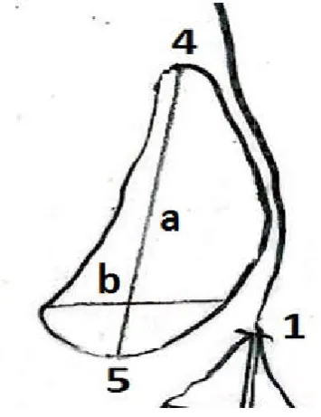
Point Sh highest point on the peripheral borders of the frontal sinus.( fig 1)
Point Sl lowest point on the peripheral borders of the frontal sinus(fig 1)
Table 2. Linear Mesurements
Co-Gn the effective length of the mandible.(fig 1)
Maximum height of frontal sinus (a) A line connecting Sh to Sl is drawn to measure the maximum height of frontal sinus.(fig 2)
[image:2.595.82.503.239.368.2]Width of frontal sinus (b) Perpendicular to above line, a line was drawn to measure the maximal width of frontal sinus. (fig 2)
Table 3. ANOVA to measure p value
df Mean square F P value Significance
Maximum heigth of Between the groups 2 73.817 1.856 0.166 Non significant
frontal sinus Within the groups 57 39.774
Total 59
Maximum width of Between the groups 2 218.064 0.879 0.421 Non significant
frontal sinus Within the groups 57 248.116
Total 59
Frontal sinus Between the groups 2 18104.617 2.139 0.127 Non significant
area Within the groups 57 8464.066
Total 59
Effective length of Between the groups 2 313.817 3.738 0.03 Significant
mandible Within the groups 57 83.949
[image:2.595.123.473.400.454.2]Total 59
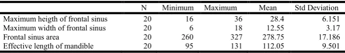
Table 4. Mean value and standard deviation for Class I
N Minimum Maximum Mean Std Deviation
Maximum heigth of frontal sinus 20 16 36 28.4 6.151
Maximum width of frontal sinus 20 6 18 12.55 3.17
Frontal sinus area 20 260 327 278.75 17.186
Table 5. Mean value and standard deviation for Class II
N Minimum Maximum Mean Std Deviation
Maximum heigth of frontal sinus 20 7 19 11.85 3.117
Maximum width of frontal sinus 20 16 40 29.05 6.337
Frontal sinus area 20 93 129 108.35 10.419
[image:3.595.130.470.168.221.2]Effective length of mandible 20 93 125 107.2 9.22
Table 6. Mean value and standard deviation for Class III
N Minimum Maximum Mean Std Deviation
Maximum heigth of frontal sinus 20 23 42 32 5.849
Maximum width of frontal sinus 20 8 18 13.55 3.426
Frontal sinus area 20 250 322 285.25 19.183
Effective length of mandible 20 108 132 115.05 8.751
Table 7. Bonferroni and Sidak methods were used for post hoc analysis
Dependent variable (I) type (J) type P value
Maximum Bonferroni Class I Class II 1
heigth of Class III 0.593
frontal Class II Class I 1
sinus Class III 0.196
Class III Class I 0.593
Class II 0.196
Sidak Class I Class II 0.919
Class III 0.483
Class II Class I 0.919
Class III 0.183
Class III Class I 0.483
Class II 0.183
Maximum Bonferroni Class I Class II 0.571
width of Class III 1
frontal Class II Class I 0.571
sinus Class III 1
Class III Class I 1
Class II 1
Sidak Class I Class II 0.469
Class III 0.864
Class II Class I 0.469
Class III 0.9
Class III Class I 0.864
Class II 0.9
Table 8. Bonferroni and Sidak methods were used for post hoc analysis (table 7 continued)
Dependent variable (I) type (J) type P value
Frontal sinus area
Bonferroni Class I Class II 0.848
Class III 0.989
Class II Class I 0.848
Class III 0.13
Class III Class I 0.989
Class II 0.13
Sidak Class I Class II 0.631
Class III 0.699
Class II Class I 0.631
Class III 0.124
Class III Class I 0.699
Class II 0.124
Effective Bonferroni Class I Class II 0.299
Length of Class III 0.915
mandible Class II Class I 0.299
Class III 0.027
Class III Class I 0.915
Class II 0.027
Sidak Class I Class II 0.27
Class III 0.664
Class II Class I 0.27
Class III 0.026
Class III Class I 0.664
Class II 0.026
[image:3.595.176.419.252.491.2] [image:3.595.176.421.517.767.2]Figure 1. Various cephalometric landmarks that are required for the tracing of lateral cephalogram. 1: Nasion (N), 2: Supspinale (point A), 3: Supramentale (point B), 4: Point Sh (Sh), 5: Point Sl
(Sl), 6: Condylion (Co), 7: Gnathion (Gn).
Figure 2. The point describe in the figure are as followed: 1: Nasion (N) 4: Point Sh (Sh), 5: Point Sl (Sl), Maximum
height(a), Maximum width(b).
Post-hoc analysis shows that the effective mandible is highest in Class III and is the shortest This signifies the correlation of the mandible classes. The maximum height, maximum width frontal sinus are not statistically significant in and Class III subjects and showed no correlation length of the mandible and the maximum width and area of frontal sinus. (p-value=>0.05)
Various cephalometric landmarks that are required for the tracing of lateral cephalogram. 1: Nasion (N), 2: Supspinale (point A), 3: Supramentale (point B), 4: Point Sh (Sh), 5: Point Sl
Condylion (Co), 7: Gnathion (Gn).
escribe in the figure are as followed:- Point Sh (Sh), 5: Point Sl (Sl), Maximum height(a), Maximum width(b).
effective length of the shortest in Class II. mandible with the different width and area of in Class I, Class II correlation between the height, maximum value=>0.05) .
DISCUSSION
The frontal sinuses are the paranasal sinuses which are superior to the eyes, in the frontal bone
part of the forehead. The development and size of frontal sinus can be crucial for diagnosing and treating various malocclusions. The origin of frontal sinus is from anterior ethmoidal cells during birth. The frontal sinus bud is present during the birth in ethmoidal region b
radiographically until the age of 5 years when it projects above the orbital rims (Harris et al., 1987
bone at the end of the first year of life The sinus grows till the age of 12 ye
annual height (stature) increments in children reached a plateau at 16 years in boys and 14 years in girls, and it was thought that these, too, were the ages at which frontal sinus enlargement ceased (Tanner, 1962).
increase in the sinus size very closely follows a growth trend similar to that of other bones. Jof
frontal sinus enlargement to be associated with prognathic subjects (Joffe, 1964; Rossouw
cephalograms are widely used to study morphologic characteristics of various malocclusions.
carried out to analyze the correlation of frontal sinus with different skeletal pattern. According to the results, we can state that there is no correlation between dimensions of frontal size with different skeletal pattern.
Although our result state there is no correlation previously studies have been carried out and it was suggested that acromegaly is associated with prominent frontal sinus overgrowth of the jawbone, and one usually finds a class III type prognathic mandible in these cases (Shafer, Hine, Levy, 1974) (Ruf and Pancherz, 1996).
Sinus enlargement to be associated with prognathic subjects (Joffe, 1964).In a similar study reported by Rossouw (1991) they had only compared the area of the frontal sinus in between adult skeletal Class III and adult skeletal Class I growth pattern cases but did not study the Class II growth pattern cases. Ruf and Pancherz(1996) suggested that the somatic maturity stage may be predicted rather accurately by analyzing Frontal Sinus development on pre
head cephalograms (Ruf and Pancherz
(Steiner, 1953) is still widely accepted as a maxillo-mandibular harmony (Jacobson, 1975).
was used to ascertain its correlation with the Frontal Sinus Area. In the present study, manual tracing was used for calculation of the maximum height, maximum width and frontal sinus area of frontal sinus. Although in some studies the digital method was used to measure these factors, the manual technique has accuracy similar to that of digital technique in this regard (Axelsson et al.,
affordability, the manual technique was used. It seems that further investigations in several centers with larger sample sizes can increase the accuracy of the obtained data and standards.
Conclusion
The importance of the frontal the normalcy is set by statistical standard values are given frontal sinus which may references.
are the paranasal sinuses which are
frontal bone, which forms the hard . The development and size of frontal sinus can be crucial for diagnosing and treating various malocclusions. The origin of frontal sinus is from anterior The frontal sinus bud is present during the birth in ethmoidal region but it is not evident radiographically until the age of 5 years when it projects above 1987). It migrates into the frontal bone at the end of the first year of life (Brown et al., 1984). The sinus grows till the age of 12 years. Tanner found that the annual height (stature) increments in children reached a plateau at 16 years in boys and 14 years in girls, and it was thought that these, too, were the ages at which frontal sinus , 1962). This suggests that the increase in the sinus size very closely follows a growth trend similar to that of other bones. Joffe, Rossouw et al found frontal sinus enlargement to be associated with prognathic Rossouw et al., 1991). The lateral cephalograms are widely used to study morphologic characteristics of various malocclusions.The present study was carried out to analyze the correlation of frontal sinus with different skeletal pattern. According to the results, we can state no correlation between dimensions of frontal size
Although our result state there is no correlation previously studies have been carried out and it was suggested that acromegaly is associated with prominent frontal sinus and overgrowth of the jawbone, and one usually finds a class III-type prognathic mandible in these cases (Shafer, Hine, Levy,
, 1996). Joffe (1964)found Frontal Sinus enlargement to be associated with prognathic subjects In a similar study reported by Rossouw et al. (1991) they had only compared the area of the frontal sinus in between adult skeletal Class III and adult skeletal Class I growth pattern cases but did not study the Class II growth ncherz(1996) suggested that the somatic maturity stage may be predicted rather accurately by analyzing Frontal Sinus development on pre-existing lateral Pancherz, 1996). TheANB Angle is still widely accepted as an indicator of mandibular harmony (Jacobson, 1975).Therefore , it was used to ascertain its correlation with the Frontal Sinus In the present study, manual tracing was used for calculation of the maximum height, maximum width and us area of frontal sinus. Although in some studies the digital method was used to measure these factors, the manual technique has accuracy similar to that of digital technique in
et al., 2004). Thus, considering its
manual technique was used. It seems that further investigations in several centers with larger sample sizes can increase the accuracy of the obtained data and
[image:4.595.74.252.422.653.2] The maximum height, maximum width and area of frontal sinus do not correlate with the effective length of mandible and also with the three skeletal types.
Acknowledgement
Authors would like to acknowledge the full staff of the Orthodontic department of Bharati Vidhyapeeth Dental College and hospital, Pune for providing the material required for the scientific research.
REFERENCES
Albarakati, S.F., Kula, K.S., Ghoneima, A.A. 2012. The reliability and reproducibility of cephalometric measurements: A comparison of conventional and digital methods. Dentomaxillofac Radiol., 41:11-7.
Atchison, K.A., Luke, L.S., White, S.C. 1991. Contribution of pretreatment radiographs to orthodontists' decision making.
Oral Surg Oral Med Oral Pathol., 71:238-45.
Axelsson, S., Storhaug, K., Kjaer, I. 2004. Post-natal size and morphology of the sella turcica. Longitudinal cephalometric standards for Norwegians between 6 and 21 years of age. Eur J Orthod., 26(6):597–604.
Brown, W.A., Molleson, T.I., Chinn, S. 1984. Enlargement of the frontal sinus. Ann Hum Biol., 11:221-6.
Devereux, L., Moles, D., Cunningham, S.J., McKnight, M. 2011. How important are lateral cephalometric radiographs in orthodontic treatment planning? Am J Orthod
Dentofacial Orthop., 139:e175-81.
Durão, A.R., Pittayapat, P., Rockenbach, M.I., Olszewski, R., Ng. S., Ferreira, A.P., et al. 2013. Validity of 2D lateral cephalometry in orthodontics: A systematic review. Prog
Orthod., 14:31.
Endo, T., Abe, R., Kuroki, H., Kojima, K., Oka, K., Shimooka, S. 2010. Cephalometric evaluation of maxillary sinus sizes in different malocclusion classes. Odontology, 98:65-72.
Harris, A.M., Wood, R.E., Nortjé, C.J., and Thomas, C.J. 1987. Gender and ethnic differences of the radiographic image of the frontal region. J Forensic Odonstostomatol,
5:51-7.
Jacobson, A. 1975. The "Wits" appraisal of jaw disharmony.
Am J Orthod., 67:125-38
Joffe, B.M. 1991. Frontal sinus enlargement associated with mandibular prognathism. J Dent Assoc S Afr 1964:127-9. Kullman, L., Eklund, E. and Grundin, R. 1990. The value of
the frontal sinuses in the identification of the unknown persons. J. Forensic Odonstostomatol, vol. 8, no. 1, p. 3-10.
Nijkamp, P.G., Habets, L.L., Aartman, I.H., Zentner, A. 2008. The influence of cephalometrics on orthodontic treatment planning. Eur J Orthod., 30:630
Rossouw PE, Lombard CJ, Harris AMP. The frontal sinus and mandibular growth prediction. Am J Orthod Dentofac
Orthop., 100:542-6
Ruf, S., Pancherz, H. 1996. Development of the frontal sinus in relation to somatic and skeletal maturity. A cephalometric roentgenographic study at puberty. Eur J Orthod.,
18(5):491-97.
Ruf, S., Pancherz, H. 1996. Frontal sinus development as an indicator for somatic maturity at puberty? Am J Orthod
Dentofac Orthop., 110:476-82.
Salehi, P., Heidari, S., Khajeh, F. 2012. Relationship between frontal sinus surface area and mandibular size on lateral cephalograms of adults. J Isfahan Dent Sch., 8:244-50. Shafer, W.G., Hine, M.K., Levy, B.M. 1974. A textbook of
oral pathology. 3rd ed. Philadelphia: WB Saunders, 606-7. Steiner, C.C. 1953. Cephalometrics for you and me. Am J
Orthod., 39:729-55.
Tanner, J.M. 1962. Growth at adolescence. 2nd ed. Oxford: Blackwell Scientific Publications, 3.