MALATHION TOXICITY ON FISH
*1
1
Department of Zoology, Gauhati University, Gopinath
2
Department of Zoology, Cotton College, Guwahati, Assam, 781001, India
ARTICLE INFO ABSTRACT
Fishes are very sensitive to the changes in their aquatic environment. For this reason, they are known as the bio
used amidst various group of pesticide in intensive ag
various pest and diseases owing to their high insecticidal property, low mammalian toxicity, low persistence and rapid biodegradability in the ecosystem. Pesticide exposure may also fatal to many non-target or
occasionally leading to the death of the fish.
organophosphate insecticides is being extensively used as dust, emulsion, and vapour variety of insect pests under different conditions.
pesticides, may induce many significant changes in fish. Present study is aimed to review the toxicological effects on haematological parame
behavioural changes, neurotoxic, histopathological alterations, respiratory responses, bioaccumulation and chromosomal changesin fishes exposed to the organophosphate pesticide Malathion.
Copyright©2016, Sanjoy Deka and Rita Mahanta.This
unrestricted use, distribution, and reproduction in any medium, provided the original work is properly cited.
INTRODUCTION
Since the removal of the organochlorine insecticide owing to its ceaseless harmful effect, organophosphate (OP) insecticides have been chosen as the most widely favoured insecticide in today’s world to make the crop free from pest for greater productivity. All OPs apparently share the common mechanism of choline esterase inhibition at nerve endings resulting excess acetylcholine in the nerve ending which
overstimulates the effector organ. Malathion (
one of the earliest organophosphate insecticides developed in 1950, was first registered for use in the United States in 1956 by the United States Department of Agriculture (USDA), which is now regulated by the United States Environmental Protection Agency (USEPA, 2006). This pesticide is being extensively used as dust, emulsion, and vapour to control wide variety of insect pests under different conditions.
one of the most effective organophosphorus insecticides used for the control of pest on greenhouses, nurseries, home and garden vegetables, field crops, fruits and domesti
other words, it is used to control aphids, mites, scale, flies, leafhoppers, leaf miner, thrips, loopers, mealy bugs, spittlebugs, corn earworms, chinch bugs, grasshoppers, armyworms, boll weevils, bollworms, lice, ticks, ants, spiders, and mosquitoes.
*Corresponding author: Sanjoy Deka,
Department of Zoology, Gauhati University, GopinathBordoloi Nagar, Guwahati, Assam, 781014, India.
ISSN: 0975-833X
Article History:
Received 07th September, 2016
Received in revised form 09th October, 2016
Accepted 25th November, 2016
Published online 30th December,2016
Key words:
Malathion, Toxicity, LC50, Fish.
Citation: Sanjoy Deka and Rita Mahanta.2016. “Malathion toxicity on fish
RESEARCH ARTICLE
MALATHION TOXICITY ON FISH - A REVIEW
1
Sanjoy Deka and
2Rita Mahanta
Department of Zoology, Gauhati University, Gopinath Bordoloi Nagar, Guwahati, Assam, 781014, India
Department of Zoology, Cotton College, Guwahati, Assam, 781001, India
ABSTRACT
Fishes are very sensitive to the changes in their aquatic environment. For this reason, they are known as the bio-indicator species to monitor the water pollution. Organophosphate pesticides are widely used amidst various group of pesticide in intensive agricultural practices to protect the crops from various pest and diseases owing to their high insecticidal property, low mammalian toxicity, low persistence and rapid biodegradability in the ecosystem. Pesticide exposure may also fatal to many target organisms like fish where it hampers its health through impairment of metabolism, occasionally leading to the death of the fish. Malathion (C
organophosphate insecticides is being extensively used as dust, emulsion, and vapour
variety of insect pests under different conditions. Malathion, one of the most extensively studied pesticides, may induce many significant changes in fish. Present study is aimed to review the toxicological effects on haematological parameters, physical parameters, biochemical parameters, behavioural changes, neurotoxic, histopathological alterations, respiratory responses, bioaccumulation and chromosomal changesin fishes exposed to the organophosphate pesticide Malathion.
This is an open access article distributed under the Creative Commons Att use, distribution, and reproduction in any medium, provided the original work is properly cited.
insecticide owing to its ceaseless harmful effect, organophosphate (OP) insecticides have been chosen as the most widely favoured insecticide in today’s world to make the crop free from pest for greater productivity. All OPs apparently share the common chanism of choline esterase inhibition at nerve endings resulting excess acetylcholine in the nerve ending which
Malathion (C10H19O6PS2),
one of the earliest organophosphate insecticides developed in tered for use in the United States in 1956 by the United States Department of Agriculture (USDA), which is now regulated by the United States Environmental This pesticide is being our to control wide variety of insect pests under different conditions. Malathion is one of the most effective organophosphorus insecticides used greenhouses, nurseries, home and garden vegetables, field crops, fruits and domestic animals.In other words, it is used to control aphids, mites, scale, flies, leafhoppers, leaf miner, thrips, loopers, mealy bugs, spittlebugs, corn earworms, chinch bugs, grasshoppers, armyworms, boll weevils, bollworms, lice, ticks, ants, spiders,
Department of Zoology, Gauhati University, GopinathBordoloi
It is applied to alfalfa, clover, pasture and range grasses, non agricultural land, cereal crops, cotton,
sugar beets, corn, beans, blueberries, stored grain, and inside homes (Malathion Fact Sheet, 2000).
Pure and Applied Chemistry (IUPAC) name for malathion is
diethyl 2-dimethoxyphosphinothioylsulfanylbutanedioate.
Chemical name is O,O-dimethyl dithiophosphate of diethyl
mercaptosuccinate. It is
(ethoxycarbonyl) ethyl O,O Structural formula ofMalathionis
Malathion can enter into surface water through spray drift, during the time of application, and runoff after rainfall. Malathion undergoes three types of metabolic modifications in animals, namely oxidative, hydrolytic and the elimination of a methyl group catalysed by glutathione S
(Toxicological Profile on Malathion, 2003
metabolite, malaoxon, is a more potent organophosphate than the parent compound, inhibit the enzyme acetyl cholinesterase and hence, prevent the hydrolysis o
acetylcholine in the central and peripheral nervous systems. Thus, Inhibition of acetyl cholinesterase results in accumulation and continuous action of the neurotransmitter
International Journal of Current Research Vol. 8, Issue, 12, pp.44120-44128, December, 2016
INTERNATIONAL
Malathion toxicity on fish - A review”, International Journal of Current Research
Bordoloi Nagar, Guwahati, Assam, 781014, India
Department of Zoology, Cotton College, Guwahati, Assam, 781001, India
Fishes are very sensitive to the changes in their aquatic environment. For this reason, they are known indicator species to monitor the water pollution. Organophosphate pesticides are widely ricultural practices to protect the crops from various pest and diseases owing to their high insecticidal property, low mammalian toxicity, low persistence and rapid biodegradability in the ecosystem. Pesticide exposure may also fatal to many ganisms like fish where it hampers its health through impairment of metabolism, C10H19O6PS2), one of the earliest
organophosphate insecticides is being extensively used as dust, emulsion, and vapour to control wide
Malathion, one of the most extensively studied pesticides, may induce many significant changes in fish. Present study is aimed to review the ters, physical parameters, biochemical parameters, behavioural changes, neurotoxic, histopathological alterations, respiratory responses, bioaccumulation and chromosomal changesin fishes exposed to the organophosphate pesticide Malathion.
is an open access article distributed under the Creative Commons Attribution License, which permits
It is applied to alfalfa, clover, pasture and range grasses, non-agricultural land, cereal crops, cotton, sunflower, soybeans, sugar beets, corn, beans, blueberries, stored grain, and inside homes (Malathion Fact Sheet, 2000). International Union of Pure and Applied Chemistry (IUPAC) name for malathion is dimethoxyphosphinothioylsulfanylbutanedioate. dimethyl dithiophosphate of diethyl
also known as S-1,2-bis
ethyl O,O-dimethyl phosphorodithioate. Structural formula ofMalathionis-
can enter into surface water through spray drift, during the time of application, and runoff after rainfall. Malathion undergoes three types of metabolic modifications in animals, namely oxidative, hydrolytic and the elimination of a d by glutathione S-transferase (GST) Toxicological Profile on Malathion, 2003). Malathion and its metabolite, malaoxon, is a more potent organophosphate than the parent compound, inhibit the enzyme acetyl cholinesterase and hence, prevent the hydrolysis of the neurotransmitter acetylcholine in the central and peripheral nervous systems. Thus, Inhibition of acetyl cholinesterase results in accumulation and continuous action of the neurotransmitter
INTERNATIONAL JOURNAL OF CURRENT RESEARCH
acetylcholine at postsynaptic sites. The water solubility of Malathion is 145 mg/L at 20°C, and its vapour pressure is 3.97× 10⁻⁵ mm Hg at 30°C. It has an octanol/water partition coefficient (log KOW) of 2.36, and bioconcentration has not been observed in majority of the studies (US EPA, 2015). Bioactivation of Malathion is necessary for it to exert its toxic effect. Bioactivation is primarily mediated by cytochrome P450 enzymes in the liver, which create the active metabolite malaoxon through oxidative sulfuration (Malathion, Technical
Fact Sheet, 2010). Although Organophosphorous pesticides
(OPs) malathion has rapid biodegradability, susceptible to
photolysis as well as hydrolysis and lesser persistency in the
environment, with 81 to 94% degradation occurring in various
non-sterile soils within ten days, yetit leaves residues in the
soil and water for several days after their application, and pose a constant threat to non-target organisms, especially fish
(Magare and Patil, 2000). Rauf, 2015 cited Martinez and
Leyhe, 2004 and stated that the half-life of malathion is up to 11 days and it degrades into malaoxon which is more toxic than the parent.
Effects on Hematological parameters
Venkataraman and Sandhya Rani, 2013 studiedacute toxicity and blood profile of freshwater fish, Clariasbatrachus (Linn.) exposed to sub-lethal concentration of 0.05, 0.25 and 0.5 ppm malathion after a time interval of 24, 48, 72 and 96 hours.
They have calculated the 96 h LC 50 of malathion as 1 ppm.
Both the researchers had noticed that with increase in the concentration and exposure period of malathion, there were significant reductions in Hb (haemoglobin), haematocrit (PCV), RBC (Red Blood Cell Count) and MCV (Mean Corpuscular Volume) levels and increase in WBC (White Blood Cell Count), MCHC (Mean Corpuscular Haemoglobin Concentration) and MCH (Mean Corpuscular Hemoglobin) levels in the malathion treated fish in contrary to the normal
values of haematological parameters in control group.
According to the records of this study, fishes exposed to 0.5 ppm (maximum concentration) for 96 h exposure of malathion exhibiteddecrease (2.36±0.04 x 106mm3) in RBC, whereas a significant risewas reported in WBC(40.91±0.20 x 103mm3), MCH (16.00±0.43 pg) andMCHC (52.83±1.86%) values. MCHC value reported with a substantial increase from 45.32 ± 0.90%(in 0.5 ppm for 24 h) to 52.83 ± 1.86% (in*/ 0.5 ppm for 96h) in treated fishes. On the other hand, fishes exposed to0. 05 ppm to 0.5 ppm malathion for 24 h to 96 h revealeda minor increase in their colorindex.
According to the researchers, acute anaemia resulting in C.
batrachus owing to a decrease in the concentration of haemoglobin in blood is due to the effect of toxic malathion on
gills, as well as decrease in oxygen. In this investigation,
haemolysis was also predicted as one of the causes for reduction in Hb, RBC and PCV values. Increase in WBC count; reflect the occurrence of leucocytosis in the treated fish samples. Both the researcher cited Sudha, 2012 and mentioned probable cause of increase in WBC count firstly as a typical defensive response of the fish against a toxic invasion and secondly asleukaemia.Alterations in MCV, MCHC, MCH were considered responsible reason by the investigators after studying the findings of Shah, 2006 to direct or feedback responses of structural damage to RBC membranes which resulted in haemolysis and impairment in haemoglobin synthesis, stress related release of RBCs from the spleen (Shah, 2006).Finally both the researchers have concluded that
malathion was moderately toxic to C. batrchus. Ahmad, 2012
in his study the effects of sub-lethal exposure of malathion on
haematological parameters of Clarias gariepinus observed
decreased value of RBC and WBC counts, hemoglobin (Hb) concentration and haematocrit (Ht) values as compared to the control fish after exposed to sublethal doses of malathion. The 96 h LC50 value was calculated to be 8.22 mg/L from which sub-lethal concentrations of 0.5, 1.0 and 2.0 mg/L malathion was exposed to the fishes for 4 weeks.
Effect on Physical Parameters
After determination of LC50as 5.8 ppm by Kabeer et al., 1981,
fish in batches of 6 were exposed to sublethal concentration of 2 ppm malathion for 48 h. Based on the data reported by
Kabeer et al. (1981), a slight but progressive decrease in the
body weight of malathion treated fish in due course of timewere observed compared with normal fish which suggested the loss of some constituents of fish body. They have not reported any change in the water content of the normal and malathion exposed fish.
Effected on Biochemical Parameters
Venkataramana et al., 2006, investigated the impact of sub
lethal concentrations (0.05, 0.25 and 0.5 ppm) of malathion on heart muscle glycogen, protein and cholesterol in freshwater gobiid fish, Glossogobius giuris with an exposure period of 24, 48, 72 and 96 hr. They have analysed the changes in the glycogen, protein and cholesterol and found that these parameters werealtered during different intervals of malathion exposure. In this study, it was evident that at 24 hr exposure time, themuscle glycogen content (1.51±0.40 mg/g) was decreased by 47.93%in comparison to the glycogen content (2.90±0.72 mg/g) of control group of fish where as it was increased subsequently at0.25 ppm (3.96±0.85 mg/g) and 0.5 ppm (4.89±0.71 mg/g) exposure to malathion. At 48 hr and 72 hr exposure time to malathion glycogen content was reported to be maximum (3.20±0.58 mg/g and 3.50±0.45 mg/g respectively) at 0.25 ppm malathion in comparison to control group (2.20±0.25 mg/g and 3.30±0.33 mg/g respectively). However, after 96 hr the quantity of muscle glycogen showed a decreasing trend reaching 20.31% and 21.26% at 0.05 ppm malathion (2.51±0.36 mg/g) and 0.5ppm malathion (2.48±0.34 gm/g) exposure respectively.
Dezwaan and Zandee, 1999 and Chaudhari, 2000 reported possible cause of depletion of glycogen content in heart muscle as glycogenolysis, resulting in anaerobic glycolysis to cope up
with the adverse condition. Venkataramana et al., 2006
observed decreasing trend of muscle protein exposed to sub lethal doses malathion for 24 hrs exposure time. The trend in terms of percentage decrease was observed to be 28.75 %( 14.32±0.98 mg/g), 23.08 % (15.46±1.45 mg/g) and 29.80% (14.11±1.41 mg/g) with 0.05, 0.25 and 0.5 ppm of malathion
treatment respectively compared to control (20.10±0.85 mg/g).
The researchers had also witnessed slight elevation of heart muscle protein exposed to 0.25 and 0.5 ppm of malathion after 48 and 72 hrs of treatment compared to both the control (22.00±1.58 mg/g and 19.10±0.13 mg/g) group. However, after 96 hrs of exposure to fish with 0.05 and 0.5 ppm of malathion, a declining trend was noticed by the investigators, attaining a minimum percentage of 29.45 (14.32±1.48 mg/g) and 32.66 (13.67±1.27 mg/g) as compared to control
cited the suggestions put forwarded by Krishnamohan et al., 1985 and Chandravathy and Reddy, 1994 as decline in the muscle protein content may perhaps be due to reduced protein synthesis, increased proteolysis and also owing to utilization
for metabolic processes under toxicity. Venkataramana et al.,
2006 also reported enhancement of muscle cholesterol level in the fish exposed to 0.05 ppm of sub lethal malathion and found a maximum level of cholesterol (1.01±0.15 mg/g)at 24h exposure time in comparison to control (0.75±0.75 mg/g). Nevertheless, after 48 hr exposure to 0.05 ppm malathion, the muscle cholesterol quantity increased by a percentage of 40.57 (0.97±0.14 mg/g) compared to control (0.69±0.14 mg/g). A similar fluctuating pattern was also observed by the researchersat 72 hr exposure period.
On the other hand, when the researchers exposed fishes to 0.5 ppm, the muscle cholesterol quantity started declining for 24,
48 and 72 hr. Yet, by 96 hr, they have noticed a decreasing
trend in heart muscle cholesterol in the fishes and recorded the
minimum percentage of 13.58 (0.70±0.15 mg/g),
14.81(0.69±0.19 mg/g) and 22.22 (0.63±0.18 mg/g) in 0.05, 0.25 and 0.5 ppm dose of malathion respectively compared to control (0.81±0.18 mg/g). Finally all the investigators concluded that fishes exposed to sub lethal concentration 0.5 ppm of malathion for 24 to 96 hrexhibited decrease in the muscle cholesterol level in the heart. Patil, and David, 2009 studied the hepatotoxic potential of malathion in the freshwater
teleost, Labeo rohita, after exposing to a sublethal
concentration (0.9 μl/L) of commercial grade malathion (50% Emulsified Concentration) for 5, 15 and 25 days. Calculated
LC50 for 96 h to the fishes for malathion was reported to be 9.0
μl/L. Sublethal dose for this study was prepared following to Sprague, 1973.Both the scholars had observed that the total, structural and soluble proteins and Acetyl Choline Esterase activity werediminished, however free amino acids, protease activity and Acetyl Choline (Ach) were found to be enhanced in the fishes exposed to sublethal dose of malathion for 5 and 15 days. Nevertheless, both of them surprisingly noticed that fish exposedtomalathion for 25 days showed values nearer to the control fish and observed to be recovering to normalcy.
Patil, and David, 2009 described that the total, structural and soluble proteins were found to be decreased owing to the occurrence ofhigh proteolytic activity, and inefficiency in the protein biosynthesis. Another reason for this decrease inprotein levels was predicted by the investigators as protein
degradation. By citing the findings of Hai et al., 1995, the
author of the paper had mentioned oxidative stress as the reason for degradation which is a typical feature of organophosphate compounds, besides their in hibitory effect on AChE. According to the researchers oxidative stress might bring changes in free radical production which in turn overwhelms the endogenous antioxidant levels causing substantial cell damage and death. Finally the researchers had concluded that exposure to sublethalmalathion affects protein metabolism and the normal neural physiology of the liver. But recovery in 25 days might be a revitalization phenomenon as all organisms make every effort to overcome stress to survive. Recovery phenomenon as stated by the author of the paper was adaptive and even strategic. Ahmad, 2012 studied the effects of sub-lethal exposure of malathion on biochemical
composition and haematological parameters of Clarias
gariepinus. and reported elevated Plasma glucose level following reduced Liver and muscle glycogen level; nevertheless, plasma protein was reported to be decreased in
fishes exposed to sublethal malathion. According to the findings of Ahmad, 2012; Alanine amino transferase (ALT), glutamate oxaloacetate transaminase (GOT) and glutamate pyruvic transaminase (GPT) activities increased in the fish exposed to sublethal malathion. But the researcher has noticed insignificant effect of malathion on magnesium and calcium ions as reduction in the concentration of Ca ions was recorded in the fish exposed to high dose of malathion and in the last period of exposure whereas Mg ions remain unchanged. According to Ahmad, 2012 significant hyperglycemia and hypo-proteinaemia was evident in the fish exposed to various doses of malathion. The investigator had witnessed that these alterations were more pronounced in the higher doses and in the last period of exposure. According to Ahmad, 2012 malfunctioning of the haematopoietic system due to Malathion exposure might be leading to reduction in different blood parameters. He believed that the elevated level of glucose reported in the blood of Malathion treated fish could be due to glycogenolysis to meet the increased energy demand. By citing
the findings Pickering, 1981 and Winkaler et al., 2007,
Ahmad, 2012 stated that stress stimulated rapid secretion of Glucocorticoids and catecholamine hormones from adrenal gland which were known to produce hyperglycemia in animals. Elevation of these hormones could be attributable to boosted gluconeogenesis response of stressed fish in their effort to satisfy their new energy demands. Ahmad, 2012 concluded that higher activities of experimented enzymes registered in his investigation might be attributed to cellular damage caused to liver by malathion.
Roopavathy et al., 2013 studied Toxicity of Malathion on
Metabolic Activities in the River Cauvery of Tilapia Fish
(Oreochromis mossambicus) by exposing sub lethal
concentration (0.5 ppm) of malathion for 24, 48, 72 and 96 hr respectively on the fishes (Four groups where another group was kept as control which was free from the pesticide exposure. when the fishes were exposed to malathion (0.5 ppm) the protein and carbohydrate content were found to have decreased in all the experimental tissues namely muscle, gills, liver and kidney. They have cited the findings of Remia et al., 2008 and stated possible reduction of protein content in the tissues as proteolysis and increased metabolism under toxicant
stress. Likewise the researchers had cited Thenmozhi et al.,
2010, and anticipated the reason for decrease in carbohydrates contents as impairment of carbohydrate metabolism due to toxic effect. Fahmy, 2012 carried out an experimental study to assess the toxic effect of the insecticide malathion on oxygen consumption and some biochemical characteristics (total protein, carbohydrate and cholesterol in liver, muscle, kidney and gills) of the tilapia fish Oreochromis niloticus by exposing a sublethal concentration of 0.5 ppm for 24,48,72, and 96 h respectively. The investigator had found that the rate of oxygen consumption and all biochemical parameters were declined on comparison with control during all the exposure periods. The result of the study showed that protein, carbohydrate and the cholesterol content were significantly declined when the fishes were exposed to malathion (0.5 ppm) in all exposure time. The researcher cited the findings of
Remia et al., 2008 to enlighten proteolysis and increased
metabolism under toxicant stress as the reason for the reduction of protein. Another reason for reduction in protein
content was cited from the findings of Venkatramana et al.,
carbohydrate metabolism due to toxic effect (Thenmozhi et al., 2010). According to the researcher the possible cause of reduction of carbohydrate content in the fish might be the active glycogenolysis and glycolytic pathway to provide excess energy in stress condition. The decrease in cholesterol was found to be high in gills and less in muscles. The reduced cholesterol level may be due to the inhibition of cholesterol biosynthesis in the liver or due to reduced absorption of dietary cholesterol (Fahmy, 2012). However, it was reported that the decline of cholesterol may be due to utilization of fatty
deposits instead of glucose for energy purpose (Kanagaraj et
al., 1993). Magar and Dube, 2013 investigated effect of Sub
Lethal Concentration of Malathion on Metabolic Profiles and Histological Studies in Heart Tissue of Channa punctatus and found that the fresh water exposed to sub lethal concentration of malathion showed reduction in the level of protein in cardiac muscle of treated group of fish during 96 hours (64.20) compared with control (76.03) whereas total lipid content declined (40.93) during 96 hours compared with control (50.10). Glycogen content in cardiac muscle also reported to be decreased (8.56) during 96 hours compared with control (14.15). In a study carried out by Borthakur, 2006 to investigate the effect of sublethal malathion on three members of hepatic MFO enzyme system namely cytochrome P450,
AHH, and XOD in Heteropneustes fossilis, a freshwater
siluroid fish, reported significantly elevated (P<0.05) hepatic cytochrome P450 activity within 24 hours of 0.1 ppm malathion exposure and progressively increased to more than 20% over control within 15th day of exposure. It was followed
by a steep rise in activity upto the 25th day (P<0.001). similar
response was reported to 0.2 ppm malathion exposed group but a highly significant decrease (P<0.05) of cytochrome P450 activity was observed in the last five days of the exposure to 0.2 ppm malathion compared to that of the 0.1 ppm exposed group.
Behavioral Changes due to malathion exposure
Behavior is regarded as a convincing tool in ecotoxicology [Drummond and Russom, 1990; Cohn and MacPhail, 1996). In toxic environments, fish showed loss of equilibrium, irregular, erratic and darting swimming movements and reason forthese changes was reported to be inhibition of the enzyme
acetylcholinesterase (AChE) activity which resulted
accumulation of acetylcholine in cholinergic synapses triggering hyperstimulation in the fishes (Mushigeri and
David, 2005). Naserabad et al., 2015 investigated acute
toxicity and behavioral changes of the Gold Fish (Carassius auratus) exposed to Malathion and Hinosan in due course of
finding LC50. The value of LC50 calculated by the researchers
for 96h duration for malathionwas found to be 4.71 mg/L. They have noticed that fishes that were exposed to malathion started exhibiting behavioralchanges and clinical symptoms at 8 mg/L after 5 hours, but these changes and symptoms appeared after 2 hours when exposed to 16 mg/L malathion. According to the investigators behavioural changes included irregular, erratic and darting swimming movements of the fishes; hyper excitability, bruise in the caudal section, loss of equilibrium and sinking of the fishes to the bottom.
Naserabad et al., 2015 also calculated and reported the value
of MAC (Maximum Allowable Concentration) for malathion at mean temperature 24±1 Cº as 0.471 mg/l, andLOEC (lowest observable effect concentration, which represents the initial toxicity threshold of a chemical) as 4 mg/lwhereas NOEC (No
Observed Effect Concentration, which represent the
concentration of toxicant that will not cause any effect) as 2 mg/l. Salwa and Ella, 2008 conducted an empirical study on Toxicity of malathion and its effect on the activity of acetylcholinesterase in various tissues of the grass carp, Ctenopharyngodon idella Val. The Acute toxicity (LC50 values) reported by them for 24 h, 48h, 72h, and 96h were 3.728 mg/l, 2.838 mg/l, 2.444 mg/l, and 2.138 mg/l respectively for grass carp. The safe concentrations of malathion was 0.0513 mg/l. The fish showed typical changes in behaviour when exposed to various concentrations of malathion. They have observed that the fishes experienced progressive lethargicity, loss of equilibrium, difficulty in respiration, exhibited convulsions, dashing against the wall of the experimental aquaria and short unpredictable bumpy body movements, settling to bottom before death. Light coloured skin was observed by the on the fishes exposed to malathion
with an excessive amount of mucus secretion over the body.
However, Control fish behaved normally according to their observation. According to the researchers, the fish exposed to malathion showed intense opercular movements due to hypoxic stress accompanied by a progressive inhibitory effect of this compound on the respiratory mechanism. Mount and Brungs, 1967 had recommended that the safe level of malathion for fishes can beat some point between 1/15 and
1/45 of the 96h LC50 concentration.
The toxicity of sub-lethal carbaryl and malathion to Clarius
batrachus was studied in a static renewal bioassay for 48 hrs and 96 hrs by Yogesh et al., 2009. The LC50 value of carbaryl for 48 hours was 13.24 ppm and 5.248 ppm respectively and for malathion 48 hours and 96 hours was 0.31 ppm and 0.25 ppm respectively. The acute toxicity was measured in terms of physical, morphological and behavioural changes for these
doses of LC50. The increase in opercular movement and bottom
to upward movement of the fish to overcome on hypoxic condition was observed in the 48 hrs and 96 hrs dose. Chronic toxicity was observed exposing the dose of 0.05 ppm malathion for 45 days and it was observed that body weight decreased in chronic dose.Resting at bottom, excess secretion of mucus, colour change, the loss of equilibrium of fish were few behavioural change observed during chronic exposure period. The results revealed that malathion is more toxic to
Clarius batrachus than carbaryl. Rauf, 2015 in his
investigation on the acute toxicity of malathion as an aquatic pollutant on the behavior and hematological indices in Indian
carp (Cirrhinus mrigala) reported LC50 values of malathion for
1, 24, 48, 72 and 96 hrson the test fish as 14.55 mg/L, 12.48 mg/L, 11.56 mg/L, 10.85 mg/L and 9.32 mg/L, respectively. During 96 hrsexposure to 9.32 mg/L of malathion, behavioral abnormalities such as hyperactivity, cough, convulsions, erratic swimming, loss of balance, rapid opercular movements, gill mucous secretion, surfacing and gulping of air were observed by him in the test fish. The hematological changes in exposed fish after 96 hrs exposure to malathion included a significant decrease in erythrocyte count, hemoglobin content, hematocrit, leukocyte count and a significant increase in neutrophils count as compared to the control fish.
Neurotoxic Effects
reported to be inhibited maximum at 24 and 48 h intervals. The increased trend of the inhibition of acetylcholinesterase activity from 12 to 24 and 48 h and decreased trend from 48 h to 96 h suggested that inhibition of the enzyme activity by malathion was dependent on the duration of exposure. The investigators had assumed that the effect of malathion decreases after 48h probably due to its degradation.The
differential inhibition (descending order) of acetyl
cholinesterase activity in the four tested tissues (brain> muscle> gill>liver) may be due to the presence of isozymes with different affinities for the substrate and the inhibitor.
Histopathological Alterations
Rosety et al., 2005 aimed to chronologically assess antioxidant response and histopathological changes in gills of gilthead seabream Sparus aurata L after acute exposure to malathion in order to assess their potential role as early-warning bio-indicators before irreversible damage occurs. Significant modifications in gill tissues exposed to a sublethal concentration of malathion (0.4 mg/l) for 24, 48, 72 and 96 h. were observed by the investigators on all of the studied detoxification enzymes (superoxide dismutase (SOD), catalase (CAT) and gluthathione peroxidase (GPX) (p<0.05). the researchers had found that catalase activity was decreased instead of increasing as it witnessed for other antioxidant enzymes. Hence, they have considered that these alterations were all time-dependent. According to the findings of these researchers, some secondary lamellae of the gill tissues showed tendency to fuse at 48 h of exposure, Whereas, histological examination exposed the clubbing and fusion of secondary lamellae, hyperplasia of the respiratory epithelium and a thickening of the basal membrane in the secondary lamellae at 72 h exposure. At 96 h, detachment of the epithelial layer triggered by strong edema at the primary lamellae and some areas in the gill tissues with comprehensive shrinkage of the secondary lamellar cells were also observed by them. Therefore, they have recommended those antioxidant enzymes as useful early-warning bioindicators of environmental pollution by malathion in the areas where it is proposed to be used in pest control activities. During the entire experimental period, they have not reported any mortality of the fishes.
In a study entitled “Malathion induced histological modifications in gills and kidney of Carassius auratus gibelio”
carried out by Cristina et al., 2008, gills and kidney tissues
were exposed to 0.05 mg/l malathion to see the histological changes for24 hours exposure period.These changes included vacuolization of cytoplasm, changes in cell and nuclear volumes, detached cubic epithelial cells lining and renal tubuli from the basal membrane. They had observed these damages continued in subsequent 48 and 72 hours exposure time to sublethal malathion. These included increased cytoplasm
acidophily in epithelial cells and appearance of
melanomacrophages aggregates following 48 hours’ exposure. In addition to these changes the researchers had also noticed necrotic renal tubuli, pycnotic and hypertrophic nuclei inside the cells. Following 72 hours of exposure, they had reported contraction of glomeruli, enlargement of Bowman’s space, penetrating lymphocytes into the renal epithelium and narrowed lumen of renal tubuli. Hence, they had assumed that the changes in the size and structure of the epithelial cells, the narrow lumen of the renal tubuli and nuclear changes indicated alteration in kidney physiology and functions. According to the finding of this research, malathion treated fishes showed
histological changes like epithelial ruptures, secondary lamellae fusion and hyperplasia of branchial epithelium in the gills structure following 48 hours exposure. After 72 hours, vascular congestion was observed by the investigators. As stated by the researchers, lamellar fusion and secondary lamellae hyperplasia possibly be protective behaviour which
might decrease gill vulnerability. The researchers assumed that
structural changes like lamellar fusion and secondary lamellae hyperplasia would have induced suffocation in the fish resulting increase in the diffusion distance between the respiratory blood and xenobiotics.
In a study carried out by Magar and Shaikh, 2013 on effect of malathion on detoxifying organ likekidney and liver , the fresh
water fish Channa punctatus exposed to sublethal
concentration of commercial grade malathion (50% EC) for period of 7 days exhibited histopathological alterations in the experimental organs. Both of them had recorded highly degenerative renal and collecting tubules with severe necrosis and spaces in Bowman’s capsule as well as shrinkage of glomeruli and renal tubules. According to the findings of this
research work, liver tissues of Channa punctatus exposed to
malathion for 4 days showed degeneration of cytoplasm and
vacuolization of hepatocytes. Pugazhvendan et al., 2009
investigated the Effect of Malathion Toxicity in the Freshwater
fish Ophiocephalus punctatus and observed histopathological
alterations on the fish. They have estimated the LC50 value of
the malathion for the freshwater fish, Ophiocephalus punctatus which was found to be 16μl/L. The upper and lower 90% confidence limits reported by the researchers were found to be 20 μl/L and 12 μl/L respectively. According to the findings of the research, the protein content in the muscle of the control fishes displayed highest activity at 7th day (10.25 ± 0.4mg/g) whereas the lower value was witnessed at 0th day i.e. (8.5 ± 0.4mg/g) in the control group. On the other hand, fishes exposed to malathion showed very lowest value of muscle protein i.e., (6.25 ± 0.1mg/g) following 7th day of experiment. The histology of brain was observed to be normal and uniformly aligned by the neural cells of control fish. The fishes exposed to malathion exhibited disintegrated and severe damaged neural cells. The broke down of neural bundles were seen. Likewise, normal architecture was reported by the investigators in the gills of control fishes. The researchers had observed severe damage, noticeable edema and active secretion of mucous in the experimental fish gill. Epithelial cells of the secondary lamellae were observed to be destructed, few lamellae were curled which finally led to congestion and haemorrhage of gills. Gills lost it natural colour following 7
dyas of exposure of the pesticide.
Pugazhvendan et al, 2009, in their studies, observed normal
histological architecture of liver in control fish but the histopathology of experimental fish liver showed proliferation of ducted cells and appearance of small spaces between hepatic cells. The liver tissue was found to be degenerative and scattered with necrosis.They had observed normal texture of ovary in control fish. In experimental fish, inhibited oocyte development, broken germinal vesicles, disappearance of yolk granules and many disturbed oogonia were observed by the investigators. Magar and Dube, 2013 investigated effect of Sub Lethal Concentration of Malathion on Metabolic Profiles and
Histological Studies in Heart Tissue of Channa punctatus and
found normal structure of heart of Channa punctatus. In
vacuolar degeneration of muscle fibrealong with the separation of Cardiac muscle fibres from each other. They have also noticed haemorrhage and haemolysis along with aggregation of inflammatory cells in the myocardial fibres of malathion treated fish.
Effect on Respiratory Responses
Magar and Shaikh, 2012, made an attempt to investigate the effect of malathion on oxygen consumption of fresh water fish Channa punctatus by exposing a sublethal dose of 0.8 ppm for a period of 24 hours, 48 hours, 72 hours and 96 hours. In this investigation total oxygen consumption in treated group of fishes was recorded to be 2.37, 1.71, 0.86 and 0.76 ml (C.C.) of O2/animal/hr during 24, 48, 72 and 96 hours respectively compared to 2.78 ml (C.C.) of O2/animal/hr recorded in control fishes. The calculated rate of oxygen consumption was found to be 0.067, 0.048, 0.024 and 0.021 ml (C.C.) of O2/gm/hr during 24, 48, 72 and 96 hours respectively which was reduced as compared with control (0.079 ml (C.C.) of O2/gm/ hr). The result indicated that the rate of oxygen consumption was reported to be decreased subsequently from 24 hours to 92 hours in the experimental fish compared to control.
Same result was reported by Fahmy, 2012. Kabeer et al., 1980, in their study Effect of malathion exposure on some physical parameters of whole body and on tissue cations of teleost, Tilapia mossambica (Peters), the rate of oxygen consumption was reported to be increased considerably during the first 24 h of exposure, but it was decreased subsequently and yet this was statistically not considered significant. According to the researchers the initial increase in the rate of oxygen consumption suggested the acceleration of oxidative metabolism which declined after 24 h when compared to normal fish. Decline in oxygen consumption was in accordance with the reduced levels of oxidative enzymes after 48 h of malathion exposure. Kabeer et al., 1980 cited Ferguson
and Goodyear, 1967 and Ferguson et al., 1966 and stated
respiratory distress as symptoms of pesticide toxicity and reported possible cause of the decreased oxygen consumption of the malathion treated fish as the absorbance of additional pesticide through the gills of the fish
Bioaccumulation of malathion
According to the Thurston Country review summary, malathion was considered to have moderate potential to bind to fish or animal fat and tissues and the octanol/water partition coefficient can indicate about this. A range of bioconcentration factors from 87 – 119 was reported from studies on freshwater fish, which indicated moderate accumulation of this pesticide in fishes. It was confirmed from the study on the fish accumulation that malathion was quickly eliminated from the fish when fishes were moved to clean water (depuration) (Thurston County Review Summary on malathion, 2012). The majority of the data available on the bioaccumulation of malathion suggested that malathion might be bioconcentrated but it is quickly metabolized or depurated from the tissue of aquatic organisms. Hence, the pesticide was not expected to be biomagnified in the food chain in such a way that it would create exposure threats to human (US EPA, 2000, Howard, 1991).
Effects on chromosome
Journal of pesticide reform, 2003 cited description of the National Institute for OccupationalSafety and Health (NIOSH) who analysed 29 laboratory studies published between 1978 and 1995 and based on the genetic damage caused by malathion and they hadstated malathion as a mutagen. These studies included tests on bacteria, fruit flies, mice, hamsters, fish, and human cell cultures. Moradi et al., 2012 conducted a
study for the evaluation of DNA damage in Cyprinus carpio
[image:6.595.64.529.357.632.2](L. 1758) exposed to malathion using Single Cell Gel Electrophoresis and found that the fish specimens exposed to diverged concentrations of malathion revealed
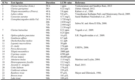
Table 1. Showing LC50 values of Malathion on fishes (compiled by Author)
Sl No Test Species Duration LC50 value Reference
1 Clarias batrachus (Linn.) 96 h 1 ppm Venkataraman and Sandhya Rani, 2013 2 Clarias gariepinus DO 8.22 mg/L Zubair Ahmad, 2012.
3 Tilapia mossambica (Peters). 48 h 5.8 ppm Kabeer et al., 1980.
4 Labeo rohita 96 h 9.0 μl/L. Vineetkumar Kallappa Patil, and Muniswamy David, 2009. 5 Carassius auratus 96 h 4.71 mg/L Saeid Shahbazi Naserabad et al., 2015.
6 Ctenopharyngodon idella Val. 24 h 3.728 mg/l,
Salwa M. and Abou El Ella, 2008. 48h 2.838 mg/l
72h 2.444 mg/l 96h 2.138 mg/l
7 Clarius batrachus 48h 0.31 ppm Yogesh et al., 2009.
96h 0.25 ppm
8 Ophiocephalus punctatus 96h 16 μl/L S.R. Pugazhvendan et al., 2009. 9 Gambusia affinis 96h 0.7 ppb
USEPA, 2006. 10 Onchorhynchus mykiss 96h 4.1 ppb
11 Lepomis macrochirus 96h 30 ppb 12 O. clarki 96h 174 ppb 13 Perca flavescens 96h 263 ppb 14 Tilapia mosambica 96h 2000 ppb 15 Cyprinus carpio 96h 6590 ppb 16 Oryzia latipes 96h 40,000 ppb
17 Ameiurus melas 96h 11.8 mg/L Martinez and Leyhe, 2004. 18 Hetereopneustes fossilis 96h 15.3 mg/L
19 Juvenile C. mrigala 96h 9.32 mg/L Rauf, 2015. 20 Coho Salmon 96h 174 μl/L
Mayer and Ellersieck, 1986. 21 Cutthroat salmon 96h 239 μl/L
significantly greater DNA damage in their blood cells than the control group. Fishes were exposed to different non-lethal concentrations (0.5, 1.5 and 3 mgL-1) of the malathion for 96 hour. The amount of DNA damage in the cells was estimated by the researchers from visual classification of cells into five type “comets” according to the tail length (Cavas, 2011; Lee and Steinert, 2003); 0: undamage, 1: low damage, 2: moderate damage, 3: high damage and 4: complete damage. Genetic Damage Index (GDI) was calculated by them using the following formula.
GDI = (n1+2n2+3n3+4n4) / (Σ / 100), where, n1: Minimum damage, n4: Maximum damage, Σ: Total number of the cells.
The DNA damage reported by the researchers was observed to be concentration dependent (P<0.05). The fish exposed to the lower concentration of malathion (0.5 mgL-1) shown relatively an insignificant GDI compared to control. Similar results were
reported by Kumar et al., 2010 on Channa punctatus.
Kushwaha et al., 2000 on C. punctatus following exposure to
malathion.
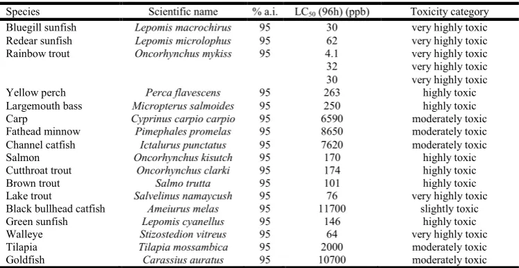
Lethal concentration (lc50) and ecotoxicity of malathion on
fishes
Lethal Concentration (LC50) and ecotoxicity of Malathion on
[image:7.595.114.488.74.267.2]different species of fishes are presented in the Table 1 and Table 2.
Acknowledgement
We are grateful to the Department of Zoology, Cotton College to provide us the latest facility to conduct different sets of experiments required to complete the aimed works.
REFERENCES
Ahmad, Z.2012. Toxicity bioassay and effects of sub-lethal exposure of malathion on biochemical composition and
haematological parameters of Clariasgariepinus.African
Journal of Biotechnology. 11(34), pp8578-8585.
ATSDR (Agency for Toxic Substances and Disease Registry). Toxicological Profile on Malathion. 2003. Retrieved from: http://www.atsdr.cdc.gov/toxpro2.html
Borthakur, M. K. R., Mahanta, R. and Goswami, U. C. 2006. Cytochrome p450, Arylhydrocarbon Hydroxylase and Xanthine oxidase activity during sublethal malathion
exposure in freshwater catfish Heteropneustes fossilis. J.
Inland Fish. Soc. India, 38(1) pp 9-14.
Cavas, T. 2011.In vivo genotoxicity evaluation of atrazine and
atrazine–based herbicide on fish Carassius auratus using
the micronucleus test and the comet assay. Food and
Chemical Toxicology, 49pp 1431-1435.
Chandravathy, V.M and Reddy, S.L. N. 1994.In vivo recovery of protein metabolism in gill and brain of freshwater fish,
Anabusscadens after exposure to lead nitrate. J. Environ.
Biol., 15pp 75-82.
Chaudhari, R.T. 2000. Impact of heavy metals on some
physiological aspects of the freshwater bivalve, Parresia
cylindrical. J. Indian Poll. Con., 18(1) pp 93-97.
Cohn J., MacPhail, R. C. 1996. Ethological and experimental approaches to behavior analysis: implications for
ecotoxicology. Environmental health perspectives, 104(2)
pp299-300.
Cristina, S. A., Cristina, M. M. and Anca, D. 2008. Malathion induced histological modifications in gills and kidney of
Carassius auratus gibelio. Lucrăristiin ńifice zootehniesi
Biotehnologii, 41(1) pp.448-453.
Dezwaan, S. and Zandee, D.I. 1999.The utilization of glycogen
and accumulation of some intermediates during
anaerobiosis in Mytilus edulis (L).Comp. Biochem.
Physiol., 43 (8) pp 47-54.
Drummond, R.A., Russom, C.L. 1990. Behavioral toxicity syndromes: A promising tool for assessing toxicity
mechanisms in juvenile fathead minnows. Environmental
toxicology and chemistry. 9 (1) pp37-46.
Fahmy, G. H. 2012. Malathion Toxicity: Effect on Some
Metabolic Activities in Oreochromis niloticus, the Tilapia
Fish. International Journal of Bioscience, Biochemistry
and Bioinformatics. 2 (1) pp 52-55.
Ferguson, D. E., & Goodyear, C. P. 1967. The pathway of
endrin entry in black bullheads, Ictalurus melas. Copeia,
1967(2), pp.467-468.
Ferguson, D. E., Ludke, J. L., & Murphy, G. G. 1966. Dynamics of endrin uptake and release by resistant and
susceptible strains of mosquito fish. Transactions of the
American Fisheries Society, 95(4), pp.335-344.
Table 2. Showing LC50 values and ecotoxicity of Malathion on fishes
Species Scientific name % a.i. LC50 (96h) (ppb) Toxicity category
Bluegill sunfish Lepomis macrochirus 95 30 very highly toxic Redear sunfish Lepomis microlophus 95 62 very highly toxic Rainbow trout Oncorhynchus mykiss 95 4.1 very highly toxic 32 very highly toxic 30 very highly toxic
Yellow perch Perca flavescens 95 263 highly toxic
Largemouth bass Micropterus salmoides 95 250 highly toxic
Carp Cyprinus carpio carpio 95 6590 moderately toxic
Fathead minnow Pimephales promelas 95 8650 moderately toxic Channel catfish Ictalurus punctatus 95 7620 moderately toxic
Salmon Oncorhynchus kisutch 95 170 highly toxic
Cutthroat trout Oncorhynchus clarki 95 174 highly toxic
Brown trout Salmo trutta 95 101 highly toxic
Lake trout Salvelinus namaycush 95 76 very highly toxic
Black bullhead catfish Ameiurus melas 95 11700 slightly toxic
Green sunfish Lepomis cyanellus 95 146 highly toxic
Walleye Stizostedion vitreus 95 64 very highly toxic
Tilapia Tilapia mossambica 95 2000 moderately toxic
Goldfish Carassius auratus 95 10700 moderately toxic
Hai, D. Q., Varga, I. S. and Matkovics, B. 1995. Effect of organophosphates on the antioxidant systems of fish tissues. Acta. Biol. Hung., 46, pp 39-50.
Howard, P.H. 1991. Handbook of environmental fate and exposure data for organic chemicals. Vol.3. Pesticides. Lewis Publishers, Chelsea, MI.
Insecticide fact sheet, Malathion. 2003.Journal of pesticide
reform/ winter. 23 (4). Retrieved from https://d3n8a8pro7
vhmx.cloudfront.net/ncap/pages/26/attachments/original/14 28423398/malathion.pdf?1428423398
Kabeer, I. A. S., Rao, K. S. J. and Rao, K. V. R. 1981. Effect of malathion exposure on some physical parameters of
whole body and on tissue cations of teleost, Tilapia
mossambica (Peters). J. Biosci., 3 (1), pp 17-21.
Kanagaraj, M. K., Ramesh, M., Sivakumari, K. and Manavalaramanujam, R. 1993.Impact of acid pollution on the serum haemolymph cholesterol of the crab, Paratelphusa hydrodromous. J.Ecotoxicol. Environ. Monit, 3(2), pp.99-102.
Krishnamohan, P. K., Indira and Rajendra, W. 1985.Protein degradation in functionally different muscles of rat during exhaustive exercise. J. Exp. Biol., 23 pp 655 - 657.
Kumar, R., Nagpure, N.S., Kwshwaha, B., Srivastava, S.K. and Lakra, W.S. 2010.Investigation of the Genotoxicity of
Malathion to Freshwater Teleost Fish Channa punctatus
(Bloch) Using the Micronucleus Test and Comet Assay.Arch Environ Contam Toxicol., (58), pp.123-130. Kushwaha, B., Srivastava, S. K., Singh, B., Nagpure, N. S. and
Ponniah, A. G. 2000. Evaluation of comet assay and
micronuclei test as genotoxic assay in Channa punctatus.
NatlAcadSciLett., (23), pp.177-179.
Lee, R. F., Steinert, S. 2003. Use of single cell gel electrophoresis/comet assay for detecting DNA damage in
aquatic (marine and freshwater) animals.Mutation
research, (544) pp 43-64.
Magar, R. S. and Shaikh, A. 2013. Effect of malathion toxicity on detoxifying organ of fresh water fish Channa punctatus. International Journal of Pharmaceutical, Chemical and Biological Sciences, 3(3), pp.723-728.
Magar, R.S. and Dube, K. V. 2013. Effect of Sub Lethal Concentration of Malathion on Metabolic Profiles and
Histological Studies in Heart Tissue of Channa punctatus.
IOSR Journal of Environmental Science, Toxicology and
Food Technology (IOSR-JESTFT). 2(6) pp 08-12. Retrieved from www.iosrjournals.org
Magar, R.S. and Shaikh, A. 2012. Effect of malathion on respiratory responses of fresh water fish Channapunctatus. Trends in Fisheries Research, 1(3) pp1-4.
Magare, S.R., Patil H. T. 2000. Effect of pesticides on oxygen consumption.Red blood cell count and metabolites of fish, Puntiusticto.Environ. Ecol., 18 pp 891-894.
Malathion Fact Sheet, 2000. Retrieved from https://www. beyondpesticides.org/assets/media/documents/pesticides/fa ctsheets/Malathion.pdf
Martinez, J. and Leyhe, J. 2004. Malathion analysis of risks to endangered and threatened Salmon and Steelhead.Office of pesticide programme, Environmental field branch, United States Environmental Protection Agency, pp108.
Martinez, J. and Leyhe, M. S. J. 2004. Malathion Analysis of Risks to Endangered and Threatened Salmon and Steelhead.M.S. Environmental Field Branch Office of Pesticide Programs.
Mayer, F. L. Jr. and Ellersieck, M. R. 1986. Manual of acute toxicity: Interpretation and data base for 410 chemicals and 66 species of freshwater animals. Washington, DC, US
Department of the Interior Fish and Wildlife Service (Resource publication 160).
Moradi, A. M., Mozdarani, H., Salimi, P. A. and Salimi, M. A.
2012.Evaluation of DNA damage in Cyprinuscarpio (L.
1758) exposed to malathion using Single Cell Gel Electrophoresis. Int. J. Mar. Sci. Eng., 2 (3) pp 185-188. Mount, D.I, and Brungs, W.A. 1967. A simplified dosing
apparatus for fish toxicological studies. Water Res.,
1(1),pp.21.
Mushigeri, S, David, M. 2005. Fenvalerate induced changes in the Ach and associated AchE activity in different tissues of
fish Cirrhinus mrigala (Hamilton) under lethal and
sub-lethal exposure period. Environmental Toxicology and
Pharmacology, 20 (1), pp.65-72.
Naserabad, S.S., Mirvaghefi, A., Gerami, M.H., and Farsani, H.G. 2015. Acute Toxicity and Behavioral Changes of the Gold Fish (Carassius auratus) Exposed to Malathion and
Hinosan. Iranian Journal of Toxicology, 8 (27) pp
1203-1208.
National Institute for Occupational Safety and Health. Registry of Toxic Effects of Chemical Substances: Succinic acid,
mercapto-, diethyl ester, S-ester with O,
Odimethylphosphorodithioate. 2002. Retrieved from
www.cdc.gov/niosh/rtecs/ wm802c80.html.27.
National Pesticide Information Centre, Oregon State University. Malathion, Technical Fact Sheet. 2010.
Retrieved from http://npic.orst.edu/factsheets/archive/
malatech.html
Patil, V. K. and David, M. 2009. Hepatotoxic potential of
malathion in the freshwater teleost, Labeorohita
(Hamilton) Veterinarski Arhiv, 79 (2) pp179-188.
Pickering, A.D. 1981. Stress and compensation in teleostean fishes: Response to social and physical factors. In: Pickering, AD (ed.) Stress and fish. Academic Press, New York, USA.295-322.
Pugazhvendan, S. R., Narendiran, N. J., Kumaran, R. G., Kumaran, S. and Alagappan, K. M. 2009. Effect of
Malathion Toxicity in the Freshwater Fish Ophiocephalus
punctatus-A Histological and Histochemical Study. World
Journal of Fish and Marine Sciences, 1 (3) pp 218-224. Rauf, A.2015. Acute toxicity and effects of malathion
exposure on behavior and hematological indices in Indian
carp, Cirrhinus mrigala (Hamilton). International Journal
of Aquatic Biology, 3(4) pp199-207.
Remia, K.M., Logaswamy, S., Logankumar, K., and
Rajmohan, D. 2008. Effect of an insecticide
(Monocrotophos) on some biochemical constituents of the fish Tilipia mossambica. Poll. Res., 27(3),pp.523-526. Reregistration Eligibility Decision (RED) for Malathion, 2006.
EPA 738-R-06-030; U.S Environmental Protection Agency, Office of Prevention, Pesticides and Toxic
Substances, Office of Pesticide Programs, U.S.
Government Printing Office: Washington, DC.
Roopavathy, J., Sukumaran, M., Rengarajan,R., Ravichelvan, R. and Narasiman, N. 2013. Toxicity of Malathion on Metabolic Activities in the River Cauvery of Tilapia Fish (Oreochromis mossambicus). International Journal of Pharmaceutical & Biological Archives, 4(5) pp 986 – 989. Rosety, M., Rosety-Rodríguez, M., Ordonez, F.J. and Rosety,
I. 2005.Time course variations of antioxidant enzyme activities and histopathology of gilthead seabream gills
exposed to malathion. Histol Histopathol., 20,
pp.1017-1020.
tissues of the grass carp, Ctenopharyngodon idella Val. Egypt. J. Aquat. Biol. & Fish., 12(2), pp.109 -117.
Shah, S.L. 2006.Hematological parameters in tench, Tinca
tinca after short term exposure to lead. J. Appl. Toxicol.,
26(3) pp 223-228.
Sprague, J. B. 1973.Measurement of pollutant toxicity of fish, utilizing and applying bioassay results.Mars. Res., 4pp 3-32.
Sudha, S. 2012. Comparative hematological studies of Clarius
batrachus in Bisalpur reservoir and Pushkar Lake. J.
Fundamental Appl. Life Sci., 2(2) pp230-233.
Thenmozhi, C., Vignesh, V., Thirumurugan, R., and Arun, S. 2010. Impacts of malathion on mortality and biochemical
changes of freshwater fish labeo rohita. Iran. J. Environ.
Health. Sci. Eng., 8(4) pp189-198.
Thurston County Review Summary on malathion, 2012. Retrieved from http://www.co.thurston.wa.us/health/ehipm/ pdf_insect/insecticide%20actives/malathion.pdf
USEPA 2005. Office of Pesticide Programs, Office of Science Coordination and Policy. Endocrine Screening Program (EDSP): Weight of evidence analysis (WOE) of potential interaction of malathion with the estrogen, androgen or thyroid pathways. Retrieved from http://www2.epa.gov/ sites/production/files/2015-06/documents/malathion-057 701_2015-06-29_txr0057180.pdf
USEPA. 2000. Malathion Reregistration Eligibility Document Environmental Fate and Effects Chapter. Office of
Pesticides Program, <http://www.epa.gov/oppsrrd1/op/
malathion/efedrra.pdf.>.
US EPA.2000. Overview of Malathion Risk Assessment. Prevention, Pesticides, and Toxic Substances, U.S.
Environmental Proection Agency.Retrieved from
<http://www.epa.gov/pesticides/op/malathion/overview.ht m>.
USEPA.2006. Reregistration eligibility decision for malathion: Case No. 0248. Office of prevention, pesticides, and toxic substances. Office of pesticide programs, United States Environmental Protection Agency, pp 196.
Venkataraman, G.V. and Sandhya Rani, P.N. 2013. Acute
toxicity and blood profile of freshwater fish, Clarias
batrachus (Linn.) exposed to Malathion.Journal of Academia and Industrial Research (JAIR), 2(3) pp 200-204.
Venkataramana, G.V., Sandhya Rani, P.N. and Murthy, P.S.2006. Impact of malathion on the biochemical
parameters of gobiid fish, Glossogobius giuris (Ham).
Journal of Environmental Biology, 27(1) pp119-122. Winkaler, E.U., Santosh, T.R.M., Machdo-Neto, J.G. and
Martinez, C.B.R. 2007.Acute lethal and sub-lethal effects of neem leaf extracts on neotrapical freshwater fish, Prochilodus lineatus. Comp. Biochem. Physiol., 145 (C) pp 236-244.
Yogesh, H. W., Yashshri, A. G., Prakash, P. A. D. E. 2009. Sublethal and Chronic Effect of Carbaryl and Malathion on
Clariu sbatrachus (Linn.).J. Appl. Sci. Environ. Manage,
13(2) pp 23 – 26.