ORIGINAL RESEARCH
FUNCTIONAL
Crossed Cerebrocerebellar Language Lateralization: An
Additional Diagnostic Feature for Assessing Atypical Language
Representation in Presurgical Functional MR Imaging
C. Méndez Orellana, E. Visch-Brink, M. Vernooij, S. Kalloe, D. Satoer, A. Vincent, A. van der Lugt, and M. Smits
ABSTRACT
BACKGROUND AND PURPOSE: Determining language dominance with fMRI is challenging in patients with brain tumor, particularly in cases of suspected atypical language representation. Supratentorial activation patterns must be interpreted with great care when the tumor is in or near the presumed language areas, where tumor tissue or mass effect can lead to false-negative fMRI results. In this study, we assessed cerebrocerebellar language fMRI lateralization in healthy participants and in patients with brain tumors with a focus on atypical language representation.
MATERIALS AND METHODS: Twenty healthy participants and 38 patients with a brain tumor underwent fMRI with a verb-generation task. Cerebral and cerebellar language lateralizations were separately classified as left-sided, right-sided, or symmetric. Electrocortical stimulation was performed in 19 patients. With the McNemar test, we evaluated the dependency between language lateralization in the cerebrum and cerebellum, and with Pearson correlation analysis, the relationship between the cerebral and cerebellar lateralization indices.
RESULTS:There was a significant dependency between cerebral and cerebellar language activation, with moderate negative correlation (Pearsonr⫽ ⫺0.69). Crossed cerebrocerebellar language activation was present in both healthy participants and patients, irrespective of handedness or typical or atypical language representation. There were no discordant findings between fMRI and electrocortical stimulation.
CONCLUSIONS: Language lateralization in the cerebellum can be considered an additional diagnostic feature to determine language dominance in patients with brain tumor. This is particularly useful in cases of uncertainty, such as the interference of a brain tumor with cerebral language activation on fMRI and atypical language representation.
ABBREVIATIONS:ECS⫽electrocortical stimulation; HP⫽healthy participant; LI⫽lateralization index
F
unctional MR imaging is a feasible diagnostic neuroimaging tool for determining hemispheric language dominance in pa-tients with brain tumor preoperatively.1Nevertheless, it has im-portant limitations when evaluating language lateralization, par-ticularly in patients with brain tumors.1-3Activation patterns must be interpreted with great care when the tumor is in or near the presumed language areas, where tumor tissue or mass effect can lead to false-negative fMRI results.2Determining language dominance is additionally challenging in left-handed patients with brain tumor. Left-handers are known to have less well-defined language-lateralization patterns, with more atypical right-sided language lateralization compared with right-handers.4-6In patients with brain tumor, the ability of fMRI to confirm Wada-established language lateralization was signifi-cantly more specific for right- than for left-handed or ambidex-trous patients, presumably due to their higher rate of typical, left-lateralized language representation.7In a study evaluating the role of fMRI confirming language dominance in patients with epi-lepsy, this technique showed higher posttest probability for lan-guage dominance in patients with typical lanlan-guage representation than in patients with atypical language representation.8
Thus far, studies assessing language lateralization have focused on the supratentorial brain. Prior fMRI studies have shown acti-vation not only in the cerebral hemispheres but also in the cere-bellum while performing specific language tasks.9-11An example Received June 21, 2014; accepted after revision August 13.
From the Departments of Radiology (C.M.O., M.V., S.K., A.v.d.L., M.S.), Neurology (C.M.O., E.V.-B), and Neurosurgery (E.V.-B., D.S., A.V.), Erasmus MC, University Medi-cal Center Rotterdam, Rotterdam, the Netherlands.
Please address correspondence to Marion Smits, MD, PhD, Department of Radi-ology (Hs-224), Erasmus MC–University Medical Center Rotterdam, PO Box 2040, 3000 CA Rotterdam, the Netherlands; e-mail: [email protected]; @MarionSmits
Indicates article with supplemental on-line tables.
is the verb-generation task, which is preferred to localize language areas in patients with tumor11-14and has been properly validated with electrocortical stimulation (ECS).15In individuals with left-hemispheric language dominance, this task has been shown to activate the right cerebellum.9Some further studies have pro-vided evidence for a so-called crossed cerebrocerebellar language lateralization pattern in healthy individuals, with both typical, left-sided and atypical, right-sided language lateralization.10,16 This crossed cerebrocerebellar language lateralization may serve as a useful additional diagnostic feature in determining language hemispheric dominance in patients with brain tumor because the cerebellar language activation is generally undisturbed by the tu-mor localized in or near the presumed supratentorial language areas. Such an additional diagnostic feature may be especially helpful in patients with potentially atypical language representa-tion, namely left-handers.
The purpose of our study was to assess cerebrocerebellar language fMRI lateralization in healthy participants and in pa-tients with brain tumor, with a focus on atypical language representation.
MATERIALS AND METHODS
Study ParticipantsWe included 20 healthy participants (HPs) whose functional data were reported in a previous study17that investigated the relation-ship between cerebral functional language lateralization and structural asymmetry of the arcuate fasciculus and 38 patients with brain tumor. Institutional review board approval was ob-tained for the inclusion of HPs and for the retrospective use of anonymized patient data. We included more left- than handed HPs to increase the probability of finding atypical right-lateralized or symmetric language representation. Handedness in healthy participants was assessed with the Oldfield Edinburgh Handedness Inventory.18Written informed consent was obtained from all HPs before participation in this study. The institutional review board waived the requirement to obtain written informed consent from patients. Patients were selected from our data base of 205 patients referred for preoperative fMRI by the neurosur-gery department at our institution between May 2004 and Sep-tember 2013. From this data base, we selected all left-handed patients(n⫽19) who had performed an fMRI language task. We then matched these with 19 right-handed patients for sex, age, and tumor location. Handedness in all patients was assessed by the neurosurgeon at presurgical neurologic examination. All patients were able to perform the language task and were native Dutch speakers except for 1 native German speaker, who was fluent in Dutch after learning this language at 37 years of age. Nineteen patients (9 left-handed) underwent an operation in an awake setting, where direct ECS was performed to identify language functions (biphasic pulse, 50-Hz frequency, 1-ms duration, 6 –12 mA).
Data Acquisition and Preprocessing
Participants performed a verb-generation task.12They were in-structed to think of a verb related to an auditorily presented noun, and in the control condition, they listened to high (2000-Hz) and low (400-Hz) tones.
Participants were scanned at 1.5T or 3T with an 8-channel head coil. Functional and structural sequences are specified in On-line Table 1. Imaging data were analyzed by using SPM8 soft-ware (http://www.fil.ion.ucl.ac.uk/spm/softsoft-ware/spm8). Func-tional images were manually aligned to the anterior commissure, realigned to correct for motion, coregistered with the individual’s T1-weighed image,19and smoothed with a 3D Gaussian full width at half maximum filter of 6⫻6⫻6 mm3.20
Anatomic images of the HPs were segmented and normalized with the functional images to the Montreal Neurological Institute standard brain space by using affine and nonlinear registration. This process resulted in resampled voxel sizes of 3⫻3⫻3 mm3 for the functional and 1⫻1⫻1 mm3for the anatomic images. Patient data were not normalized, in line with routine presurgical analysis, but were resliced with preservation of the acquired voxel size.
Data Analysis
Statistical activation maps were generated with a general linear model, by using a boxcar function convolved with a hemody-namic response function, corrected for temporal autocorrelation and filtered with a high-pass filter of 128-second cutoff. Motion parameters were included as regressors of no interest to reduce potential confounding effects of motion. Individual t-contrast images of language activation (verb-generation⬎tones) were generated for all participants. Different from the analysis per-formed with the HPs in our previous study,17individual lateral-ization indices (LIs) were determined by ROIs known to be in-volved in language processing: the inferior frontal gyrus, superior temporal and middle temporal gyri, angular and supramarginal gyri, and the cerebellum. Furthermore, in this study, the number of activated voxels within these ROIs was calculated by using a threshold-independent method.21,22 The LI was defined as (LH⫺RH)/(LH⫹RH), whereLHandRHare the number of activated voxels in the left and right hemispheres, respectively. Activation was classified for the cerebrum and cerebellum sepa-rately as left-lateralized for LI values between 0.1 and 1.0, right-lateralized for LI values between⫺0.1 and⫺1.0, or symmetric for LI values between or equal to⫺0.1 and 0.1.21
Patients’ individual t-contrast images, thresholded individu-ally but at a minimum t-value of 3.2, were assessed qualitatively by a neuroradiologist with 11 years’ experience in presurgical fMRI who was blinded to the handedness of patients and ECS findings. Language activation was assessed in the same predetermined lan-guage areas as those quantitatively assessed in the HPs. For each region, activation was categorized as left-lateralized, right-lateral-ized, symmetric, or no activation, on the basis of which an overall assessment of supratentorial language representation was made. Cerebellar activation was not taken into account when determin-ing overall language lateralization.
or vice versa, we defined this relationship as crossed cerebrocer-ebellar language activation.23
Statistical Analysis
Statistical analysis was performed by using SPSS (Version 20; IBM Armonk, New York). Age differences between left- and right-handed participants were examined by using an independent-samplesttest. Sex differences between both groups of patients were examined by using the Fisher exact test. To test whether the lateralizations in the cerebrum and in the cerebellum were inde-pendent of each other, we performed a McNemar test in HP and patient data. In HPs, the possible relationship between the LIs of the cerebrum and of the cerebellum was investigated with a scat-terplot and was tested with Pearson correlation analysis. We used a significance level of␣⫽.05 for all analyses.
RESULTS
Participant Characteristics
From the 20 HPs included in the study (mean age, 32.8 years; range, 25–54 years, 9 men), 13 were classified18as left-handed and 7, as right-handed (Table). There were no significant differences between the left- and right-handed HPs for age (t[18]⫽ ⫺0.416, P⫽.682) or sex (P⫽.374, Fisher exact test). Patient characteris-tics are shown in On-line Table 2. There were no significant dif-ferences between the left- and right-handed patients for age (t[36]⫽ ⫺0.723,P⫽.474) or sex (P⫽.728, Fisher exact test). In 24 patients, tumors were reported as low-grade, and in 13 pa-tients, as high-grade on histopathologic examination. Thirty-two of the 38 patients underwent surgery. ECS was performed in 19 of these, and language regions were identified in 5.
Cerebrocerebellar Language Lateralization in HPs The cerebral and cerebellar language lateralizations and LIs in HPs are presented in Fig 1 and the Table. We found a crossed cerebrocerebellar activation in 60% (12/20) of HPs. None of the participants showed language lateralization toward the same
hemisphere in both the cerebrum and cerebellum. Of the 13 left-handed participants, 6 showed typical and 7, atypical language representation: right-lateralized cerebral activation in 5 and sym-metric cerebral activation in 2 participants. Crossed cerebrocer-ebellar language activation was observed in 7 left-handed par-ticipants (5/6 with typical and 2/7 with atypical language representation). Of the remaining 6 participants, cerebellar acti-vation was symmetric in 4 and lateralized in 2. All 7 right-handed participants showed typical language representation, of whom 5 showed a crossed cerebrocerebellar language activation. The remaining 2 participants showed symmetric activation in the cerebellum.
There was a significant dependency between the cerebral and cerebellar language lateralization patterns with2(3,n⫽20)⫽ 8.533,P⫽.036. The scatterplot (Fig 2) indicated a negative direc-tion of this dependency, with a moderate negative correladirec-tion between the LIs of the cerebrum and the cerebellum (Pearson r(20)⫽ ⫺0.69,P⫽.001).
Cerebrocerebellar Lateralization in Patients with Tumor The cerebral and cerebellar language lateralizations in patients are presented in Fig 1 and in On-line Table 2. We found crossed cerebrocerebellar activation in 76% (29/38) of patients. None of the patients showed language lateralization toward the same side in both the cerebrum and cerebellum.
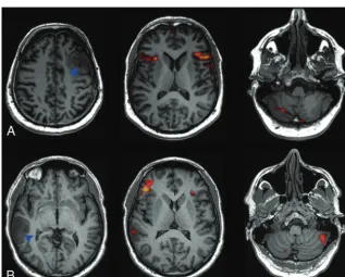
Of the 19 left-handed patients, 13 showed typical, and 6, atyp-ical language representation. Crossed cerebrocerebellar language activation was observed in 12/13 left-handed patients with typical language representation. Of the 6 patients with atypical language representation, cerebral language activation was right-lateralized in 1 and symmetric in 5. Of these, 1 patient with symmetric cere-bral language activation showed right-lateralized activation in the cerebellum (Fig 3A). His tumor was localized in the presumed language areas in the left hemisphere, which could have reduced language activation, resulting in a potentially false symmetric lan-guage-activation pattern. In addition, this patient was a bilingual speaker whose mother tongue was German. Although he was flu-ent in Dutch, he performed the verb-generation task in his mother tongue while the task was presented in Dutch. It is known that language activation in bilingual patients may be more symmet-ric,24and his performing the task with the interference of both languages may have contributed to the unexpected language-ac-tivation pattern we observed. The other patients showed symmet-ric (3 patients) or no activation (1 patient) in the cerebellum.
Of the 19 right-handed patients, 17 showed typical and 2, atyp-ical language representation. Crossed cerebrocerebellar language activation was observed in 16 of the 17 patients with typical lan-guage representation. Of the 2 patients with atypical lanlan-guage representation, one showed right-lateralized cerebral activation with crossed cerebrocerebellar language representation (Fig 3B) and the other showed symmetric cerebral and cerebellar language activation.
There was a significant dependency between the lateralization in the cerebrum and in the cerebellum [2(6,n⫽38)⫽42.06,
P⫽.000].
There were no discordant findings between fMRI and ECS, which positively identified language representation in the same
Healthy participant demographics and lateralization indices
Age (yr) Sex
EHI
Scorea CerebrumLI CerebellumLI LateralizationCerebrum LateralizationCerebellum
25 M ⫺100 0.42 ⫺0.36 L R
30 M ⫺100 ⫺0.44 0.34 R L
33 F ⫺100 ⫺0.27 0.03 R S
27 F ⫺100 0.34 ⫺0.41 L R
28 F ⫺100 0.02 ⫺0.43 S R
35 F ⫺100 0.24 ⫺0.19 L R
53 M ⫺90 ⫺0.18 0.13 R L
36 F ⫺90 ⫺0.28 0.07 R S
30 M ⫺80 0.13 ⫺0.22 L R
29 M ⫺78 0.32 ⫺0.40 L R
28 F ⫺20 ⫺0.04 ⫺0.13 S R
31 M ⫺20 0.48 0.00 L S
43 M 0 ⫺0.18 0.00 R S
30 F 100 0.21 ⫺0.30 L R
34 F 100 0.16 ⫺0.04 L S
34 M 100 0.32 ⫺0.12 L R
34 F 100 0.31 ⫺0.46 L R
29 F 100 0.42 ⫺0.30 L R
32 M 100 0.33 ⫺0.09 L S
28 F 100 0.43 ⫺0.25 L R
Note:—L indicates left-sided; R, right-sided; S, symmetric; EHI, Oldfield Edinburgh Handedness Inventory.
a
hemisphere in 5 patients. In one of these patients, fMRI language activation was found to be present in both hemispheres, whereas it was only identified on the operated side with ECS (the con-tralateral hemisphere was not assessed with ECS).
DISCUSSION
We found a significant dependency between language lateraliza-tion in the cerebrum and in the cerebellum, both in HPs and in patients with brain tumor, in line with previous studies in healthy left- and right-handers with typical language representation.10,23 Furthermore, we found a moderate inverse correlation of cere-brocerebellar lateralization; in other words, the more strongly language was lateralized toward a cerebral hemisphere, the more strongly it was lateralized to the contralateral cerebellar hemi-sphere. In almost all cases in which activation in the cerebellum was lateralized, there was a crossed cerebrocerebellar lateraliza-tion pattern, irrespective of whether language representalateraliza-tion was typical or atypical. This means that as a rule of thumb, in cases of clear cerebellar lateralization, cerebral language lateralization can be assumed contralateral. Language lateralization in the cerebel-lum may thus serve as an additional diagnostic feature for deter-mining hemispheric language dominance in individuals with ei-ther typical or atypical language representation. Cerebellar
activation was found to be symmetric in a minority of HPs and patients. In these cases, there was no clear correlation with cere-bral language representation; in some, cerecere-bral language was also symmetric, but in others, cerebral activation was clearly lateral-ized. In cases in which assessment of cerebral lateralization is hin-dered by tumor effects and cerebellar activation is symmetric, another examination such as the Wada test or ECS is thus still required to determine language dominance.1,8
Neuroimaging studies of the verb-generation task indicated that the cerebellum is involved in generating or maintaining ar-ticulatory representation,25even though no speech motor output was required. Both lesion and functional neuroimaging studies suggest that the cerebellum contributes to diverse cognitive lan-guage components and aspects of lanlan-guage production,25,26and cerebellar activation has been reported not only in productive tasks but also in speech-comprehension tasks.27The mechanism underlying the observed crossed cerebrocerebellar activation re-mains to be elucidated. Connectome analyses could be used to attempt to find white matter pathways that may be responsible for these findings.
Irrespective of handedness or the side of lateralization, crossed cerebrocerebellar activation was observed in most cases and
[image:4.594.55.524.47.417.2]ticularly in all patients, except one who was bilingual, in whom cerebellar activation was lateralized. This finding is of particu-lar clinical use in cases in which hemispheric language domi-nance cannot be assessed because of the interference of
lan-guage activation due to tumor2,28and in left-handed patients in whom diag-nostic uncertainty is greater due to the increased prevalence of atypical lan-guage representation.
To the best of our knowledge, this is the first study confirming crossed cere-brocerebellar activation in a large group of HPs and patients with brain tumor with a high prevalence of atypical lan-guage representation. To assess the cor-relation between cerebral and cerebellar lateralization quantitatively, we used a threshold-independent method, which is less prone to within-subject variability than threshold-dependent LI calcula-tion and generates LIs that are more in agreement with clinical findings.21For the assessment of language lateralization in patients with brain tumor, we chose to assess language lateralization qualita-tively to remain as close to the clinical routine as possible. Studies comparing quantitative with qualitative assessment of language lateralization have shown that visual inspection by an experienced rater is reliable for presurgical assess-ment of language lateralization.29-31 This qualitative approach provided a clinically applicable assessment of the cerebrocellebellar relationship in pa-tients with tumor.
The main limitation of our study, as in many studies assessing functional lan-guage lateralization presurgically,1,32is the relative lack of a criterion standard. Next to techniques such as the Wada test and ECS that are commonly considered criterion standards, transcranial mag-netic stimulation,33 magentoencepha-lography,34and fMRI are currently be-ing used as presurgical methods to evaluate language lateralization. In our study, in combination with fMRI, ECS was performed in most of our patients, but in many, no language area was iden-tified. ECS assessment is limited to just the brain region surrounding the tumor, and language areas at some distance from the tumor are thus not identified. Even when a language area is identified, we cannot know for certain whether this is the sole, dominant hemisphere: Bilat-eral language representation cannot be assessed with certainty. While we found no discordance between ECS and fMRI, the number of patients in whom this could be assessed with certainty was small. A minor limitation of our study is that patients were scanned on several scanners, consistent with
FIG 2.Scatterplot of the healthy participant lateralization indices of the cerebrum and cerebellum.
[image:5.594.59.370.46.321.2] [image:5.594.56.374.361.616.2]daily clinical practice. Nevertheless, we used a standardized imag-ing protocol for presurgical fMRI evaluations, maintainimag-ing simi-lar image resolution across our MR imaging systems and stan-dardized image analysis. Another minor limitation was the difference in assessment of handedness: In healthy participants, the standardized Oldfield Edinburgh Handedness Inventory was used while in patients, handedness information was obtained from the preoperative neurosurgical evaluation. Finally, patients with both high- and low-grade gliomas were included in this study. Neurovascular decoupling is a potential limitation of fMRI in high-grade tumors. Our study population was too small to allow a formal comparison or a distinction among these tumor grades.
CONCLUSIONS
Cerebellar activation may provide an additional diagnostic fea-ture to assess hemispheric language dominance, both in typical and atypical language representations. This is particularly useful in left-handed patients with brain tumor, in whom language rep-resentation is commonly atypical, resulting in diagnostic uncer-tainty, especially when there is potential interference of the tumor with language activation. When cerebellar activation is found to be lateralized, we can, as a rule of thumb, assume that there is contralateral hemispheric language dominance. This crossed cerebrocerebellar pattern of activation could be included as a di-agnostic tool in future guidelines of clinical fMRI examinations, which should further specify that a language task that is known to involve the cerebellum is used.
Disclosures: Aad van der Lugt—UNRELATED:Grants/Grants Pending: General Elec-tric Healthcare*;Payment for Lectures (including service on Speakers Bureaus): General Electric Healthcare.* Marion Smits—UNRELATED:Grants/Grants Pending: Erasmus MC,* Coolsingel Foundation*;Payment for Lectures (including service on Speakers Bureaus): European Society for Magnetic Resonance in Medicine and Biol-ogy*;Travel/Accommodations/Meeting Expenses Unrelated to Activities Listed: Radiological Society of North America, European Congress of Radiology, European Society for Magnetic Resonance in Medicine and Biology. *Money paid to the institution.
REFERENCES
1. Stippich C, Rapps N, Dreyhaupt J, et al.Localizing and lateralizing language in patients with brain tumors: feasibility of routine pre-operative functional MR imaging in 81 consecutive patients. Radi-ology2007;243:828 –36
2. Smits M.Functional magnetic resonance imaging (fMRI) in brain tumour patients.Eur Assoc NeuroOncol Mag2012;2:123–28 3. Petrella JR, Shah LM, Harris KM, et al.Preoperative functional MR
imaging localization of language and motor areas: effect on thera-peutic decision making in patients with potentially resectable brain tumors.Radiology2006;240:793– 802
4. Knecht S, Dra¨ger B, Deppe M, et al.Handedness and hemispheric language dominance in healthy humans.Brain2000;123(pt 12): 2512–18
5. Szaflarski JP, Binder JR, Possing ET, et al.Language lateralization in left-handed and ambidextrous people: fMRI data.Neurology2002; 59:238 – 44
6. Tzourio-Mazoyer NN, Josse G, Crivello F, et al.Interindividual vari-ability in the hemispheric organization for speech.Neuroimage
2004;21:422–35
7. Dym RJR, Burns JJ, Freeman KK, et al.Is functional MR imaging assessment of hemispheric language dominance as good as the Wada test? A meta-analysis.Radiology2011;261:446 –55
8. Medina LS, Bernal B, Ruiz J.Role of functional MR in determining
language dominance in epilepsy and nonepilepsy populations: a Bayesian analysis.Radiology2007;242:94 –100
9. Binder JR, Frost JA, Hammeke TA, et al.Human brain language areas identified by functional magnetic resonance imaging.J Neu-rosci1997;17:353– 62
10. Hubrich-Ungureanu PP, Kaemmerer NN, Henn FAF, et al. Lateral-ized organization of the cerebellum in a silent verbal fluency task: a functional magnetic resonance imaging study in healthy volun-teers.Neurosci Lett2002;319:91–94
11. Frings M, Dimitrova A, Schorn CF, et al.Cerebellar involvement in verb generation: an fMRI study.Neurosci Lett2006;409:19 –23 12. Smits M, Visch-Brink EG, Schraa-Tam CK, et al.Functional MR
imaging of language processing: an overview of easy-to-implement paradigms for patient care and clinical research. Radiographics
2006;26:S145–58
13. Wise RJ, Chollet F, Hadar U, et al.Distribution of cortical neural networks involved in word comprehension and word retrieval.
Brain1991;114(pt 4):1803–17
14. Ojemann JG, Ojemann GA, Lettich E.Cortical stimulation mapping of language cortex by using a verb generation task: effects of learn-ing and comparison to mapplearn-ing based on object namlearn-ing.J Neuro-surg2002;97:33–38
15. Bizzi A, Blasi V, Falini A, et al.Presurgical functional MR imaging of language and motor functions: validation with intraoperative elec-trocortical mapping.Radiology2008;248:579 – 89
16. FitzGerald DB, Cosgrove GR, Ronner S, et al.Location of language in the cortex: a comparison between functional MR imaging and elec-trocortical stimulation.AJNR Am J Neuroradiol1997;18:1529 –39 17. Vernooij MW, Smits M, Wielopolski PA, et al.Fiber density
asym-metry of the arcuate fasciculus in relation to functional hemi-spheric language lateralization in both right- and left-handed healthy subjects: a combined fMRI and DTI study.Neuroimage
2007;35:1064 –76
18. Oldfield RC.The assessment and analysis of handedness: the Edin-burgh inventory.Neuropsychologia1971;9:97–113
19. Friston KJ, Holmes AP, Poline JB, et al.Analysis of fMRI time-series revisited.Neuroimage1995;2:45–53
20. Friston KJ, Zarahn E, Josephs O, et al.Stochastic designs in event-related fMRI.Neuroimage1999;10:607–19
21. Suarez RO, Whalen S, Nelson AP, et al.Threshold-independent functional MRI determination of language dominance: a validation study against clinical gold standards. Epilepsy Behav 2009;16: 288 –97
22. Branco DM, Suarez RO, Whalen S, et al.Functional MRI of memory in the hippocampus: laterality indices may be more meaningful if calculated from whole voxel distributions. Neuroimage 2006; 32:592– 602
23. Jansen A, Flo¨el A, Van Randenborgh J, et al.Crossed cerebro-cere-bellar language dominance.Hum Brain Mapp2005;24:165–72 24. Abutalebi J.Neural aspects of second language representation and
language control.Acta Psychologica2008;128:466 –78
25. Marie¨n P, Ackermann H, Adamaszek M, et al.Consensus paper: language and the cerebellum: an ongoing enigma.Cerebellum2014; 13:386 – 410
26. O’Halloran CJ, Kinsella GJ, Storey E.The cerebellum and neuropsy-chological functioning: a critical review.J Clin Exp Neuropsychol
2012;34:35–56
27. Londei A, D’Ausilio A, Basso D, et al.Sensory-motor brain network connectivity for speech comprehension.Hum Brain Mapp2010; 31:567– 80
28. Ulmer JL, Hacein-Bey L, Mathews VP, et al.Lesion-induced pseudo-dominance at functional magnetic resonance imaging: implica-tions for preoperative assessments.Neurosurgery2004;55:569 –79; discussion 580 – 81
30. Jones SE, Mahmoud SY, Gonzalez-Martinez J, et al.Application of a computerized language lateralization index from fMRI by a group of clinical neuroradiologists. AJNR Am J Neuroradiol 2013;34: 564 – 69
31. Gutbrod K, Spring D, Degonda N, et al.Determination of language dominance: Wada test and fMRI compared using a novel sentence task.J Neuroimaging2012;22:266 –74
32. Jones SE, Mahmoud SY, Phillips MD.A practical clinical method to
quantify language lateralization in fMRI using whole-brain analy-sis.Neuroimage2011;54:2937– 49
33. Ro¨sler J, Niraula B, Strack V, et al.Language mapping in healthy volunteers and brain tumor patients with a novel navigated TMS system: evidence of tumor-induced plasticity.Clin Neurophysiol
2014;125:526 –36