1-Vinyl-1
H
-indole-3-carbaldehyde
S. Selvanayagam,a* B. Sridhar,bK. Ravikumar,b S. Kathiravancand R. Raghunathanc
a
Department of Physics, Kalasalingam University, Krishnankoil 626 190, India, b
Laboratory of X-ray Crystallography, Indian Institute of Chemical Technology, Hyderabad 500 007, India, andcDepartment of Organic Chemistry, University of Madras, Guindy Campus, Chennai 600 025, India
Correspondence e-mail: [email protected]
Received 19 May 2008; accepted 22 May 2008
Key indicators: single-crystal X-ray study;T= 293 K; mean(C–C) = 0.002 A˚; Rfactor = 0.045;wRfactor = 0.127; data-to-parameter ratio = 17.6.
In the title compound, C11H9NO, the C and O atoms of the
attached carbaldehyde group deviate by just 0.052 (2) and 0.076 (1) A˚ , respectively, from the mean plane of the indole ring system. In addition to van der Waals forces, the molecular packing is stabilized by C—H O hydrogen bonds, which form a C(7) chain motif, and – interactions (centroid– centroid distance 3.637 A˚ ) between the pyrrole and benzene rings of the indole ring system.
Related literature
For related literature, see: Padwaet al.(1999); Mathiesenet al.
(2005); Grinev et al. (1984); Gadaginamath & Patil (1999); Rodriguezet al.(1985); Karthicket al.(2005); Selvanayagamet al.(2005); Sonaret al.(2005). For bond-length data, see: Allen
et al.(1987).
Experimental
Crystal data
C11H9NO
Mr= 171.19
Monoclinic,P21=n a= 8.3200 (5) A˚
b= 8.1490 (5) A˚
c= 13.1620 (7) A˚ = 99.952 (1)
V= 878.95 (9) A˚3
MoKradiation = 0.08 mm1
0.240.220.20 mm
Data collection
Bruker SMART APEX CCD area-detector diffractometer Absorption correction: none 9730 measured reflections
2072 independent reflections 1823 reflections withI> 2(I)
Rint= 0.020
Refinement
R[F2> 2(F2)] = 0.044
wR(F2) = 0.127
S= 1.05 2072 reflections
118 parameters
H-atom parameters constrained max= 0.21 e A˚3
[image:1.610.47.237.519.716.2]min=0.19 e A˚3
Table 1
Hydrogen-bond geometry (A˚ ,).
D—H A D—H H A D A D—H A
C9—H9 O1i
0.93 2.51 3.390 (2) 159
Symmetry code: (i)x1 2;yþ
1 2;zþ
1 2.
Data collection:SMART(Bruker, 2001); cell refinement:SAINT (Bruker, 2001); data reduction:SAINT; program(s) used to solve structure:SHELXS97(Sheldrick, 2008); program(s) used to refine structure: SHELXL97 (Sheldrick, 2008); molecular graphics: ORTEP-3 (Farrugia, 1997) and PLATON (Spek, 2003); software used to prepare material for publication:SHELXL97and PARST (Nardelli, 1995).
SS thanks the Vice Chancellor and management of Kala-salingam University, Anand Nagar, Krishnankoil, for their support and encouragement.
Supplementary data and figures for this paper are available from the IUCr electronic archives (Reference: BT2714).
References
Allen, F. H., Kennard, O., Watson, D. G., Brammer, L., Orpen, A. G. & Taylor, R. (1987).J. Chem. Soc. Perkin Trans. 2, pp. S1–19.
Bruker (2001).SAINTandSMART. Bruker AXS Inc., Madison, Wisconsin, USA.
Farrugia, L. J. (1997).J. Appl. Cryst.30, 565.
Gadaginamath, G. S. & Patil, S. A. (1999).Indian J. Chem. B,38, 1070–1074. Grinev, A. N., Shevdov, V. L., Krichevskii, E. S., Romanova, O. B., Altukkhova, L. B., Kurilo, G. N., Andreeva, N. I., Golovina, S. M. & Mashkovskii, M. D. (1984).Khim. Farm. Zh.18, 159–163.
Karthick, S., Selvanayagam, S., Velmurugan, D., Ravikumar, K., Arumugam, N. & Raghunathan, R. (2005).Acta Cryst.E61, o1780–o1782.
Mathiesen, J. M., Ulven, T., Martini, L., Gerlach, L. O., Heinemann, A. & Kostenis, E. (2005).Mol. Pharmacol.68, 393–402.
Nardelli, M. (1995).J. Appl. Cryst.28, 659.
Padwa, A., Brodney, M. A., Liu, B., Satake, K. & Wu, T. (1999).J. Org. Chem. 64, 3595–3607.
Rodriguez, W. G., Temprano, F., Esteban-Calderon, C., Martinez-Ripoll, M. & Garcia-Blanco, S. (1985).Tetrahedron,41, 3813–3823.
Selvanayagam, S., Chandak, M. S., Velmurugan, D., Ravikumar, K. & Raghunathan, R. (2005).Acta Cryst.E61, o3122–o3123.
Sheldrick, G. M. (2008).Acta Cryst.A64, 112–122.
Sonar, V. N., Parkin, S. & Crooks, P. A. (2005).Acta Cryst.C61, o78–o80. Spek, A. L. (2003).J. Appl. Cryst.36, 7–13.
Structure Reports
Online
supporting information
Acta Cryst. (2008). E64, o1163 [doi:10.1107/S160053680801547X]
1-Vinyl-1
H
-indole-3-carbaldehyde
S. Selvanayagam, B. Sridhar, K. Ravikumar, S. Kathiravan and R. Raghunathan
S1. Comment
Indoles and their derivatives have been interest for many years, since large number of natural products contain indole
systems and they are found in a number of pharmaceutical products, fragrances and dyes (Padwa et al., 1999). Indole
derivatives are identified as interfering with a G protein-independent signaling pathway of the CRTH2 receptor
(Mathiesen et al., 2005). These derivatives possess antidepressant (Grinev et al., 1984), anti-microbial (Gadaginamath &
Patil, 1999) and anti-inflammatory (Rodriguez et al., 1985) activities. In view of its importance, we have undertaken the
single-crystal X-ray diffraction study and report here its results.
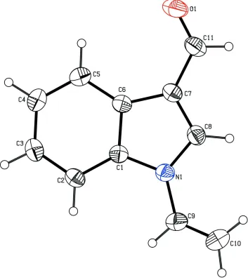
The X-ray study confirmed the molecular structure and atomic connectivity for (I), as illustrated in Fig. 1. The geometry
of the indole ring system is comparable to those reported for other indole derivatives (Karthick et al., 2005;
Selvanayagam et al., 2005; Sonar et al., 2005). The bond length of C9—C10 [1.284 (2) Å] confirms the double bond
character (Allen et al., 1987). The sum of the angles at N1 of the indole ring (360°) is in accordance with sp2
hybridization.
The indole ring is planar with a maximum deviation of 0.017 (1) Å for atom C8. The carbaldehyde group atoms C11
and O1 deviate 0.052 (2) and 0.076 (1) Å, respectively from the best plane of the indole ring.
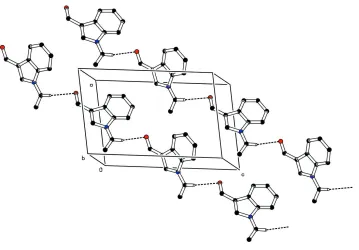
In addition to the van der Waals forces, the molecular packing is stabilized by intermolecular C—H···O hydrogen bond
(Table 2). Atom H9 of C9 forms a intermolecular hydrogen bond with oxygen atom O1 forming a C(7) chain motif of C
—H···O hydrogen bond along the diagonal of ac plane (Fig. 2). In addition to this a weak π···π interaction between the
pyrrole ring (N1/C1/C6—C8) at (x,y,z) and benzene ring (C1—C6) at (1 -x, -y), -z) stabilizes the molecular packing. The
centroid-to-centroid distance is 3.637Å.
S2. Experimental
A mixture of N-vinylindole (0.05 mol) and DMF (0.15 mol) was stirred with POCl3 (32.3 ml). The reaction mixture was
poured into ice water (300 ml) and stirred for 30minutes at less than 10° C. The precipitated solid was collected by
filtration and washed well water (100 ml). In order to get the diffraction quality crystals, the compound was recrystallized
from ethyl acetate.
S3. Refinement
The H atoms were positioned geometrically with C—H distances of 0.93 Å and were included in the refinement in the
Figure 1
The structure and atom-numbering scheme for the title compound; displacement ellipsoids are drawn at the 30%
Figure 2
Molecular packing of the title compound viewed down the b axis; H-bonds are shown as dashed lines. For clarity, H
atoms, not involved in hydrogen bonds, have been omitted.
1-Vinyl-1H-indole-3-carbaldehyde
Crystal data
C11H9NO
Mr = 171.19 Monoclinic, P21/n Hall symbol: -P 2yn
a = 8.3200 (5) Å
b = 8.1490 (5) Å
c = 13.1620 (7) Å
β = 99.952 (1)°
V = 878.95 (9) Å3
Z = 4
F(000) = 360
Dx = 1.294 Mg m−3
Mo Kα radiation, λ = 0.71073 Å Cell parameters from 4554 reflections
θ = 2.1–23.6°
µ = 0.08 mm−1
T = 293 K Block, colourless 0.24 × 0.22 × 0.20 mm
Data collection
Bruker SMART APEX CCD area-detector diffractometer
Radiation source: fine-focus sealed tube Graphite monochromator
ω scans
9730 measured reflections 2072 independent reflections
1823 reflections with I > 2σ(I)
Rint = 0.020
θmax = 28.0°, θmin = 3.0°
h = −10→10
k = −10→10
l = −17→16
Refinement
Refinement on F2 Least-squares matrix: full
R[F2 > 2σ(F2)] = 0.044
wR(F2) = 0.127
S = 1.05
2072 reflections 118 parameters 0 restraints
map
Hydrogen site location: inferred from neighbouring sites
H-atom parameters constrained
where P = (Fo2 + 2Fc2)/3 (Δ/σ)max < 0.001
Δρmax = 0.21 e Å−3 Δρmin = −0.19 e Å−3
Special details
Geometry. All e.s.d.'s (except the e.s.d. in the dihedral angle between two l.s. planes) are estimated using the full covariance matrix. The cell e.s.d.'s are taken into account individually in the estimation of e.s.d.'s in distances, angles and torsion angles; correlations between e.s.d.'s in cell parameters are only used when they are defined by crystal symmetry. An approximate (isotropic) treatment of cell e.s.d.'s is used for estimating e.s.d.'s involving l.s. planes.
Refinement. Refinement of F2 against ALL reflections. The weighted R-factor wR and goodness of fit S are based on F2, conventional R-factors R are based on F, with F set to zero for negative F2. The threshold expression of F2 > σ(F2) is used only for calculating R-factors(gt) etc. and is not relevant to the choice of reflections for refinement. R-factors based on F2 are statistically about twice as large as those based on F, and R- factors based on ALL data will be even larger.
Fractional atomic coordinates and isotropic or equivalent isotropic displacement parameters (Å2)
x y z Uiso*/Ueq
O1 0.81283 (15) 0.20464 (18) −0.13362 (9) 0.0826 (4)
N1 0.41791 (12) 0.24005 (13) 0.07004 (7) 0.0469 (3)
C1 0.54641 (14) 0.14623 (13) 0.12226 (8) 0.0424 (3)
C2 0.56155 (16) 0.06833 (16) 0.21713 (9) 0.0505 (3)
H2 0.4800 0.0748 0.2572 0.061*
C3 0.70276 (18) −0.01899 (17) 0.24918 (10) 0.0581 (3)
H3 0.7166 −0.0736 0.3121 0.070*
C4 0.82544 (17) −0.02764 (17) 0.18966 (11) 0.0594 (4)
H4 0.9197 −0.0870 0.2139 0.071*
C5 0.80997 (15) 0.04980 (16) 0.09564 (10) 0.0527 (3)
H5 0.8925 0.0434 0.0563 0.063*
C6 0.66781 (14) 0.13815 (13) 0.06061 (8) 0.0431 (3)
C7 0.60762 (15) 0.23003 (15) −0.03148 (9) 0.0480 (3)
C8 0.45684 (16) 0.28725 (16) −0.02188 (9) 0.0503 (3)
H8 0.3901 0.3497 −0.0712 0.060*
C9 0.27187 (17) 0.27412 (19) 0.10596 (12) 0.0615 (4)
H9 0.2701 0.2521 0.1751 0.074*
C10 0.14066 (19) 0.3327 (2) 0.05257 (15) 0.0779 (5)
H10A 0.1365 0.3567 −0.0169 0.094*
H10B 0.0498 0.3511 0.0834 0.094*
C11 0.68162 (19) 0.2565 (2) −0.12120 (11) 0.0621 (4)
H11 0.6233 0.3191 −0.1742 0.075*
Atomic displacement parameters (Å2)
U11 U22 U33 U12 U13 U23
O1 0.0720 (7) 0.1185 (10) 0.0649 (7) 0.0021 (6) 0.0336 (6) 0.0075 (6)
N1 0.0474 (5) 0.0518 (6) 0.0428 (5) 0.0032 (4) 0.0110 (4) −0.0026 (4)
C1 0.0457 (6) 0.0418 (5) 0.0396 (5) −0.0022 (4) 0.0075 (4) −0.0069 (4)
C2 0.0595 (7) 0.0521 (7) 0.0417 (6) −0.0035 (5) 0.0136 (5) −0.0023 (5)
C4 0.0562 (7) 0.0544 (7) 0.0641 (8) 0.0080 (6) 0.0011 (6) 0.0007 (6)
C5 0.0475 (6) 0.0527 (7) 0.0591 (7) 0.0000 (5) 0.0129 (5) −0.0070 (5)
C6 0.0472 (6) 0.0418 (6) 0.0411 (5) −0.0053 (4) 0.0102 (4) −0.0072 (4)
C7 0.0543 (7) 0.0486 (6) 0.0425 (6) −0.0046 (5) 0.0124 (5) −0.0023 (5)
C8 0.0566 (7) 0.0513 (7) 0.0427 (6) 0.0017 (5) 0.0079 (5) 0.0021 (5)
C9 0.0564 (8) 0.0736 (9) 0.0586 (8) 0.0089 (6) 0.0209 (6) −0.0013 (6)
C10 0.0580 (9) 0.0892 (12) 0.0894 (12) 0.0170 (8) 0.0205 (8) 0.0036 (9)
C11 0.0670 (8) 0.0743 (9) 0.0481 (7) −0.0059 (7) 0.0185 (6) 0.0041 (6)
Geometric parameters (Å, º)
O1—C11 1.2079 (19) C5—C6 1.3933 (17)
N1—C8 1.3610 (16) C5—H5 0.9300
N1—C1 1.3949 (15) C6—C7 1.4390 (17)
N1—C9 1.4055 (16) C7—C8 1.3645 (18)
C1—C2 1.3869 (16) C7—C11 1.4393 (18)
C1—C6 1.4023 (16) C8—H8 0.9300
C2—C3 1.3760 (19) C9—C10 1.284 (2)
C2—H2 0.9300 C9—H9 0.9300
C3—C4 1.392 (2) C10—H10A 0.9300
C3—H3 0.9300 C10—H10B 0.9300
C4—C5 1.3751 (19) C11—H11 0.9300
C4—H4 0.9300
C8—N1—C1 108.24 (10) C5—C6—C1 119.20 (11)
C8—N1—C9 126.55 (12) C5—C6—C7 134.39 (11)
C1—N1—C9 125.17 (11) C1—C6—C7 106.40 (10)
C2—C1—N1 129.57 (11) C8—C7—C6 106.95 (10)
C2—C1—C6 122.48 (11) C8—C7—C11 123.72 (13)
N1—C1—C6 107.94 (10) C6—C7—C11 129.29 (12)
C3—C2—C1 116.91 (12) N1—C8—C7 110.46 (11)
C3—C2—H2 121.5 N1—C8—H8 124.8
C1—C2—H2 121.5 C7—C8—H8 124.8
C2—C3—C4 121.60 (12) C10—C9—N1 126.30 (15)
C2—C3—H3 119.2 C10—C9—H9 116.8
C4—C3—H3 119.2 N1—C9—H9 116.8
C5—C4—C3 121.31 (12) C9—C10—H10A 120.0
C5—C4—H4 119.3 C9—C10—H10B 120.0
C3—C4—H4 119.3 H10A—C10—H10B 120.0
C4—C5—C6 118.49 (12) O1—C11—C7 125.68 (15)
C4—C5—H5 120.8 O1—C11—H11 117.2
C6—C5—H5 120.8 C7—C11—H11 117.2
C8—N1—C1—C2 −178.34 (12) N1—C1—C6—C7 −0.32 (12)
C9—N1—C1—C2 −0.3 (2) C5—C6—C7—C8 178.96 (13)
C8—N1—C1—C6 0.77 (13) C1—C6—C7—C8 −0.24 (13)
C9—N1—C1—C6 178.86 (12) C5—C6—C7—C11 1.3 (2)
C1—C2—C3—C4 0.56 (19) C9—N1—C8—C7 −179.00 (12)
C2—C3—C4—C5 −0.6 (2) C6—C7—C8—N1 0.73 (14)
C3—C4—C5—C6 0.0 (2) C11—C7—C8—N1 178.60 (12)
C4—C5—C6—C1 0.47 (17) C8—N1—C9—C10 11.7 (3)
C4—C5—C6—C7 −178.65 (12) C1—N1—C9—C10 −166.00 (16)
C2—C1—C6—C5 −0.47 (17) C8—C7—C11—O1 −178.09 (15)
N1—C1—C6—C5 −179.66 (10) C6—C7—C11—O1 −0.7 (3)
C2—C1—C6—C7 178.87 (10)
Hydrogen-bond geometry (Å, º)
D—H···A D—H H···A D···A D—H···A
C9—H9···O1i 0.93 2.51 3.390 (2) 159