i
Development of a novel CD52
functionalised nanoparticle for
the targeting of Chronic
Lymphocytic Leukaemia
Submitted by
Laura Christine Kickham
A thesis submitted for the degree of Doctor of
Philosophy
Under the supervision and direction of
Dr Adriele Prina-Mello,
Dr Anthony McElligott,
Professor Yuri Volkov,
Professor Paul Browne
Department of Clinical Medicine
Trinity College, The University of Dublin
ii
Declaration
I declare that this thesis has not been submitted as an exercise for a degree at this or any other university and it is entirely my own work. I have read, and I understand the plagiarism provisions in the General Regulations of the University Calendar for the current year, found at: http://www.tcd.ie/calendar.
I agree to deposit this thesis in the University’s open access institutional repository or allow the Library to do so on my behalf, subject to Irish Copyright Legislation and Trinity College Library conditions of use and acknowledgement.
I consent to the examiner retaining a copy of the thesis beyond the examining period, should they so wish (EU GDPR May 2018).
iii
Acknowledgements
I would like to thank the Celtic Alliance for Nanohealth, MULTIFUN and Science
Foundation Ireland for the funding for this research.
I would like to express my sincere thanks to my supervisor’s, Dr Adriele
Prina-Mello, Dr Tony McElligott, Professor Yuri Volkov and Professor Paul Browne,
for the opportunity to undertake this research. Together they provided me with
constant support, guidance, encouragement and most importantly freedom of
research; and I will be forever grateful to them.
I would like to thank Professor Elisabeth Vandenberghe for the provision of
patient-derived cells, and Dr Fiona Quinn of the Cancer Molecular Diagnostics
Laboratory, St James’s Hospital, for kindly providing patient cytogenetic data.
To all members of the Nanomedicine and Molecular Imaging group, both past
and present, for all of their assistance and support over the years: Dr Ciarán
Maguire, Dr Dania Movia, Dr Omar Mahfoud, Dr Tatsiana Rakovich, Dr Kieran
Crosbie-Staunton and Mr Gareth Clarke. Massive thank you to Dr Sarah
Brophy of the John Durkan Leukaemia laboratory for all of her assistance and
support, and most importantly friendship.
To all of my friends in the 0.72 reading room: Dr Ayokunmi Ajetunmobi, Mr
Melad Aswisi, Dr Sarah Brophy, Mr Gareth Clarke, Dr Kieran
Crosbie-Staunton, Dr Susan Heavey, Dr Aidan Toner and Ms Alexandra Tuzova, thank
you for being such wonderful office buddies and for the memories I will never
forget.
Special thanks to my wonderful boyfriend Ayokunmi, without whom I would
iv
shoulder when times became tough. Thank you to your lovely family who have
always been there to cheer me on.
I would like to thank all of my family and friends, in particular Mary and Patrick,
who have always provided me with a true home away from home.
And last, but certainly not least, thank you to my amazing family: Mom, Dad,
Michelle (and Java), for always being there, showing unwavering support and
unconditional love. Thank you for always encouraging me to follow my dreams
- I hope that I will always continue to do you all proud.
I dedicate this thesis to my late grandfather, Maurice “Mossie” Kickham – a true
v
Abstract
Nanomedicine has been recognised as a next-generation technology for the
treatment and management of many disease states, including blood cancers.
The term “nanotechnology” was first coined in 1974 and, is at the interface
of biology, chemistry and physics nanomedicine has transformed over the past
two decades to a $40 billion market. Chronic lymphocytic leukaemia (CLL) is a
highly heterogeneous B cell malignancy, with a variable clinical course;
dependent on patient characteristics and the presence of relevant prognostic
markers. Though there has been significant progress in the clinical translation
of therapies for the management of CLL, frontline therapies consist of
chemoimmunotherapies targeting CLL cell surface markers such as CD20 and
CD52. CD52 is a long-established CLL therapeutic target; with the anti-CD52
monoclonal antibody alemtuzumab being approved as a targeted therapy in
the management of CLL in 2001. Here, we propose the development of a novel,
anti-CD52 functionalised nanoparticle for the treatment of CLL as a
proof-of-concept therapeutic model. We propose CD52 as a suitable cell surface marker
for therapeutic targeting in CLL, and utilising characterisation techniques such
as Nanoparticle Tracking Analysis (NTA), scanning electron microscopy (SEM)
and flow cytometry demonstrate successful functionalisation and
characterisation of Fe2O3 nanoparticles with anti-CD52 monoclonal antibody
(Fe2O3@CD52). Qualitatively and quantitatively, we demonstrate enhanced
interaction and uptake of Fe2O3@CD52 nanoparticles in CLL cells when
compared to normal B cells. The data suggests clathrin-mediated endocytosis
as a possible mechanism of uptake of nanoparticles by CLL cells. Additionally,
vi
treatment with nanoparticles to endothelial monolayers under
physiologically-relevant fluid shear flow conditions. Finally, we demonstrate that my novel
nanoparticle can significantly induce apoptosis in patient-derived CLL cells over
8 and 24 hours, when compared to non-functionalised (Fe2O3@APTES)
nanoparticles. We report that Fe2O3@CD52 is equally potent in inducing
apoptosis in cells with favourable or poor prognostic markers. Additionally, we
demonstrate that the inclusion of the purine analogue, fludarabine; or the
addition of an additional targeting monoclonal antibody (yielding
double-functionalised nanoparticles) to the surface of Fe2O3@CD52 does not enhance
its activity.
In conclusion, this thesis aims to bridge the gap between haemato-oncology
and nanotechnology to provide some of the insights required to address the
unmet need for a targeted nanotherapeutic in the management of blood
cancers, and CLL in particular. Using multiple validated technologies, here we
present the findings of a robust study with the potential to contribute to the
ever-expanding fields of both nanotherapeutics and haemato-oncology by providing
the opportunity for the further progress and development of novel, targeted
vii
Table of Contents
Declaration ... ii
Acknowledgements... iii
Abstract ... v
Table of Contents ... vii
List of figures ... xii
List of tables ... xvii
List of abbreviations ... xix
Communications ... xxii
1. Introduction... 1
1.1 Nanotechnology and applications in medicine... 2
1.1.1 Nanotechnology and nanoparticles ... 2
1.1.2 Nanomedicine ... 5
1.1.3 Nanoparticles in cancer ... 8
1.1.4 Nanoparticles and the tumour microenvironment ... 9
1.2 Nanoparticles and their biomedical applications ... 11
1.2.1 Iron (III) oxide nanoparticles (SPIONs) ... 12
1.2.1.1 Synthesis of SPIONs ... 15
1.2.1.2 Surface functionalisation of SPIONs ... 18
1.2.2 Nanoparticle characterisation techniques, protocols and methodologies ... 19
1.3 B lymphocytes and Chronic Lymphocytic Leukaemia ... 22
1.3.1 The role of the B cell ... 26
1.3.2 The surface of the B cell ... 27
1.3.3.1 CD19 ... 29
1.3.3.2 CD5 ... 30
1.3.3.3 CD20 ... 31
1.3.3.4 CD38 ... 32
1.3.3.5 CD52 ... 33
1.4 Chronic Lymphocytic Leukaemia ... 35
1.4.1 The importance of the microenvironment in CLL ... 39
1.4.2 Current approaches and outlook in the treatment of CLL ... 44
viii
1.4.2.1.1 Anti-CD20 monoclonal antibodies .. 50
1.4.2.1.2 Anti-CD19 monoclonal antibodies .. 53
1.4.2.1.3 Anti-CD52 monoclonal antibodies .. 54
1.4.3 Nanotherapeutic approaches in haematological malignancies ... 57
1.4.3.1 Recent advancements in novel nanotherapeutics for CLL ... 64
1.5 Hypothesis ... 65
2. Materials and Methods ... 67
2.1 Materials ... 68
2.2 Methods ... 73
2.2.1 Nanoparticle functionalisation ... 73
2.2.1.1 Preparation of amino-terminated silanised nanoparticles (Fe2O3 @ APTES) ... 74
2.2.1.2 Preparation of antibody functionalised nanoparticles using APTES and EDC coupling (Fe2O3 @ CD19/CD20/CD52) ... 74
2.2.1.3 Preparation of fludarabine functionalised nanoparticles (Fe2O3 @ fludarabine) ... 76
2.2.1.4 Preparation of fludarabine - antibody functionalised nanoparticles using APTES and EDC coupling (Fe2O3 @ fludarabine – CD19/CD20/CD52) 76 2.2.1.5 Preparation of fluorescent pH sensitive doped Fe2O3 nanoparticles (Fe2O3@ STP ester) ... 77
2.3 Nanoparticle characterisation ... 78
2.3.1 Nanoparticle Tracking Analysis ... 78
2.3.2 Scanning electron microscopy ... 81
2.3.3 Dynamic light scattering ... 82
2.4 Biological investigations of nanoparticles ... 85
2.4.1 Ethical approval……… .85
2.4.2 Cell culture ... 85
2.4.3 Whole Blood Flow Cytometry Diagnostic Panel ... 89
2.4.4 Determination of appropriate cell surface marker for targeting of nanoparticles ... 90
2.4.4.1 Gating strategy ... 91
2.4.5 Determination of successful functionalisation of Fe2O3with anti-CD52 antibody and theoretical loading of antibody to nanoparticles ... 92
2.4.6 Measuring the interaction and uptake of nanoparticles by CLL cell line, I-83, and patient derived malignant B CLL cells and normal B cells. ... 93
2.4.7 Preparation of poly L-lysine coated borosilicate glass coverslips. ... 94
ix
2.4.9 Qualitative assessment of the interaction of Fe203
nanoparticles with normal and malignant B cells using laser
scanning confocal microscopy. ... 96
2.4.10 Qualitative assessment of the interaction of Fe203 nanoparticles with normal and malignant B cells using epifluorescence microscopy. ... 97
2.4.11 Assessment of the mechanism of internalisation of functionalised nanoparticles ... 98
2.4.12 Imaging and recording the interactions of Fe2O3 nanoparticles with normal and malignant B lymphocytes, utilising helium ion microscopy ... 99
2.4.12.1 The effects of pre-processing on the morphology of the B lymphocyte as investigated with Helium Ion Microscopy ... 100
2.4.13 Morphology of B lymphocytes as investigated using He Ion Microscopy ... 102
2.4.14 Effects of Fe2O3 nanoparticles on the morphology of B Lymphocytes, as investigated using Helium Ion Microscopy ... 104
2.4.15 Determination of toxicity of non-functionalised and functionalised Fe2O3 nanoparticles on malignant B lymphocyte cell line I-83 using ethidium bromide-acridine orange staining and flow cytometric analysis of Annexin V and Propidium Iodide staining ... 104
2.4.16 The effects of shear stress on B lymphocytes and their interactions with endothelial cells in a shear flow environment. ... 108
2.4.16.1 Comparison of toxicity profiles of malignant B lymphocytes following treatment with nanoparticles under static and shear conditions. ... 110
2.4.16.2 Adherence of I-83 cells and CLL patient samples to endothelial cells, and the effect of nanoparticles, in a shear flow environment. ... 109
2.4.17 Statistical analysis. ... 114
3. Characterisation and assessment of the effects of bare Fe2O3 nanoparticles on B cells ... 115
3.1 Introduction ... 116
3.2 Results ... 127
3.2.1 Nanoparticle Tracking Analysis .. ... 127
3.2.2 Qualitative assessment of the interaction of Fe2O3 nanoparticles with the I-83 cell line using epifluorescence microscopy. ... 129
3.2.3 Qualitative assessment of the interaction of Fe2O3 nanoparticles with the I-83 cell line, normal B cells and CLL patient samples, using laser scanning confocal microscopy. ... 132
3.2.4 Optimisation of fixation and preservation of B lymphocytes for Helium ion microscopy.. ... 137
x
3.2.6 Determination of toxicity of bare Fe2O3 nanoparticles on
the I-83 cell line ...145
3.3 Discussion ...152
4. Development, characterisation and investigation of functionalised Fe2O3 nanoparticles in patient-derived CLL cells and the I-83 cell line ...163
4.1 Introduction ...164
4.2 Results ...171
4.2.1 Nanoparticle Tracking Analysis of APTES-coated Fe2O3 nanoparticles ...171
4.2.2 Scanning electron microscopy ...174
4.2.3 Determination of the expression of CD5, CD19, CD20 and CD52 on B cells and patient-derived CLL cells ...175
4.2.4 Confirmation of successful functionalisation of Fe2O3@APTES nanoparticles ...182
4.2.5 Qualitative assessment of the interaction of CD52-functionalised Fe203 nanoparticles (Fe2o3@CD52) with B lymphocytes ...184
4.2.6 Assessment of the mechanism of internalisation of Fe2O3 nanoparticles by the I-83 cell line ...186
4.2.7 Quantitative assessment of the interaction of malignant cells with fluorescent pH-sensitive doped Fe2O3 nanoparticles ...188
4.2.8 Determination of toxicity of Fe2O3@CD52nanoparticles on the I-83 cell line, using ethidium bromide-acridine orange staining and flow cytometric analysis of Annexin V – PI staining ...191
4.3 Discussion ...196
5. Functional effects of Fe2O3@CD52 on patient-derived CLL cells ...209
5.1 Introduction ...210
5.2 Results ...217
5.2.1 The effect of nanoparticles on the adherence of CLL patient samples to endothelial cells under shear flow conditions ...217
5.2.2 Determination of toxicity of Fe2O3@CD52nanoparticles on patient-derived CLL cells ...221
5.3 Discussion ... 233
6. General discussion and future work ...243
6.1 General discussion and summary ...244
6.1.1 Summary ...244
6.1.2 General discussion ...246
6.2 Future directions ...257
7. References ...258
8. Appendices ...286
xi
8.2 BET analysis of Sigma Fe2O3 nanoparticles ... 288
8.3 Monoclonal antibody elution from the surface of Fe2O3
nanoparticles following covalent linkage ... 289 8.4 The effects of a range of functionalised nanoparticles on the I-83
cell line ... 290 8.5 The effect of Fe2O3 nanoparticle treatment on the adherence of
CLL cells to the endothelial monolayer ... 291 8.6 The effects of shear flow rate on the viability of the I-83 cell line
following treatment with 25µg/mL Fe2O3 nanoparticles for 30
minutes. ... 293 8.7 The effect of Fe2O3@CD52 on CLL cell adhesion to the HDLEC
monolayer under a shear flow rate of 0.08 Dynes/cm2. ... 294 8.8 The effect of treatment with the novel, early-stage,
xii
List of figures
Chapter 1: Introduction
Figure 1.1: Nanotechnology is widely used across a variety of sectors. This field continues to advance, with economic impact growing
annually………..……..4
Figure 1.2: Nanomedicine is at the epicentre of the convergence of biology, physics, and chemistry, allowing for a multi and
inter-disciplinary collaborative approach to be taken towards new medicinal projects………...………...5
Figure 1.3: Schematic of a multi-layered magnetic nanoparticle for use in personalised medicine……….17
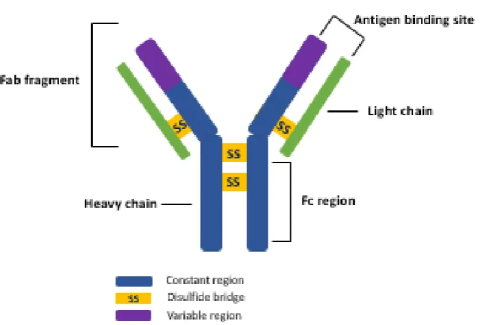
Figure 1.4: Schematic illustrating the basic structure of
immunoglobulin……….…23
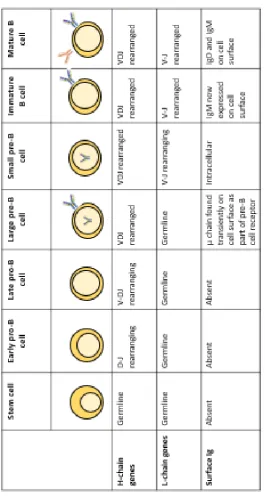
Figure 1.5: Development of B cells from stem cell and the importance of VDJ rearrangement in the successful development and expression of surface immunoglobulins………25
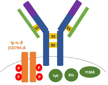
Figure 1.6: Schematic of the B cell receptor (BCR) and the other
components involved in its upstream signalling………..28
Figure 1.7: Simple schematic illustrating the importance of the
microenvironment and external signalling cues in CLL………..43
Chapter 2: Materials and Methods
Figure 2.1: The Malvern NS500-NTA system, which tracks and records the movement of nanoparticles under Brownian motion and
subsequently calculates the mean and mode particle size, generates particle size distribution graphs and calculates nanoparticle per ml concentration……….……….….80
Figure 2.2: Gating strategy undertaken for flow cytometric analysis within this thesis………..91
Figure 2.3: Image shows set-up of the confocal microscopy
workspace……….……97
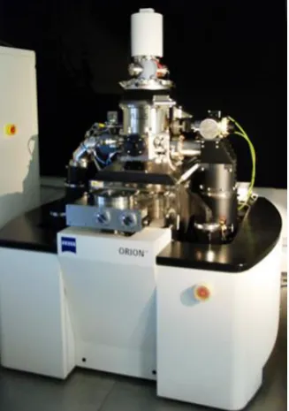
Figure 2.4: Image shows Zeiss Orion Helium Ion
Microscope………..……..…….103
xiii
Figure 2.6: Schematic highlighting the variety of implicating factors in the generation of a microfluidics system in the study of complex
multifactorial diseases such as CLL………110
Figure 2.7: Representative image of CLL cells under shear flow…..110
Chapter 3: Characterisation and assessment of the effects of bare Fe2O3 nanoparticles on B cells
Figure 3.1: NTA analysis of bare Fe2O3 nanoparticles in ultra-pure water………..…...…….128
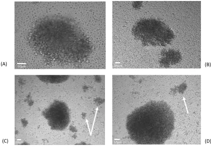
Figure 3.2: The I-83 cell line demonstrates considerable aggregation when cultured in suspension, and treatment with bare Fe2O3
nanoparticles effects cell aggregation………131
Figure 3.3: B lymphocytes interact with bare Fe2O3 nanoparticles
following 24 hour incubation……….134
Figure 3.4: The I-83 cell line interacts with bare Fe2O3 nanoparticles following 24 hour incubation ………135
Figure 3.5: Patient-derived CLL cells will interact and internalise bare Fe2O3 nanoparticles following 24-hour incubation………...133
Figure 3.6: Helium ion micrographs showing the effects of
pre-processing on normal B lymphocytes……….140
Figure 3.7: Normal B cells isolated from healthy donors were
determined as ≥95% CD19+………....141
Figure 3.8: Helium Ion Micrographs of normal B cells……….143
Figure 3.9: Patient-derived CLL cells appear as a smoothened surface morphology, with a spherical shape………144
Figure 3.10: Treatment with bare Fe2O3 nanoparticles significantly effects the viability of I-83 cells, as measured by ethidium bromide-acridine orange staining………145
Figure 3.11: Treatment with bare Fe2O3 nanoparticles induces significant levels of apoptosis/necrosis in I-83 cells, as measured by annexin V-PI staining……….………….. 147
Figure 3.12: Treatment with bare Fe2O3 nanoparticles induces
significant levels of apoptosis/necrosis in normal B cells, as measured by annexin V-PI staining…..……….148
Figure 3.13: Treatment with bare Fe2O3 nanoparticles induces
significant levels of apoptosis/necrosis in CLL patient sample cells, as measured by annexin V-PI
staining………...………..…….……..150
xiv
cells, normal B cells and patient-derived CLL cells with 100µg/mL
Fe2O3 nanoparticles………...151
Chapter 4: Development, characterisation and investigation of functionalised Fe2O3 nanoparticles in patient-derived CLL cells and the I-83 cell line
Figure 4.1: NTA analysis of 50nm Fe2O3 nanoparticles following
coating with APTES, in ultra-pure water……….172
Figure 4.2: Fe2O3@APTES nanoparticles visualised by scanning
electron microscopy………...174
Figure 4.3: Determination of a suitable CLL cell surface marker, using flow cytometric analysis………....176
Figure 4.4: Flow cytometry scatter plots of MFI of anti-CD52-AlexaFluor647 across the I-83 cell line, normal B cells and patient-derived CLL cells………..………...………..177
Figure 4.5: Flow cytometry scatter plots of MFI of
anti-CD5-AlexaFluor647 across the I-83 cell line, normal B cells and patient-derived CLL cells……….……….…….178
Figure 4.6: Flow cytometry scatter plots of MFI of anti-CD19-AlexaFluor488 across the I-83 cell line, normal B cells and patient-derived CLL cells……….……… ……….179
Figure 4.7: Flow cytometry scatter plots of MFI of anti-CD20-AlexaFluor488 across the I-83 cell line, normal B cells and patient-derived CLL cells……….……….…….180
Figure 4.8: Relative expression of CD52 in patient-derived CLL
cells………...………...181
Figure 4.9: Fe2O3@APTES nanoparticles can be successfully
functionalised with anti-CD52 monoclonal antibody……….183
Figure 4.10: Fe2O3@CD52 interacts with patient-derived CLL cells at an increased level to that of [email protected]
Figure 4.11: Chlorpromazine significantly affects the levels of uptake of
Fe2O3@CD52 in the I-83 cells……….187
Figure 4.12: Amiloride significantly effects the levels of uptake of
Fe2O3@CD52 in the I-83 cells……….187
Figure 4.13: Fe2O3@CD52 functionalised nanoparticle are internalised in I-83 cells………..…190
xv
Figure 4.15: I-83 cell viability is significantly affected by treatment with Fe2O3@CD52 nanoparticles, as measured by ethidium
bromide-acridine orange staining………....192
Figure 4.16: Treatment with Fe2O3@CD52 nanoparticles induces significant levels of apoptosis/necrosis in I-83 cells, as measured by Annexin V-PI staining and flow cytometric analysis over 8
hours………..………..193
Figure 4.17: Treatment with Fe2O3@CD52 nanoparticles induces significant levels of apoptosis/necrosis in I-83 cells, as measured by Annexin V-PI staining and flow cytometric analysis over 24
hours………194
Figure 4.18: Representative flow cytometric density plots of annexin V-propidium iodide staining of I-83 cells following treatment with
Fe2O3@CD52 nanoparticles over 8 hours and 24
hours………...…………...……..195
Chapter 5: Functional effects of Fe2O3@CD52 on patient-derived CLL cells
Figure 5.1: Patient-derived CLL cell adherence to the endothelial
monolayer increases with time……….…220
Figure 5.2: Treatment with 25µg/mL Fe2O3@CD52 30 minutes prior results in significantly less adhesion of patient-derived CLL cells to HUVEC cells at a fluid shear flow rate of 0.5 Dynes/cm2…………....220
Figure 5.3: Representative image of CLL patient sample whilst under a shear flow rate of 0.5 Dynes/cm2, through a sample chamber coated with an endothelial monolayer of HUVECs………221
Figure 5.4: Treatment with Fe2O3@CD52 nanoparticles over 8 hours induces significant apoptosis/necrosis in patient-derived CLL
cells………..226
Figure 5.5: Treatment with Fe2O3@CD52 nanoparticles over 8 hours induces significant apoptosis/necrosis in patient-derived CLL
cells………..227
Figure 5.6: Responsiveness to treatment with Fe2O3@CD52 in
patient-derived CLL patient samples………..228
Figure 5.7: Responsiveness of patient-derived CLL cells to treatment with 50µg/mL Fe2O3@CD52 as categorised by Binet Stage, IGHV mutational status and CD38 status……….229
xvi
xvii
List of tables
Chapter 1: Introduction
Table 1.1: List of FDA-approved nanomedicines detailing material, indication(s), advantages gained from using each product and the year(s) each material was approved………...6
Table 1.2: Summary of some of the most commonly utilised
nanotherapeutics in haematological malignancies……….59
Table 1.3: Summary of some available nanoparticles used in the
diagnosis and monitoring of leukaemia and lymphoma……….…63
Chapter 2: Materials and Methods
Table 2.1: List of antibodies used in this work……….68
Table 2.2: Details of the clinical characteristics of CLL patient samples used within this study………..71
Table 2.3: Details a quick reference guide as to the chronic
lymphocytic leukaemia patient sample used in each experiment within this study………..……….…72
Table 2.4: Chronic B Lymphoproliferative flow cytometry
Panel………...………..90
Table 2.5: Shows the maximum binding potential of anti-CD52
antibody to the surface of Fe2O3 nanoparticles as calculated using the amount of anti-CD52 used in each functionalisation
reaction……….93
Table 2.6: Shows fluorescently doped, pH sensitive functionalised nanoparticles used in uptake and interaction studies and their
individual purpose within this study………..95
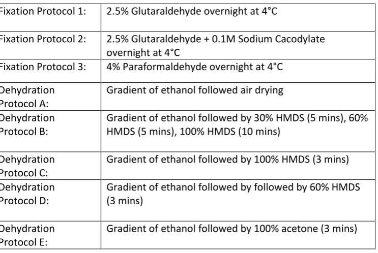
Table 2.7: Shows differing fixation protocols and dehydration protocols which can be utilised to prepare samples for electron
microscopy……….………..100
Table 2.8: Sets out steps for the careful preparation of biological samples for Helium ion
microscopy……….………….103
Table 2.9: Table demonstrates the variety of controls implemented in each flow cytometric analysis of Annexin V-PI staining for the study of apoptotic events in cell samples………..107
Chapter 3: Characterisation and assessment of the effects of bare Fe2O3 nanoparticles on B cells
Table 3.1: Shows the various parameters recorded during NTA
xviii
Chapter 4: Development, characterisation and investigation of functionalised Fe2O3 nanoparticles in patient-derived CLL cells and the I-83 cell line
Table 4.1: The various parameters recorded during NTA analysis of Fe2O3
nanoparticles………..173
Table 4.2: The percentage of positivity for monoclonal
CD52-AlexaFluor647 cell surface antigen across eight patient-derived CLL cell………..…………..178
Table 4.3: Illustrates the monoclonal antibody used in the generation of functionalised nanoparticles for these uptake studies and the purpose of including each of these antibodies in this body of
work………..………189
Chapter 5: Functional effects of Fe2O3@CD52 on patient-derived CLL cells
xix
List of abbreviations
µg, microgram
µL, microlitre
µm, micrometer
µs, microsecond
ADCC, antibody-dependent cellular cytotoxicity
Akt, protein kinase B
ALL, acute lymphoblastic leukaemia
AML, acute myeloid leukaemia
APTES, (3-aminopropyl) triethoxysilane,
ATM, ataxia-telangiectasia-mutated
BCR, B cell receptor
BET, Brunauer, Emmett and Teller theory
BMSC, bone marrow stem cells
BTK, Bruton’s Tyrosine Kinase
CAP, cyclophosphamide/ doxorubicin/ prednisone combination
CD, cluster of differentiation
CDC, complement-dependent cytotoxicity
CHOP, cyclophosphamide/ doxorubicin/ vincristine/ prednisone combination
CLL, chronic lymphocytic leukaemia
DLS, dynamic light scattering
DNA, deoxyribonucleic acid
dPBS, Dulbecco’s phosphate-buffered saline
EDC, 1-ethyl-3-(-3-dimethylaminopropyl) carbodiimide hydrochloride
EDTA, ethylenediaminetetraacetic acid
EGFR, epidermal growth factor receptor
EMA, European Medicines Agency
EPR, enhanced permeability and retention
FBS, foetal bovine serum
xx
FDA, Food and Drug Administration
FL, follicular lymphoma
GC, germinal centre
HDLEC, human dermal lymphatic endothelial cell
HER2, human epidermal growth factor receptor 2
HEV, high endothelial venules
HIV, human immunodeficiency virus
HMDS, hexamethyldisilazane
HSC, haematopoietic stem cells
HUVEC, human umbilical vein endothelial cell
Ig, immunoglobulin
IGHV, immunoglobulin variable region heavy chain
IR, infrared
ITAM, immunoreceptor tyrosine-based activation motif
ITIM, immunoreceptor tyrosine-based inhibition motif
ITP, immune thrombocytopenia
kDa, kilodalton
kV, kilovolts
MAC, membrane attack complex
MASP 1/2, mannose-associated serine protease 1
MBL, mannose-binding lectin
MDS, myelodysplastic syndromes
MHC, major histocompatibility complex
mL, millilitre
MNP, magnetic nanoparticle
MRI, magnetic resonance imaging
mRNA, messenger RNA
MUC-1, mucin 1, cell surface associated
NF- κB, nuclear factor kappa-light-chain-enhancer of activated B cells
NHL, Non-Hodgkin lymphoma
xxi
NLS, nuclear localisation signal
nm, nanometer
nM, nanomolar
NTA, nanoparticle tracking analysis
ORR, objective response rate
pA, picoampere
PBMC, peripheral blood mononuclear cell
PBS, phosphate-buffered saline
PCR, polymerase chain reaction
PDT, photodynamic therapy
PEG, poly(ethylene) glycol
PFS, progression-free survival
PI3K, phosphoinositide-3 kinase
PS, phosphotidylserine
PTK, protein-tyrosine kinase
PTT, photothermal therapy
QELS, quasi-electric light scattering
RGD, arginylglycylaspartic acid
RNA, ribonucleic acid
SEM, scanning electron microscopy
SHP-1, Src homology region 2 domain-containing phosphatase-1
SPION, super paramagnetic iron oxide nanoparticle
TD, T cell dependent
TNF, tumour necrosis factor
VCAM, vascular cell adhesion protein 1
xxii
Communications
Publications
Hutchinson D, Müller J, McCarthy JE, Gun’ko YK, Verma NK, Di Cristo L, Kickham L, Movia D, Prina-Mello A, Volkov Y. Cadmium nanoparticles citrullinate cytokeratins within lung epithelial cells: cadmium as a potential cause of citrullination in chronic obstructive pulmonary disease. Int J Chron Obstruct Pulmon Dis. 2018; 13:441-449.
Kickham, L.C., McElligott A.M., Vandenberge, E.A., Volkov. Y., Browne, P.V. “Interrogating the Interaction of CD52 functionalised Metallic Nanoparticles with Malignant B Lymphocytes” Blood 2015; 126(23):A4437.
Oral and poster presentations
“An Interrogation into the Interaction of Metallic Nanoparticles with Malignant B Lymphocytes” – Laura Kickham, Anthony M. McElligott, Adriele Prina-Mello, Yuri Volkov and Paul Browne. InterReg 4A Irish Presidential Meeting, Swansea University, October 2014.
Invited Speaker - “An Interrogation into the Interaction of Metallic Nanoparticles with Malignant B Lymphocytes” – Laura Kickham, Anthony M. McElligott, Yuri Volkov and Paul Browne. Haematological Association of Ireland, Clarion Hotel Sligo, October 2014.
“Nanoparticle uptake by Malignant B Cells: The Potential of Nanomedicine in the Diagnosis and Treatment of Haematological Malignancies” – Laura Kickham, Anthony M. McElligott, Adriele Prina – Mello, Elisabeth Vandenberghe, Yuri Volkov and Paul Browne. Haematological Association of Ireland Annual Meeting, Europa Hotel Belfast, 18-19 Oct 2013.
“Applications of Nanomaterials in the Diagnostics of Haematological Malignancies” – Laura Kickham, Omar Kazem Mahfoud, Dania Movia, Tony McElligott, Paul Browne, Adriele Prina – Mello and Yuri Volkov. Euronanoforum, Convention Centre Dublin, 18-20 June 2013.
1
2
1.1 Nanotechnology and applications in medicine
1.1.1 Nanotechnology and nanoparticles
Nanotechnology has been defined as the “intentional design,
characterisation, production and applications of materials, structures,
devices and systems by controlling their size and shape in the
nanoscale range (1-100nm)” though this term has also been used in
reference to materials up to 500nm and rarely exceeding 700nm (Mody
et al., 2010). The European Commission first adopted the definition of a
nanomaterial in 2011 and has since then reviewed several times to
ensure reflection of the evolving landscape of these materials. They can
be defined as “A natural, incidental or manufactured material containing
particles, in an unbound state or as an aggregate or as an agglomerate
and where, for 50% or more of the particles in the number size
distribution, one or more external dimensions is in the size range 1 nm
- 100 nm” (European Commission, 2011).
A nanoparticle can be classed as such if it is of a single dimension and
on the nanoscale in size (Powers et al., 2007). Within a biological
context, they are comparable in size to viruses (20-300 nm) and
significantly smaller than cells (30 to 300 µm). Upon reaching the
nanoscale, the properties of the bulk material can become altered. This
is as a result of nanoparticles having a greater surface area to volume
than larger particles, meaning their small diameter provides a large and
available surface area, often causing the nanoparticle to be more
reactive than the bulk material (Mahdavi, et al., 2013). The importance
3
influence the physicochemical properties of a material, such as surface
charge and dispersion stability (Duan & Li, 2013). Nanotechnology is by
no means a recent advancement in the scientific field, having been
presented by Nobel Laureate Richard P. Feynman in 1959 during a talk
entitled “There is plenty of room at the bottom” (Feynman, 1960), though
its potential for the advancement of the scientific field is only more
recently becoming clear. During this talk, Feynman described how in
order to fully understand “big science”, scientists should also understand
the scientific phenomena on the smaller scale (Feynman, 1960). The
term “nanotechnology” was then first coined by Norio Taniguchi in 1974,
(Taniguchi, 1974) for which the European Society for Precision
Engineering and Nanotechnology awarded him the Lifetime
Achievement Award in 1999.
Nanotechnology has demonstrated its implications across a variety of
branches of science and industries, including: biotechnology and
medicine, the food industry, agriculture, textiles environmental science
and electronics (Figure 1.1). The crossover of the use of
nanotechnology has resulted in the significant advancement of
manufacturing processes, engineering and healthcare, to name but a
few. As of 2018, the global nanotechnology market value was estimated
at approximately $40 billion, with a projected growth rate of 17%,
meaning that by 2021 this market could be worth over $90 billion globally
(Koshovets & Ganichev, 2016) (McWilliams, 2016) (Markets, 2018). The
use of nanotechnology for the advancement of medicine and healthcare
4
5
1.1.2 Nanomedicine
Nanomedicine forms the epicentre of the convergence of physics,
chemistry and biology (Figure 1.2). This convergence of technologies
has allowed for the combination of differing approaches from scientists
of various backgrounds to tackle problems directly related to the medical
and biological fields.
Figure 1.2: Nanomedicine is at the epicentre of the convergence of biology, physics, and chemistry, allowing for a multi and inter-disciplinary collaborative approach to be taken towards new medicinal projects.
Interestingly, the application of nanoparticles, as carriers or vectors, has
been shown to be of great potential in the field of targeted therapies for
a range of disordered and diseased states, and there are now more than
[image:27.595.190.516.257.567.2]6
(Sanna et al., 2014) (Tran, DeGiovanni et al., 2017)(European
Medicines Agency, 2018). A selection of the most commonly used FDA
[image:28.595.61.457.204.787.2]approved nanoparticles is summarised in Table 1.1 below.
Table 1.1: List of FDA-approved nanomedicines detailing material, indication(s), advantages gained from using each product and the year(s) each material was
approved. Adapted from Bobo et al. 2016, European Medicines Agency and Tran et
al. 2017.
Name Material
description
Advantage Indication(s) Year(s)
approv ed
Nanotherm® (MagForce)
Iron oxide Allows for cellular uptake and introduces superparamagnetism
Glioblastoma 2010
Feraheme™/ ferumoxytol (AMAG pharmaceutical s) Ferumoxytol SPION with polyglucose sorbitol carboxymethyl ether Magnetite suspension allows for prolonged release, decreasing dose number Anaemia in chronic kidney disease 2009 Venofer® (Luitpold Pharmaceutical s)
Iron sucrose Allows for increased dosage Anaemia in chronic kidney disease 1999 INFeD® (Sanofi Avertis) Iron dextran (low MW)
Allows for increased dosage Anaemia in chronic kidney disease 1957 Ferrlecit® (Sanofi Avertis) Sodium ferric gluconate
Allows for increased dosage Iron deficiency in chronic kidney disease 1999 Feridex®/Endor em® (AMAG pharmaceutical s) SPION coated with dextran Introduces superparamagnetism Imaging agent 1996/ 2008 GastroMARK™; umirem® (AMAG pharmaceutical s) SPION coated with silicone Introduces superparamagnetism Imaging agent 2001/ 2009 DepoDur® (Pacira Pharmaceutical s) Visudyne® (Bausch and Lomb) Liposomal Verteporfin
Increased delivery to site of diseased vessels;
photosensitive release
Macular degeneration , It age-related; myopia; ocular histoplasmos is 2000 Doxil®/Caelyx ™ (Janssen) Liposomal doxorubicin
Improved delivery to site of disease; decrease in systemic toxicity of free drug
7
Estrasorb™ (Novavax)
Micellar Estradiol
Controlled delivery of therapeutic Menopausal therapy 2003 Abraxane®/ABI-007 (Celgene) Albumin-bound paclitaxel nanoparticles Improved solubility; improved
delivery to tumour
Breast cancer, NSCLC, pancreatic cancer 2005/ 2012/ 2013 MM-398 (Onivyde) Lipsomal 5-fluorouracil and leucovorcin Increased potency allows for reduced dosage
Metastatic pancreatic cancer
2015
Fulphila PEGylated
filgrastim
Decreased elimination allows for fewer treatments Neutropenia infection control in non-myeloid malignancy patients 2018
Although enormous advances have been made in nanotechnology,
many of these have not yet achieved either an industrial development
or translation to an available therapy. Some nanoparticles have been
shown to potentially improve the pharmacokinetics and
pharmacodynamics of drugs; they may provide a platform in which to
deliver an increased concentration of a therapeutic agent than would
normally be permissible, and they can improve the affinity of the drug
within the blood stream through evasion of the reticuloendothelial
system (RES) and providing the ability to solubilise hydrophobic drugs.
Nanoparticle evasion of the RES is a highly attractive feature in many
applications, and attributed to the lack of serum proteins on the surface
of functionalised nanoparticles, resulting in minimal uptake by
macrophages and subsequent increased longevity in the bloody stream
(Bae & Park, 2012) (Ferrari, 2008). Additionally, nanoparticles provide
the opportunity for controlled, directed release of drugs at a site or target
using environmental or an external signal, and importantly possess the
ability to accumulate in tumours through the phenomenon known as the
enhanced permeability and retention (EPR). (Onoue et al., 2014) (Islam,
8
1.1.3 Nanoparticles in cancer
There are several limitations to conventional cancer therapies, such as
low to moderate therapeutic efficacy, drug resistance and poor delivery
resulting in low concentrations of the therapeutic at the site of action
(Lammers et al. , 2012). This has resulted in a call for novel and
combined cancer therapeutics, with nanoparticles being extensively
studied to fulfil this purpose. First-generation nanoparticles have proven
relatively easy to synthesise and were designed for use in diagnostics
and imaging solely. A newer generation of nanoparticles have
multi-functional characteristics and are designed for combined therapeutic
and diagnostic applications, known as theranostics (Li, Nejadnik, &
Daldrup-Link, 2017) (Gobbo et al. , 2015). This means that the
characteristics of nanoparticles that may have been originally developed
for solely diagnostic purposes or therapeutic purposes have been
combined, resulting in a nanoparticle suitable for both diagnostics and
therapeutics. This has been successfully demonstrated with
magneto-polymeric nanohybrids for both the detection and the targeted treatment
of breast cancer (Yang et al., 2007).
Iron oxide nanoparticles have been used for several years as an
imaging contrast agent for MRI. This allows healthcare practitioners to
image the specific area where the tumour resides within the body, with
magnetic particles combined with MRI scanning resulting in greater
imaging contrast (Ebrahimi et al. , 2016) (Gobbo et al. , 2015). In
addition to their proven use as contrast agents, magnetic iron oxide
nanoparticles have also shown the potential in drug delivery. These
9
suitable for such drug conjugation and delivery. Surface
functionalisation relates to the modification of the surface of a
nanoparticle using one or more of a vast array of ligands, proteins,
functional groups or compounds which can enhance the properties of
the bare material. The use of iron oxide nanoparticles as a drug delivery
system can help to overcome the issues of conventional
chemotherapeutics, and potentially even immunotherapeutics by
offering increased specificity, crossing biological barriers and offering
controlled release. In addition, a controlled and specific drug delivery
utilising techniques such as MRI targeting or a tissue/cell surface marker
targeting ligand could increase the retention of the drugs at the site of
the tumour cells, potentially avoiding over-dosage and decreasing the
notable chemotherapeutic side effects (Li, Nejadnik, & Daldrup-Link,
2017) (Lima-Tenorio et al., 2015). In addition to their use as a contrast
agent or drug delivery system, iron oxide nanoparticles are also used in
magnetic hypothermia therapy, for solid tumour treatment. For this,
nanoparticles are localised to the solid tumour tissue and energy
absorption by the nanoparticles in alternating magnetic field results in
localised heating of the cells inducing cell death (Askari et al.,
2016)(Kossatz, et al., 2015). A more detailed discussion of the uses of
nanoparticles and their biomedical applications can be found in section
1.2.
1.1.4 Nanoparticles and the tumour microenvironment
Control in particle size, in combination with appropriate surface coating
may also allow a stealth-effect against the body’s immune system.
10
may severely inhibit efficiency and these barriers must be overcome in
order to fully exploit the theoretical potential of nanoparticles. (Tiwari, et
al., 2012) For instance, the endothelial cells which line the interior of the
entire vascular system represent a major barrier for therapeutic agents
travelling from the bloodstream to the target tissues. These endothelial
gaps can vary in size considerably, dependent on the tumour type, and
therefore result in non-uniform extravasation into the tumour site.
Additionally, nanoparticle extravasation can also be governed by
particle size, with smaller nanoparticles demonstrating enhanced
penetration of the tumour matrix compared to larger nanoparticles. In an
attempt to address this, several groups have shown that a degradable
nanoparticle with the ability to reduce in size upon extravasation results
in an increased accumulation of nanoparticles at the desired site (Tong
et al., 2012) (Tong et al., 2013). However, the mononuclear phagocyte
system (MPS), consisting of monocytes and macrophages will often
sequester the majority of administered nanoparticles, where the cells
will then process and degrade components of the administered
nanoparticle, often rendering them non-functional (Parambath, 2018).
This approach has as of yet not been investigated clinically. Over 40,000
studies published in the last 10 years have focused on nanoparticle
targeting strategies and substantial progress has been made toward our
understanding of how nanoparticles interact with cells and tissues
(Rosenblum et al., 2018)(Li, et al., 2016)
In order to maximise the functionality of the nanoparticle, it is therefore
desirable to develop a therapeutic that can passively or actively target
11
major advances in the use of nanomaterials in medicine, including the
use of magnetic nanoparticles such as iron oxide, gold and silver
nanoparticles, nanoshells and nanocages. Liposomes have also been
shown to be of great benefit in the field of nanomedicine (Bozzuto &
Molinari, 2015). One of the more exciting and important developments
in the realm of nanomedicine was the introduction of antigen-specific
functionalised nanoparticles. This involves the conjugation of an
appropriate ligand that demonstrates a specific binding activity towards
the targeted cell type (Bozzuto & Molinari, 2015).
1.2 Nanoparticles and their biomedical applications
Nanoparticles have demonstrated their role in healthcare and medicine
for many years, and as a result have been extensively studied as both
a direct therapeutic agent and as a carrier for biologically relevant
agents (Bobo et al., 2016) (Wagner et al., 2006) (Caruso et al., 2012)
(Sanvicens & Pilar Marco, 2008). Although nanoparticles are ubiquitous
in the environment, prior to the release of any new
nanoparticle-containing product, each constituent and product must undergo intense
scrutiny and study in order to assess their safety and efficacy,
dependent on the desired function of the biomedical nanoparticle.
Because of their small size, many nanoparticles can easily enter the
body and cross many barriers, potentially gaining access to many
tissues and organs, where they have the potential to cause a number of
different issues. It has been postulated that nanoparticles less than
10nm in size have the potential to act similarly to gases, and separate
12
ingested can reach the liver, heart, spleen, gastrointestinal tract, lungs
and even the brain (Bahadar et al., 2016). Targeted therapeutics
employing the use of nanoparticles have been shown to have the
potential to revolutionise healthcare, and the way in which we approach
the management of different disease states (Revia & Zhang, 2016)
(Yong et al., 2009) (Zhang, et al., 2008). The properties and use of
nanoparticles, including those that contribute to the use of nanoparticles
in biomedical applications are discussed in further detail below.
1.2.1 Iron (III) oxide nanoparticles (SPIONs)
Iron (III) oxide (Fe2O3) is an inorganic compound of paramagnetic
nature. Superparamagnetic iron oxide nanoparticles, or SPIONs, have
generated a large amount of interest as a potential candidate in
nanomedicine, both as potential therapeutics and as diagnostic tools,
due to their inherent biocompatibility and magnetic properties (Gobbo et
al., 2015)(Senapati et al., 2018). Superparamagnetism refers to the
nanoparticles ability to randomly switch, or flip, direction when under the
influence of temperature, and where the typical time between two flips
is known as the Neel relaxation time. When no magnetic field is applied,
and the time taken to measure the magnetisation of the nanoparticles is
longer than the Neel relaxation time (where the average value of
magnetisation appears as zero), the nanoparticles are said to be in the
superparamagnetic state. While in this state, an external field can
magnetise the nanoparticles (Marghussain, 2015). The concept of
superparamagnetism was first proposed and described by Frenkel and
Doefman in 1930 (Frenkel & Doefman, 1930). Some of the benefits of
13
nanoparticles do not retain any net magnetisation following removal of
the external magnetic field, making them easily controllable in MRI, and
retention of stability upon immersion into fluid, making them ideal for use
in biological settings (Kolhatkar et al., 2013).
The potential uses of Fe2O3 nanoparticles as MRI contrast agents has
been an area of intense interest for many years and was a pivotal step
towards the realisation of these materials for biomedical use. In 2009,
this was accelerated with the FDA approval of ferumoxytol, the first
Fe2O3 nanoparticle approved for clinical use, indicated for the treatment
of iron deficiency anaemia in adults with chronic kidney disease
(McCullough et al., 2012). SPIONs have since demonstrated their use
in various applications such as malignant hyperthermia, imaging and
magnetic separation techniques such as those used for cellular
separation and DNA sequencing (Sabale, et al., 2017). As therapeutic
agents, SPIONs have drawn increasing attention due to their potential
in gene therapy and targeted drug delivery (Gupta & Gupta, 2005)
(Mody et al., 2010). This wide range of potential bio-applications of
SPIONS is heavily influenced by the particles physical, chemical and
magnetic properties, in addition to its shape and size (Patil, et al., 2018).
The inherent characteristics of SPIONs may be exploited for
image-guided therapies and multimodal theranostics, where more than one
treatment strategy is employed, such as with photothermal therapy
(PTT), photodynamic therapy (PDT), magnetic hyperthermia and
immunotherapy (Saeed et al., 2018). PTT is a non-invasive cancer
treatment approach, utilising visible of infrared (IR) light where, in the
14
to the disease area, the laser energy is converted into thermal energy
which can then be used for tumour ablation (Revia & Zhang, 2016). PDT
is a technique that destroys cancer cells using the combination of
photo-sensitisers, such as SPIONs, and a laser source. This combination
results in the release of reactive oxygen species, along with sufficient
cellular oxygen, which stimulates apoptosis within the tumour cells
(Revia & Zhang, 2016) (Gobbo et al., 2015). However, an as of yet
insurmountable drawback of both PDT and PTT is the irrevocable
damage to the normal tissue types in their surrounding environment
resulting from the penetration of photosensitiser beams. Whilst this is an
area which has drawn much attention and resulted in the use of highly
size-specific photosensitiser beams, it still remains a concern (Gobbo et
al., 2015).. As previously discussed, magnetic hyperthermia is an
approach which exploits the ability to heat SPIONs when placed in an
external alternating magnetic field. The resultant heat then acts as a
tumour ablator. This approach is one of the older multimodal
approaches in the treatment of tumours using SPIONS, having been first
demonstrated intracellularly in 1979 by Gordon et al. (Gordon et al.,
1979) (Saeed et al., 2018). The use of PDT, PTT and magnetic
hyperthermia has demonstrated great potential in the treatment of solid
tumours, though the scalability of these technologies remain to be seen.
Another important use of iron oxide nanoparticles is their use in
nano-based immunotherapy. The proven biocompatibility and adaptable
nature of iron oxide nanomaterials have identified them as useful tools
in the generation of novel immunotherapies. A detailed discussion as to
15
within this section. The method employed in the generation of these
SPIONs can however be pivotal to ensuring their suitability for use.
1.2.1.1 Synthesis of SPIONs
There are a wide variety of methods in which to develop SPIONs of the
correct size and shape for their intended use. These include sol-gel
synthesis, electrospray synthesis, hydrothermal reactions, hydrolysis
and co-precipitation of iron salts. Co-precipitation is an extremely
common method in the synthesis of SPIONs as this technique results in
a high nanoparticle yield. This technique, however, allows the producer
very limited control over the size distribution, meaning that there may be
additional purification steps following synthesis, dependent on the
intended use of the SPIONs. The size distribution of SPIONs, and the
synthesis of uniform-sized SPIONs is very important as the behaviour
and properties depend largely on the dimensions of the particle.
(Mahdavi, et al., 2013) (Mody et al., 2010) However, another
characteristic of nanoparticles such as SPIONs is their large
surface-to-volume ratio with high surface energies, resulting in their common
aggregation and tendency to accumulate in one location. Additionally,
as with their bulk material, SPIONs have a high reactivity tendency and
are therefore easily oxidised, resulting in a loss of magnetism and
dispersal. For these reasons, it is vital in the successful generation of
SPIONs for biomedical purposes to provide the most suitable surface
coating for the SPIONs, in order to maximise the abilities of this
materials in biomedical applications (Mahdavi, et al., 2013). SPIONs can
be used as scaffolds for the conjugation of a range of biologically and
16
antibodies and nucleotides. The release and distribution/diffusion
mechanism, bioavailability and biodegradation rate are important
factors in the development of these so-called functionalised SPIONs. In
order to generate these functionalised nanoparticles, SPIONs first need
to be coated with a surfactant, polymer or conjugation material. This
may consist of oleic acid, dextran, chitosan or poly(ethylene glycol)
(PEG), or they may need to be silanised using (3-aminopropyl)
triethoxysilane, referred to herein as APTES (Mahdavi, et al., 2013).
APTES is an aminosilane commonly used in the process of the
generation of functionalised nanoparticles, including SPIONs, and it acts
as a bonding agent for the adhesion of appropriate molecules to the
surface of materials. A representative illustration of a multi-layered
functionalised SPION is shown in Figure 1.3. SPIONs are categorised
into two main categories based on their hydrodynamic diameter:
standard SPIONs (50 to 150nm) and ultrasmall SPIONs (<50nm in size).
Most studies looking at the use of SPIONs for biomedical use focus on
the use of SPIONs in the range of 10 to 100nm. SPIONs with a
hydrodynamic radius of >200nm or <10nm tend to be sequestered by
the spleen, or removed through renal clearance (Singh et al., 2010)
(Flias & Tsourkas, 2009).
However, the potential toxicity of SPIONs towards normal cells has
somewhat hindered their successful implementation as a clinically
approved therapeutic agent. Whilst the nanoscale properties
demonstrated by SPIONs have proven to be of great potential benefit in
the furthering of nanomedicine, these same properties (a large surface
17
propensity to diffuse across biological membranes) can potentially result
in an increase in cytotoxicity (Singh et al., 2010). In addition to the direct
toxicity and clinical responses of the biological system requiring intense
scrutiny, the degradation and accumulation of SPIONs following
administration is a key point of study (Patil, et al., 2018). Exposure to
SPIONs has been associated with toxic effects such as inflammation,
apoptosis, impaired mitochondrial function, the generation of reactive
oxygen species (ROS) and an increase in micronuclei (an indicator of
significant chromosomal damage, and hence an indicator of
genotoxicity) (Singh et al., 2010). However, SPIONs have been shown
to demonstrate low toxicity in vivo (Hussain et al., 2005) (Kim, et al.,
2006). Additionally, a large study comparing the toxicity of several
SPIONs in vitro demonstrated that Fe2O3 were safe and non-cytotoxic
below 100µg/mL (Karlsson et al., 2008). The precise mechanism of
toxicity is strongly influenced by the size and shape of the SPION, the
surface coating; and the mechanism of uptake of the SPION by the cell
type.
Figure 1.3: Schematic of a multi-layered magnetic nanoparticle for use in
personalised medicine. Figure depicts iron oxide core for use as a carrier or for
18
toxicity and a targeting ligand, often an antibody, for cellular targeting. Reproduced with permission from Handbook of Clinical Nanomedicine: Nanoparticles, Imaging, Therapy and Clinical Applications (Movia D, Poland C, Tran L, Volkov Y, Prina-Mello A. Multi-layered Nanoparticles for Personalized Medicine: Translation into Clinical Markets).
1.2.1.2 Surface functionalisation of SPIONs
One of the most notable uses of nanoparticles in biomedical applications
is their ability to be used as a “scaffold” or “carrier” for other biologically
relevant components. This involves a strategy known as surface
functionalisation and involves the incorporation of material(s) on to the
surface of the nanoparticle (briefly discussed in section 1.2.1.1). Whilst
there are a number of methods in which this end goal can be achieved,
the “core-shell” approach is very commonly utilised. This usually
involves surface functionalisation as a separate step to the synthesis of
the SPION itself (Wu et al., 2015). This “core-shell” approach to surface
functionalisation can be achieved through a number of bioconjugation
strategies including covalent conjugation and electrostatic conjugation
(El-Boubbou, 2018).
Organic materials such as dextran, PEG, chitosan and hyaluronic acid
are amongst some of the most commonly used surface modifiers in the
development of SPIONs for biomedical applications. SPIONs with such
a surface coating have been developed for potential use as MRI agents,
magnetic cell separation, magnetic recording and gene delivery
(Uthaman et al., 2015) (Wu et al., 2015).
One of the more intricate methods of surface functionalisation includes
the addition of particular functional groups to the surface of the SPION.
This can include the addition of groups such as hydroxyl groups (-OH),
19
The addition of such groups renders the surface of the SPION easily
modifiable, allowing for the easy attachment of differing biologically
relevant molecules. Aminosilane is a small molecule that is often used
to modify the surface of SPIONs, resulting in available functional groups
(i.e. amine groups) on the surface of the SPION, which are then
available for further functionalisation APTES is an aminosilane which is
commonly used in the process of silanisation, and results in the
anchoring of the -NH2 group to the surface of the SPION (Wu et al.,
2015). Following on from silanisation of SPIONs, it is then possible to
attach a drug/molecule or antibody to the surface of these SPIONs using
a carbodiimide compound such as 1-ethyl-3-(-3-dimethylaminopropyl)
carbodiimide hydrochloride (EDC) (El-Boubbou, 2018). This process
acts through the activation of the carboxyl groups, allowing for their
direct conjugation to the available -NH2 groups. Such techniques have
resulted in the generation of anti-EGFR SPIONs, anti-HER2 SPIONs,
prostate-specific antigen SPIONs and MUC-1 SPIONs, amongst many
others in generation (Mu, et al., 2015) (Mu, et al., 2015) (Bates et al.,
2014) (Aghanejad et al., 2018). As mentioned previously, the majority of
these SPIONs have been generated as diagnostic or imaging agents,
but there is considerable research ongoing in the field of
biologically-active nanomedicines (El-Boubbou, 2018).
1.2.2 Nanoparticle characterisation techniques, protocols and
methodologies
Nanoparticles, including those with a potential for use in nanomedicine,
display novel physicochemical properties (Lin et al., 2014)(Gioria, et al.,
20
attractive choice for novel medicinal approaches, but also means that
the advancement of their use as a novel nanomedicinal requires a
step-wise and thorough characterisation. These novel physicochemical
properties have the potential for implications on their physiological
interactions, but due to the novelty of these technologies, and the rapid
advancement in this field, there is a lack of standardised
characterisation methods for nanoparticles and nanomedicines (Lin et
al., 2014). These physiological interactions may be influenced by a
number of factors, including: the biodistribution of the nanomedicine and
the phagocytosis and endocytosis of the nanodrug. There are a number
of nanoparticle parameters that may need to be considered such as pH
and solubility. Additionally, there is a special emphasis placed on
nanoparticle parameters such as size, surface area, shape, charge and
aggregation state (Prina-Mello et al., 2013). To date, there have been
no stringent guidelines developed for the characterisation of
nanoparticles for diagnostic or therapeutic use. However, the US Food
and Drug Administration (US FDA) has issued guidelines for the
characterisation of materials for the food and cosmetic industries,
including the characterisation of drug products, including biological
products that contain nanomaterials (Food and Drug Administration
(FDA), 2017). Over the past two years, efforts have been made in
instigating official legislation for the use of nanomaterials in biomedical
applications. To date, these materials are currently subject to the same
legislation as chemical substances and require applicable safety and
21
In the production and characterisation of nanoparticles, size is one of
the most critical factors. This characteristic is responsible for the
regulation and circulation of nanomaterials in the bloodstream, their
penetration across physiological barriers, localisation and the induction
of cellular response. (Lin et al., 2014) (Ferrari, 2008) (Jiang et al., 2008).
It has been reported that the response of a nanoparticle to its
surrounding environment is largely size dependent, due to the size of
the surface area interacting with the surrounding environment (Jiang et
al., 2008) (Tsao et al., 2011). There are a number of different methods
that may be employed in the characterisation of the size of
nanoparticles. These include nanoparticle tracking analysis (NTA),
scanning electron microscopy (SEM), transmission electron microscopy
(TEM), Helium Ion Microscopy (HIM) and dynamic light scattering
(DLS). Below is a detailed description of NTA technology and DLS. A
detailed discussion surrounding microscopy techniques in the context of
nanoparticle investigations can be found in Chapter 2.3. Within this
chapter, we will discuss the use of NTA, SEM and DLS as a means to
confirm the size, dispersion state and aggregation rate of Fe2O3
22
1.3 B lymphocytes and Chronic Lymphocytic Leukaemia
In 1890, Emil von Behring first established the functional importance in
circulating antitoxins in the immunity to diphtheria and tetanus (Von
Behring & Kitasato, 1890) (Kaufmann, 2017). From this evolved the first
acknowledgement of cells known as the B cell where, in 1967, Paul
Ehrlich proposed that these “antitoxins” were produced by antibody
receptors. He proposed that an immune cell bearing several different
antibody receptors could be stimulated by the binding of an antigen to
produce and release this receptor which was complementary to the
antigen (Ehrlich, 1967) (Cooper, 2015). Over the past century, there has
been significant knowledge garnered in the understanding of what we
now know as the B cell, including the understanding of the
antibody-antigen relationship in the 1930s (Tiselius & Kabat, 1939) and the
determination that antibody production correlates with immunisation
(Fagraeus, 1948). It was not until 1965 that the distinct lineages of the
B cell and the T cell became apparent (Cooper et al., 1965) (Cooper,
2015).
Early B cell development is the ordered rearrangement of the
immunoglobulin (Ig) H and L chain loci, with Ig proteins playing a vital
role in the regulation of this error-prone process (LeBien & Tedder,
2008). In addition to the expression of specific cell surface and
cytoplasmic markers, V(D)J recombination distinguishes the
subpopulations of developing B cells. Ig are heterodimeric proteins that
are composed of four chains: two heavy (H) chain and two light (L)
chains. Functionally, these chains can be separated further again:
23
constant (C) domains that will specify effector functions like complement
or the binding to Fc receptors (Figure 1.4) (Schroeder & Cavacini, 2013).
B cell development begins in the foetal liver but continues in the bone
marrow. At this point in the B cell lineage, the immature cell is easily
killed upon contact with an antigen, and therefore must develop into a
mature cell. B cell development begins as the lymphoid progenitor cells
receive signals from the bone marrow stromal cells. The somatic
recombination process responsible for the joining of these V, D and J
[image:45.595.173.524.164.401.2]24
by two recombinases encoded by the recombination activating genes –
RAG1 and RAG2 (Eibel, 2015). Cells then undergo D-J joining on the H
chain, becoming early pro-B cells where they then begin to express
CD45 and MHC Class II molecules. The late pro-B cell stage is then
complete upon the attachment of the V segment to the D-JH component.
From here, the pro-B cell will then become a pre-B cell upon the
expression of the membrane Ig light chains in their pre-B cell receptor.
Within this pre-B cell receptor complex are Igα Igβ signal transduction
molecules, containing ITAMs (immunoreceptor tyrosine activation
motifs) which will become phosphorylated in response to antigen
binding with the B cell receptor (BCR) (more can be found about the B
cell receptor in Chapter 1.3.2). It is at this point in the B cell lineage that
division occurs, with recombination being halted and B cell clones being
produced all producing the same Ig chains, and the B cell being known
as the large pre-B cell. A schematic depicting B cell development can
be found in Figure 1.5 (LeBien & Tedder, 2008). Small pre-B cells are B
cells which are no longer dividing. These cells undergo V-J joining on
the L chain expressing the Ig chain on the cell surface, where it is known
as an immature B cell. These cells will express the Ig δ chain and
membrane IgD. At this point in the B cell lineage, the cell is known as a