Original Article
Frequency of activating mutations in
FGFR2
exon 7 in
bladder tumors from patients with early-onset and
regular-onset disease
Christine Spiegelberg1, Johannes Giedl1, Nadine T Gaisa2, Anja Rogler1, Marc-Oliver Riener1, Thomas
Filbeck3, Maximilian Burger4, Petra Ruemmele5, Arndt Hartmann1, Robert Stoehr1
1Institute of Pathology, University Hospital Erlangen, Friedrich-Alexander-University Erlangen-Nuremberg, 2Institute
of Pathology, RWTH Aachen University, 3Department of Urology, University Hospital Erlangen,
Friedrich-Alexander-University Erlangen-Nuremberg, 4St. Josef Medical Centre, Department of Urology, University of Regensburg, 5Institute of Pathology, University of Regensburg, Germany
Received January 20, 2014; Accepted January 20, 2014; Epub March 15, 2014; Published April 1, 2014
Abstract: The FGF/FGFR-system plays an important role in embryogenesis, tissue homeostasis and carcinogenesis. Mutational activation of FGFR2 resulting in aberrant FGFR2 signaling activation is known from both hereditary germ line alterations and somatic mutations in various malignancies (e.g. breast, gastric or ovarian cancer). FGFR2 mu-tations are mainly located within the hinge between Ig-like domains (exon 7), around the 3rd Ig-like domains and within the kinase domain. For bladder cancer only sparse data on FGFR2 mutations are available. Most interestingly a case of early-onset papillary carcinoma of the bladder showing a FGFR2 p.Pro253Arg mutation in exon 7 in a pa-tient with Apert Syndrome was reported recently. To further evaluate the importance of FGFR2 exon 7 alterations in bladder cancer a cohort of 254 bladder tumors (cohort 1: unselected cases: n=139; cohort 2: early-onset bladder cancer cases (age at time of diagnosis ≤45 years): n=115) was analyzed. Sections from formalin-fixed, paraffin-embedded bladder tumors were used for DNA isolation. After precise microdissection exon 7 of the FGFR2 gene was analyzed by direct Sanger sequencing. All cases could be analyzed successfully. Mutations in exon 7 of FGFR2 could not be detected in any of the cases. All tumors showed wild type sequence. Our data demonstrate that the recently reported association between early-onset papillary carcinoma of the bladder with germ line FGFR2 p.Pro253Arg mutation could not be found in our cohorts of sporadic bladder tumors. These results indicate that FGFR2 gene mutations might only play a minor role in bladder carcinogenesis.
Keywords: FGFR2, mutation, bladder cancer, early-onset, Apert Syndrome, sequencing
Introduction
Aberrant expression or uncontrolled mutational activation of multiple FGF family members and their four corresponding receptors (FGFR) are found in multiple cancers. FGFRs are trans-membrane glycoproteins with a conserved str- ucture comprising an extracellular part with two to three immunoglobulin-like domains (IgI-III), a transmembrane domain and an intracel-lular split tyrosine-kinase domain. Signal
trans-duction of the FGF/FGFR system is quite
com-plex. Complexity levels are built up by different
affinity of FGFRs for specific FGFs and the fact
that FGFRs are subjected to alternative splicing [1, 2].
Deregulated FGF/FGFR signaling is also known in tumors of the genito-urinary system, espe-cially in bladder, prostate (PCa) and renal cell carcinoma (RCC). For PCa and RCC mainly over-expression of FGFRs was reported, activating mutations seem to play no important roles [1-5].
In bladder cancer very high frequencies of alter -ations in FGFR3 were reported. Activating
muta-tions have first been reported more than twelve
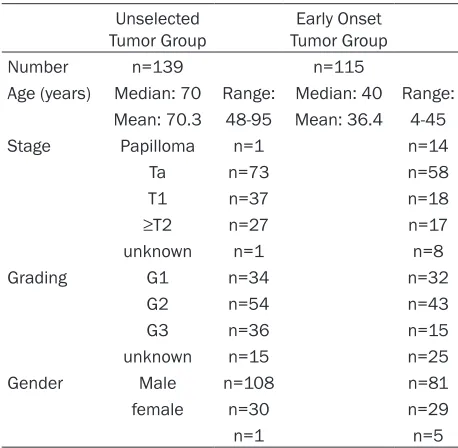
under-Table 1. Characteristics of the study cohort Unselected
Tumor Group Tumor GroupEarly Onset
Number n=139 n=115
Age (years) Median: 70 Range: Median: 40 Range: Mean: 70.3 48-95 Mean: 36.4 4-45
Stage Papilloma n=1 n=14
Ta n=73 n=58
T1 n=37 n=18
≥T2 n=27 n=17
unknown n=1 n=8
Grading G1 n=34 n=32
G2 n=54 n=43
G3 n=36 n=15
unknown n=15 n=25
Gender Male n=108 n=81
female n=30 n=29
n=1 n=5
lying molecular mechanisms still remain unc- lear. Recently, constitutively activated fusion genes generated via chromosomal
re-arrange-ments were identified as a new mechanism of
uncontrolled FGFR3 activation [1, 7].
Interestingly, a case of early-onset low-grade papillary bladder cancer in a patient with Apert Syndrome and a germ line FGFR2 p.Pro253Arg
mutation was described for the first time recently [8]. The specific mutation was also
detectable in the bladder tumor with
concur-rent FGFR3 wild type sequence. In general, only
sparse data on FGFR2 mutation analysis in bladder cancer are available. Ricol and col-leagues investigated the entire coding region of FGFR2 in 33 bladder tumors. Two tumors sh- owed deletions (exon 13 or exon 17), but no single point mutations could be detected [9]. This lack of comprehensive data prompted us to perform a mutation analysis of FGFR2 exon 7 containing codon 253 in a large series of bladder tumors from patients with both early-onset and regular-early-onset disease.
Materials and methods
Bladder cancer tissue samples
Overall, 254 archival formalin-fixed,
paraffin-embedded bladder tumors (cohort 1: unselect-ed cases: n=139; cohort 2: early-onset bladder
cancer cases (age at time of diagnosis ≤45
years): n=115) achieved by transurethral
resec-pathologist (AH, JG). Non-malignant tissue was removed from the section by scraping off with a sterile needle. Tumor tissue was then collected with a separate sterile needle and transferred into a sterile tube. DNA isolation was performed using the High Pure PCR Template Preparation Kit (Roche Diagnostics GmbH, Mannheim, Germany) according to the manufacturer’s inst- ructions.
FGFR2 exon 7 mutation analysis
Exon 7 of the FGFR2 gene was amplified by
PCR using primers (sense: 5’- GGT CTC TCA TTC TCC CAT CCC -3’; antisense: 5’- CCC AGT TGT GGG TAC CTT TAG -3’) obtained from Metabion (Martinsried, Germany) in a total volume of 25 µl containing approx. 100 ng DNA, 0.2 mM dNTP (Promega), 0.18 µM primers and 0.0025
U/µl GoTaq (Promega, Mannheim, Germany).
The thermal cycling conditions were as follows: initial denaturation for 3 min at 94°C, 45 cycles of denaturation at 94°C for 1 min, annealing at 61°C for 1 min, elongation at 72°C for 1 min
and final primer extension at 72°C for 10 min. Gradient PCR was used for optimization of cycling conditions. After amplification, PCR-products (size: 280 bp) were purified using the
Qiagen Dye Ex 2.0 TM Spin Kit according to the
manufacturer’s conditions. Sequence analysis
was performed (primers: sense: 5’- TCA TTC TCC CAT CCC CAC -3’; antisense: 5’- CCC AGT TGT GGG TAC CTT TAG -3’) using Applied Biosystems Big Dye Terminator v1.1 Cycle
tions of the bladder were analyzed. The
tumors were diagnosed according to the
WHO classification of bladder tumors and
staged according the TNM system [10, 11]. Clinical and histopathological characteris-tics of patients and tumors are shown in Table 1. Prior IRB approval was obtained for the study.
Microdissection and DNA isolation
DNA was extracted from paraffin-embed -ded tumor tissues as described previously [12]. In brief, 5 µm histologic tissue
sec-tions were deparaffinized and stained with
[image:2.612.89.318.83.307.2]Sequencing Kit and an Applied Biosystems ABI 3500 Genetic Analyzer.
Results
Specificity and sensitivity of the sequencing
analysis
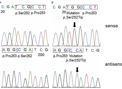
As no positive control containing the FGFR2 p.Pro253Arg mutation was available we aimed
the testing for the specificity and sensitivity of
our methodical approach before screening the tumor samples. For this reason we used DNA from the endometrium adenocarcinoma cell line MFE-280 with a known FGFR2 p.Ser252Trp mutation [13]. This mutation is adjacent to the FGFR2 p.Pro253Arg mutation reported in the bladder tumor from the patient with Apert Syndrome. The expected FGFR2 mutation could clearly be detected in the MFE-280 cells (Figure 1B) using our sequencing method. In
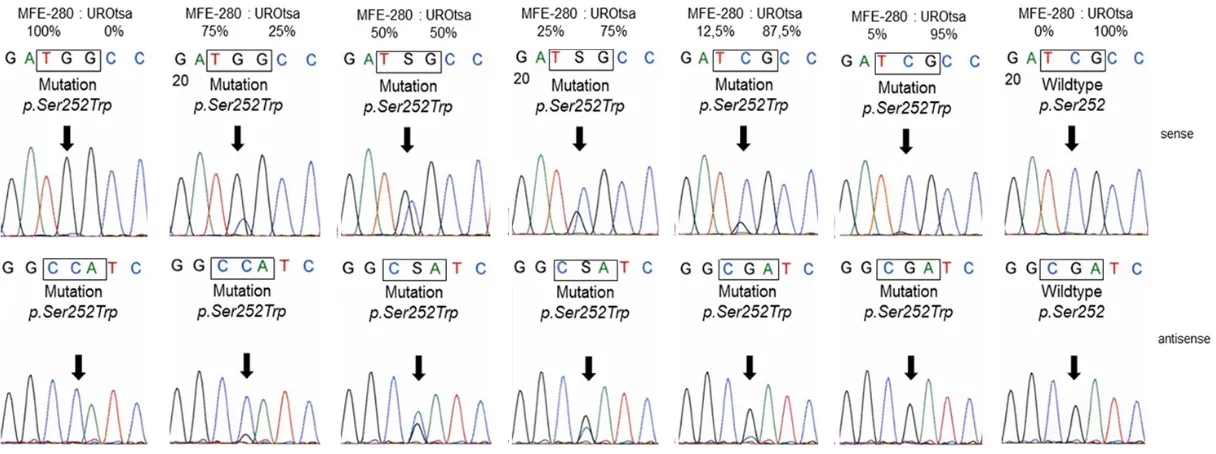
order to test the sensitivity of our method we spiked DNA from MFE-280 cells with DNA from the cell line UROtsa representing normal
uro-thelium without any alterations [14]. We were able to detect the FGFR2 p.Ser252Trp muta-tion in DNA from MFE-280 cells containing more than 85% wild type DNA from UROtsa cells (Figure 2).
FGFR2 exon 7 sequence analysis in bladder
tumors
All 254 bladder tumors could be analyzed suc -cessfully. Mutations in exon 7 of the FGFR2 gene were found in 0/254 tumors. All cases
showed a wild type sequence (Figure 1A).
Discussion
In the presented study we analyzed exon 7 of the FGFR2 gene to evaluate the frequency of
[image:3.612.94.509.70.372.2]mutations could be detected at all. These data indicate that exon 7 mutations play no role in bladder tumorigenesis in general.
One explanation for missing the mutation could be working with material highly contaminated with wild-type stromal cells. Great care was taken during microdissection for enrichment of tumor cells. In addition, testing the sensitivity of our method showed that a mutation could clearly be detected in a background of >80% wild type DNA. Therefore we do not think that methodical problems hampered our analysis.
FGFR2 mutations were frequently found in
other cancers (e.g. breast cancer, gastric can-cer, endometrial cancer) [15]. Most of these mutations clustered around the hinge region and the third Ig-like domain of FGFR2. Only in endometrial cancer FGFR2 mutations clus-tered within the kinase domain [16]. Therefore we did not expand our analyses to further exons as the probability for discovering mutations outside the known hot spots was low. Moreover, four studies presenting whole exome or whole
genome sequencing data from bladder tumors
were published most recently [17-20]. Overall 204 tumors were investigated in these studies. None of the tumors showed a FGFR2 mutation
which strengthens our findings.
The general role of FGFR2 in bladder cancer is still not clear. The splice variant FGFR2-IIIb is expressed in normal urothelium. About 30% of
analyzed tumors showed a decreased FGFR2
expression, and this decreased expression on both mRNA and protein level was associated
with poor prognosis (tumor-specific death) [21].
The chromosomal region of the FGFR2 gene
(chromosome 10q25.3-26) was described as affected by loss of heterozygosity in approx. 27% of analyzed cases without any correlation
to decreased FGFR2 expression. Re-expression of FGFR2 resulted in lower proliferation rates and a decreased tumorigenic potential in nude mice [9]. Based on these data FGFR2 appears to act as a tumor-suppressor with protective properties in bladder cancer but conclusive data are still missing.
In conclusion, the results of our study on FGFR2
exon 7 sequencing analysis in combination
with previously data suggest no role of muta-tional activation of FGFR2 in bladder carcino-genesis. Expression levels of FGFR2 might
influence both tumor aggressiveness and
pat-ient’s outcome but the underlying mechanisms
need to be clarified in more detail.
Acknowledgements
The present work was performed in fulfillment of the requirements for obtaining the degree
“Dr. med.” (M.D.). We acknowledge support by Deutsche Forschungsgemeinschaft and Fri- edrich-Alexander-University Erlangen-Nuremb- erg within the funding program Open Access Publishing. Verena Popp, Claudia Knoll, Nina Oks and Karina Dresel are thanked for excel-lent technical assistance.
Disclosure of conflict of interest
None.
Address correspondence to: Dr. Robert Stoehr, Institute of Pathology, University Hospital Erlangen, Friedrich-Alexander-University Erlangen-Nuremberg, Krankenhausstr. 8-10, 91054 Erlangen, Germany. Tel: +49-9131-8543610; Fax: +49-9131-8524745; E-mail: [email protected]
References
[1] Acevedo VD, Ittmann M, Spencer DM. Paths of FGFR-driven tumorigenesis. Cell Cycle 2009 Feb 15; 8: 580-588.
[2] Di Martino E, Tomlinson DC, Knowles MA. A de-cade of FGF receptor research in bladder can-cer: past, present, and future challenges. Adv Urol 2012 Jul 31; 2012: 429213.
[3] Tsimafeyeu I, Demidov L, Stepanova E, Wynn N, Ta H. Overexpression of fibroblast growth factor receptors FGFR1 and FGFR2 in renal cell carcinoma. Scand J Urol Nephrol 2011 Feb 18; 45: 190-195.
[4] Koufou S, Lunz JC, Borchardt A, Keck B, Kneitz B, Gaisa NT, Hafner C, Giedl C, Rau TT, Rogler A, Wieland WF, Hartmann A, Stoehr R. Muta-tional activation of FGFR3 is not involved in the development of prostate cancer. Pathobiology 2010 Nov 29; 77: 249-252.
[5] Stoehr CG, Stoehr R, Hartmann A, Hofstaedter F, Junker K, Blaszyk H, Wieland WF, Otto W, Denzinger S, Walter B. Mutational activation of FGFR3: no involvement in the development of renal cell carcinoma. J Cancer Res Clin Oncol 2012; 138: 359-361.
[7] Williams SV, Hurst CD, Knowles MA. Oncogenic FGFR3 gene fusions in bladder cancer. Hum Mol Genet 2013 Feb 15; 22: 795-803. [8] Andreou A, Lamy A, Layet V, Cailliez D, Gobet F,
Pfister C, Menard M, Frebourg T. Ealry-onset low-grade papillary carcinoma of the bladder associated with apert syndrome and a germ-line FGFR2 mutation (Pro253Arg). Am J Med Genet A 2006 Oct 15; 140: 2245-2247. [9] Ricol D, Capellen D, El Marjou A,
Gil-Diez-di-Medina S, Girault JM, Yoshida T, Ferry G, Tuck-er G, Poupon MF, Chopin D, ThiTuck-ery JP, Radvanyi F. Tumour suppressive properties of fibroblast growth factor receptor 2-IIIb in human bladder cancer. Oncogene 1999; 18: 7234-7243. [10] Mostofi FK, Davis CJJ and Sesterhenn IA. Histo
-logical typing of urinary bladder tumours. In: World Health Organization International Histo -logical Classification. New York: Springer, 1999.
[11] Sobin LH, Wittekind C. TNM Classification of Malignant Tumours. Hoboken, New Jersey: John Wiley & Sons, 2002.
[12] Stoehr R, Zietz S, Burger M, Filbeck T, Denz -inger S, Obermann EC, Hammerschmied C, Wieland WF, Knuechel R, Hartmann A. Dele-tions of chromosomes 9 and 8p in histologi-cally normal urothelium in patients with blad-der cancer. Eur Urol 2005; 47: 58-63.
[13] Krakstad C, Birkeland E, Seidel D, Kusonmano K, Petersen K, Mjos S, Hoivik EA, Wik E, Halle MK, Oyan AM, Kalland KH, Werner HMJ, Trovik J, Salvesen H. High-throughput mutation profil -ing or primary and metastatic endometrial cancers identifies KRAS, FGFR2 and PIK3CA to be frequently mutated. PLoS One 2012; 7: e52795.
[14] Petzoldt JL, Leigh IM, Duffy PG, Sexton C, Mas -ters JR. Immortalisation of human urothelial cells. Urol Res 1995; 23: 377-380.
[15] Katoh M. FGFR abnormalities underlie a spec-trum of bone, skin, and cancer pathologies. J Invest Dermatol 2009 Apr 29; 129: 1861-1867.
[16] Katoh M. Cancer genomics and genetics of FGFR2. Int J Oncol 2008; 33: 233-237. [17] Guo G, Sun X, Chen C, Wu S, Huang P, Li Z,
Dean M, Huang Y, Jia W, Zhou Q, Tang A, Yang Z, Li X, Song P, Zhao X, Ye R, Zhang S, Lin Z, Qi M, Wan S, Xie L, Fan F, Nickerson ML, Zou X, Hu X, Xing L, Lv Z, Mei H, Gao S, Liang C, Gao Z, Lu J, Yu Y, Liu C, Li L, Fang X, Jiang Z, Yang J, Li C, Zhao X, Chen J, Zhang F, Lai Y, Lin Z, Zhou F, Chen H, Chan HC, Tsang S, Theodorescu D, Li Y, Zhang X, Wang J, Yang H, Gui Y, Wang J, Cai Z. Whole-genome and whole-exome se-quencing of bladder cancer identifies frequent alterations in genes involved in sister chroma-tid cohesion and segregation. Nat Genet 2013; 45: 1459-1463.
[18] Balbás-Martínez C, Sagrera A, Carrillo-de-San -ta-Pau E, Earl J, Márquez M, Vazquez M, Lapi E, Castro-Giner F, Beltran S, Bayés M, Carrato A, Cigudosa JC, Domínguez O, Gut M, Herranz J, Juanpere N, Kogevinas M, Langa X, López-Knowles E, Lorente JA, Lloreta J, Pisano DG, Richart L, Rico D, Salgado RN, Tardón A, Cha-nock S, Heath S, Valencia A, Losada A, Gut I, Malats N, Real FX. Recurrent inactivation of STAG2 in bladder cancer is not associated with aneuploidy. Nat Genet 2013; 45: 1464-1469. [19] Ross JS, Wang K, Gay LM, Al-Rohil RN, Nazeer
T, Sheehan CE, Jennings TA, Otto GA, Donahue A, He J, Palmer G, Ali S, Nahas M, Young G, Labrecque E, Frampton G, Erlich R, Curran JA, Brennan K, Downing SR, Yelensky R, Lipson D, Hawryluk M, Miller VA, Stephens PJ. A high fre-quency of activating extracellular domain ERBB2 (HER2) mutations in micropapillary urothelial carcinoma. Clin Cancer Res 2014 Jan 1; 20: 68-75. DOI: 10.1158/1078-0432. CCR-13-1992.
[20] Gui Y, Guo G, Huang Y, Hu X, Tang A, Gao S, Wu R, Chen C, Li X, Zhou L, He M, Li Z, Sun X, Jia W, Chen J, Yang S, Zhou F, Zhao X, Wan S, Ye R, Liang C, Liu Z, Huang P, Liu C, Jiang H, Wang Y, Zheng H, Sun L, Liu X, Jiang Z, Feng D, Chen J, Wu S, Zou J, Zhang Z, Yang R, Zhao J, Xu C, Yin W, Guan Z, Ye J, Zhang H, Li J, Kristiansen K, Nickerson ML, Theodorescu D, Li Y, Zhang X, Li S, Wang J, Yang H, Wang J, Cai Z. Frequent mu -tations of chromatin remodelling genes in tran-sitional cell carcinoma of the bladder. Nat Genet 2011; 43: 875-879.