MECHANISMS REGULATING BONE RESORPTION
IN VITRO
A thesis submitted in partial fulfilment of the degree of Doctor of Philosophy, University of London.
Peter Alexander Hill
B.Sc.,M.Sc.,B.D.S.,F.D.S.R.C.PS., D.D.O., M.Orth.R.C.S.
1994
Department of Cell and Molecular Biology Strangeways Research Laboratory
Worts Causeway Cambridge
and
Department of Orthodontics Institute of Dental Surgery
Eastman Dental Hospital Gray's Inn Road
ProQuest Number: U539184
All rights reserved
INFORMATION TO ALL USERS
The quality of this reproduction is dependent upon the quality of the copy submitted. In the unlikely event that the author did not send a complete manuscript and there are missing pages, these will be noted. Also, if material had to be removed,
a note will indicate the deletion.
uest.
ProQuest U539184
Published by ProQuest LLC(2016). Copyright of the Dissertation is held by the Author. All rights reserved.
This work is protected against unauthorized copying under Title 17, United States Code. Microform Edition © ProQuest LLC.
ProQuest LLC
789 East Eisenhower Parkway P.O. Box 1346
ABSTRACT
This thesis encompasses two separate aspects of the biology of osteoclasts. Firstly, proteins were purified from bone matrix that modulate osteoclast formation and function. The effects of two of the most abundant growth factors insulin-like growth factors-l (IGF-I) and -II (IGF-II) on osteoclast differentiation and function were also studied. The aim of the second area of research was to determine the roles of individual members of the cysteine proteinase (CP) and matrix metalloproteinase (MMP) family on bone resorption using selective enzyme inhibitors.
Bone matrix is a rich source of polypeptide growth factors and evidence suggests that osteoclasts do not mature functionally until they reach bone. Since bone resorption occurs at discrete sites throughout the skeleton, it is likely that an osteoclast activating factor(s) could be sequestrated in bone matrix where it may regulate osteoclast formation from haemopoietic precursors.
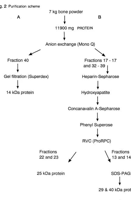
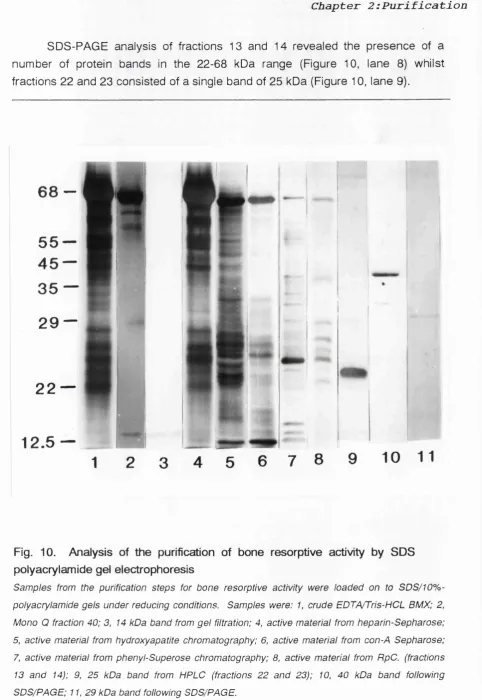
Matrix proteins were extracted from bovine cortical bone with EDTA/Tris-HGI under non-dissociative conditions at neutral pH. Four distinct bone resorptive proteins of 14, 25, 29, and 40 kDa were purified and partially characterized using
an in vitro neonatal calvarial assay. The 14 and 25 kDa proteins possessed
insulin-like growth factor (IGF) and transforming growth factor- 6 activity respectively, whilst the 40 kDa protein enhanced the formation of osteoclast-like cells in a mouse bone marrow àssay. The 29 kDa protein stimulated bone resorption in an isolated osteoclast assay, suggesting a direct action on osteoclasts. This is the first report of (i) a protein isolated from bone matrix that stimulates osteoclast differentiation and (ii) a protein that directly stimulates osteoclast function.
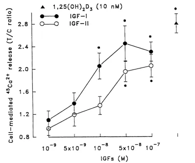
IGF-I and -II were found to enhance both osteoclast differentiation and function but they were less effective than 1,25 Dihydroxyvitamin D3. The stimulatory effects of IGFs on osteoclast activity were mediated via an interaction with the type I IGF receptor present on osteoblastic cells.
Osteoclasts solubilize mineralized bone by means of acid pH generation and proteinase action, notably enzymes of the métallo and cysteine classes. To determine the roles and sites of action of the individual proteinases in bone resorption both natural and synthetic enzyme inhibitors were utilized.
both calvarial and osteoclast lacunar resorption, whilst CA074 was without effect. These results suggest that cathepsin L and/or S act extracellularly and possibly intracellularly whilst cathepsin B acts intracellularly perhaps through the activation of other proteinases involved in bone collagen degradation.
Six MMP inhibitors were used: initially the tissue inhibitors of metalloproteinases (TIMP) -1 and -2 were tested followed by four synthetic inhibitors. These were CT435, a general MMP inhibitor; CT543 and CT1166, compounds that are concentration dependent selective inhibitors of gelatinases-A and -B; and Ro 31-7467, a concentration-dependent selective inhibitor of collagenase. All the compounds produced a dose-dependent inhibition in calvarial bone resorption. However, CT1166 and C T543 only produced a 40% inhibition and Ro 31-7467 a 50% inhibition at concentration selective for the inhibition of gelatinase and collagenase respectively. The compounds also produced a partial reduction in the resorptive activity of interleukin-1 -stimulated osteoclast cultures.
ACKNOWLEDGMENTS
I am indebted to Professor M.C.Meikle, Dr J.J.Reynolds and Dr G.Murphy for allowing me to carry out this research at Strangeways Research Laboratory and for their guidance throughout the project, I am also grateful to the M RC for the Clinical Training Fellowship award. I would like to thank all the members of the Cell and Molecular Biology Department for their continued help and advice in particular Gary Dew for general scientific advice, Robin Ward for my initiation in protein purification and Barry Halls and Christine Riches for their unstinting work in the animal house. Thanks are extended to Professors A.Boyde and S.J.Jones and their team at UCH (introduction to isolated osteoclast assay and use of their Confocal Laser Microscope), Dr A.Docherty, Celltech and Dr K.Bottomley, Roche Industries (MMP inhibitors), and Dr D.Buttle, SRL for CP inhibitors and Dr J.Compston for the Image Analysis facility.
ABBREVIATIONS
aM E M alpha-Minimal Essential Medium
BSA Bovine Serum Albumin
BGJ Biggers Gwatkin Judah
cAMP cyclic Adenosine 3'5'-Monophosphate
ConA Concanavalin-A
CSF Colony Stimulating Factor
DMEM Dulbecco's Modified Eagle's Medium
DPM Disintegrations Per Minute
EDTA Ethylene Diamine Tetra-acetic Acid
PCS Fetal Calf Serum
FPLC Fast Performance Liquid Chromatography
igG Immunoglobulin G
IL-1a Interleukin-1 alpha
LDH Lactate Dehydrogenase
M MP Matrix Metalloproteinase
MNC Multinucleated Cell
PAGE Polyacrylamide Gel Electrophoresis
PBS Phosphate Buffered Saline
PTH Parathyroid Hormone
SDS Sodium Dodecyl Sulphate
TIM P Tissue Inhibitor of Metalloproteinases
TAB Tris Azide Buffer
TRAP Tartrate Resistant Acid Phosphatase
LIST OF CONTENTS
ABSTRACT 2
Acknowledgments 4
Abbreviations 5
CHAPTER 1 11
GENERAL IN TR O D U C TIO N 11
OSTEO CLAST FORM ATIO N 14
M ECHANISM OF BONE R ESO RPTIO N 15
Mineral Dissolution 16
Organic Matrix Degradation 17
Lysosomal cysteine proteinase 17
Matrix metalloproteinases 18
SYSTE M IC FACTORS INFLUENCING OSTEOCLAST ACTIVITY 22
Parathyroid hormone 22
1,25 dihydroxyvitamin D3 23
Calcitonin 24
LOCAL FACTORS INFLUENCING OSTEOCLAST A CTIVITY 25
Prostaglandins 25
Interleukin-1 25
Tumour necrosis factor 26
Interleukin-6 26
Interferon-gamma 27
Leukemia inhibitory factor 27
Transforming growth factors 27
Fibroblast growth factors 28
Platelet-derived growth factor 28
Insulin-like growth factors 29
Reactive oxygen species and Nitric oxide 29
Colony stimulating factors 30
Integrins 34
M ETHODS FOR STUDYING BONE RESORPTIO N 37
Bone organ cultures 37
Isolated osteoclast assay 37
Use of monocyte-macrophages as osteoclast surrogates 39
Human osteoclasts 39
Assays for osteoclast formation 39
CHAPTER 2: Purification of bone matix polypeptides from bone matrix 42
Materials and Methods 43
Results 49
Discussion 64
CHAPTER 3: IGF-I and -II stimulate osteoclast formation and function 67 via the type I IG F receptor
Materials and Methods 68
Results 71
Discussion 79
C HAPTER 4: Inhibition of bone resorption by cysteine proteinase inhibitors 83
Materials and Methods 84
Results 87
Discussion 98
C HAPTER 5: Inhibition of bone resorption by TIMP-1 and TIM P -2 100
Materials and Methods 101
Results 101
Discussion 107
CHAPTER 6 : Inhibition of bone resorption by selective inhibitors 109
of collagenase and gelatinase
Materials and Methods 110
Results 114
Discussion 123
CHAPTER 7: Inhibition of bone resorption by selective M M P inhibitors 126 and immunolocalization of MMPs and TIMP-1 in isolated osteoclasts
Materials and Methods 127
Results 131
Discussion 141
CHAPTER 8 : Directions for future research 147
R EFE R E N C E S 147
LIST OF TABLES
Table 1 Table 2 Table 3 Table 4 Table 5
Table 6
Table 7 Table 8
Table 9
Table 10
Table 11
Table 12
Table 13
Table 14
Table 15 Table 16
Table 17
Table 18 Table 19
Table 20 Table 21 Table 22
20
36 55 57 61 Main subgroups of matrix metalloproteinasesModulators of bone resorption
Purification of bone resorptive activity from bone matrix Purification of growth factor activity from bone matrix Effect of 29 kDa protein on lacunar resorption by isolated osteoclasts
Effect of 40 kDa protein on (A) the formation of TRAP-positive MNCs and (B) lacunar resorption in an osteoclast assay Effects of IGF-I and IGF-II on osteoclast lacunar resorption Effects of IGF-I and IGF-II on lacunar resorption by osteoclasts co-cultured with osteoblasts
Recovery from the inhibitory effects of CP inactivators on PTH-stimulated release of "^^Ca from mouse calvarial bones. Effects of CP inactivators on pH]-thymidine uptake into DMA pH]-proline incorporation into proteins in calvarial bones
Effect of CP inactivators on the release of 8 -glucuronidase, N-acetyl B-glucosaminidase and LDH from calvarial bones Effects of CP inactivators on the surface area and volume of osteoclast resorption lacunae
Effects of CP inactivators on bone resorption using an in vivo/ in vitro assay
Recovery from the inhibitory effects of TIMP-1 and T IM P -2 on stimulated bone resorption in calvarial bones
Effects of TIMP-1 and TIM P -2 on DMA and protein synthesis Effects of TIMP-1 and TIM P -2 on release of 8-glucuronidase and LDH from calvarial bones
Recovery from the inhibitory effects of C T 1 166 and Ro 31 -7467 on stimulated bone resorption
Effects of C T 1 166 and Ro 31 -7467 on DMA and protein synthesis 117 Effects of C T 1 166 and Ro 31 -7467 on glycolysis in rat osteoclast 121 and mouse osteoblast cultures
Recovery from the inhibitory effects of Ct435 and C T543 on stimulated bone resorption
Effects of CT435 and CT543 on DMA and protein synthesis Effects of CT435 and CT543 on the release of "^^Ca^+ from osteoid-free calvarial bones by chicken osteoclasts
List of Figures
Figure 1 Hypothetical pathways of osteoclast formation 31
Figure 2 Purification scheme for the isolation of bone resorptive 50 proteins
Figure 3 Mono Q anion-exchange chromatography of BMX 51
Figure 4 Gel filtration of fraction 40 from Mono Q chromatography 52
Figure 5 Heparin-Sepharose chromatography of BMX 53
Figure 6 Hydroxyapatite chromatography of BMX 54
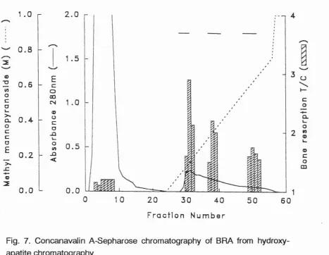
Figure 7 Concanavalin A-Sepharose of BMX 56
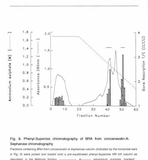
Figure 8 Phenyl-Superose chromatography of BMX 58
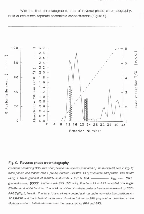
Figure 9 Reverse phase chromatography of BMX 59
Figure 10 Analysis of the purification of BRA by 60
SDS PAGE
Figure 11 Effects of IGF-I and IGF-II on ^^Ca^+ release
from calvariae 71
Figure 12 Effects of IGF-I and IGF-II on rat osteoclast 73 lacunar resorption after a prolonged sedimentation time
Figure 13 Effect of alR -3 on IGF-I and IGF-II stimulated 75 lacunar resorption by osteoclast-osteoblast co-cultures
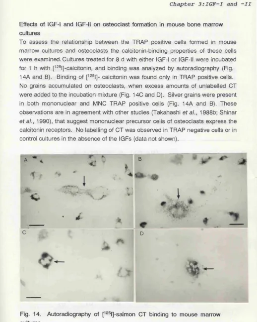
Figure 14 Autoradiography of p^®l]-salmon CT binding to
mouse marrow culures 76
Figure 15 Effects of IGF-I and IGF-II on the formation of 77 TRAP positive MNCs
Figure 16 Scanning electron micrograph of resorption lacunae on 78 ivory by mouse marrow cells cultured with IGF-II
Figure 17 Structures of the CP inactivators 87
Figure 18 Effects of the CP inactivators on basal and stimulated 88 "^^Ca^+ and pH]-proline release from calvarial bones
Figure 19 Effects of CP inactivators at different concentrations on 89 stimulated release of pH]-proline from calvariae
Figure 20 Time-course of the effects of the CP inactivators on bone 90 resorption in calvarial explants
Figure 21 Scanning electron micrograph of (A) osteoclast lacunae 93 and (B) a chicken osteoclast
Figure 22 Effects of the CP inactivators on osteoclast pit formation 94
Figure 23 Effects of the CP inactivators on the volume of 96
Figure 24 Effects of TIMP-1 a n d -2 on basal and stimulated 102 release of "^^Ca^+ and pH]-proline from calvariae
Figure 25 Effects of TIMP-1 and -2 at different concentrations on 103 "^^Ca^+ release from calvariae
Figure 26 Time course of the effects of TIMP-1 and -2 on 104
"^^Ca^+ release from calvarial bones
Figure 27 Structures of C T 1166 and Ro 31 -7467 114
Figure 28 Effects of C T 1166 and Ro 31 -7467 at different concentrations 115 on stimulated releases of p-H]-proline from calvarial bones
Figure 29 Time course of the effects of C T1166 and Ro 31 -7467 on 116 pH]-proline releases from calvarial bones
Figure 30 . Effects of C T 1166 and Ro 31 -7467 on the degradation of 118 type I collagen films by mouse osteoblasts
Figure 31 Effects of C T 1166 and Ro 31 -7467 on rat osteoclast pit 119 number
Figure 32 Effects of C T 1166 and Ro 31 -7467 on rat osteoclast pit 120 volume
Figure 33 Zymogen analysis of ivory and calvarial bone extracts 122
Figure 34 Structures of CT435 and CT543 131
Figure 35 Effects of CT435 and CT543 at different concentrations 132
on stimulated release of "^^Ca^+ from calvarial bones
Figure 36 Effects of CT435 and CT543 on basal and stimulated release of 133 "^^Ca^+ and pH]-proline release from calvariae
Figure 37 Effects of CT435 and CT543 on rat osteoclast lacunar resorption 137
Figure 38 Immunolocalization of MMPs and TIMP-1 in isolated 139
Chapter 1 :
Introduction
CHAPTER 1.
GENERAL INTRODUCTION
Bone is a highly specialized form of connective tissue the functions of which are (a) structural, providing a rigid internal skeleton due to the tensile strength of collagen and the compressive strength of hydroxyapatite and (b) physiological, playing a role in calcium and phosphate homeostasis.
At the macroscopic level there are two major forms of bone; compact or cortical bone, and trabecular, or cancellous bone. The former is found in the diaphyses of the long bones, which surround the marrow cavities whilst spongy bone is limited to the metaphyseal region of long bones and within the confines of the flat bones and vertebrae. Cortical bone appears dense with smooth periosteal and endosteal surfaces. The cancellous bone comprises networks of fine interlacing bone spicules, known as trabeculae giving it a spongy appearance. The trabeculae follow a definite pattern dictated by the mechanical stresses placed upon the bone in situ.
At the microscopic level two bone types can be distinguished: woven and lamellar. Woven bone is the first to appear in embryonic development or in the repair of fractures, it is composed of loosely and randomly arranged coarse collagen bundles. It is usually more cellular, and the osteocytes which lie in lacunae are large and irregular in shape and size. Immature bone is usually replaced by lamellar bone. The latter bone is characterized by a highly ordered arrangement of both its collagen bundles and cells. Lamellar bone is laid down in concentric layers on the periosteal and endosteal surfaces. Internal to the lamellar bone, Haversian systems predominate. The blood vessels that nourish bone run in the canals of the Haversian systems and these are surrounded by a number of osteocytes which communicate with each other through cytoplasmic processes within canaliculi. The surfaces of bone are covered with non-mineralized layers of connective tissue called periosteum on the external surface and endosteum on the internal surface i.e. lining both the marrow cavities and Haversian canals.
Chapter 1 :
Introduction
of 33 KDa is the most abundant non-collagenous organic component of bone. It may have a role to play in the mineralization process since it binds calcium (Bolander et al., 1988). Osteocalcin a 5.8 KDa protein is secreted by osteoblasts, odontoblasts and chondrocytes (Owen et al., 1990; Li an et a!., 1993). Osteocalcin binds tightly to hydoxyapatite via carboxy g I utam i c acid residues and may regulate crystal growth. Osteopontin and bone sialoprotein are phosphorylated
calcium binding proteins of 32 and 33 KDa respectively. They contain the tripeptide sequences Arg-Gly-Asp (ROD) typically found in proteins with cell binding properties via a class of receptors known as integrins (Hynes, 1992). Osteopontin synthesis is stimulated by the bone resorptive hormone 1,25 dihydroxyvitamin D3 (Oldeberg et al., 1989) and osteopontin may assist in the resorptive process by anchoring osteoclasts to bone via vitronectin receptors (Reinholt ef a/., 1990).
There are four different types of cells in bone. Osteoblasts, lining cells and osteoclasts are present on bone surfaces and osteocytes in the interior. Osteoclast progenitors are mononuclear cells which arise in haemopoietic tissue and travel to the bone microenvironment via the circulation, where they differentiate and fuse to form osteoclasts. Osteoblasts, lining cells and osteocytes are derived from local mesenchymal cells called osteoprogenitor cells (Marks and Popoff, 1988).
Osteoblasts are plump lining cells characteristically found in a layer one cell thick closely applied to the surface of developing bone, or in Haversian systems, the site of bone remodelling. The active osteoblast is columnar in shape (20-30 /L/m) with the nucleus at the opposing end of the cell to the bone surface. Osteoblasts are characterized by extensive rough endoplasmic reticulum (RER) and mitochondria whilst the Golgi apparatus is poorly developed (Scott, 1967). The cell displays an irregular contour due to the large number of cytoplasmic processes that terminate on the bone surface (Scott, 1967) or penetrate the osteoid layer (Dudley and Spiro, 1961). Osteoblasts synthesize and secrete type I collagen, proteoglycan, cytokines such as transforming growth factor and glycoproteins into a region of unmineralized matrix (osteoid) between the cell body and mineralized matrix. Some active osteoblasts become surrounded by matrix and are then transformed into osteoid osteocytes.
Chapter 1 : Introduction
apparatus. The osteocytes lie within separate lacunae within the bone and their network of interlacing processes pass through the bone in small canaliculi. This extensive syncytium facilitates communication between the osteocytes and cells lining the bone surface.
Osteoclasts are the principal resorptive cells of the skeleton being typically large (up to 200,000 ^m^) and multinucleate (2-100 nuclei). Transmission electron micrographs show osteoclasts to possess several distinctive features (Holtrop and King, 1977). They have an extensive Golgi apparatus but little endoplasmic reticulum. There are many mitochondria and lysosomes but few ribosomes. Osteoclasts are usually found closely apposed to bone surfaces. The cell creates an acidic environment adjacent to the bone surface by which mineral dissolution occurs. This results in the formation of small areas of resorption called Howship's lacunae. The cell surface associated with bone has several distinctive features; the plasma membrane is apposed tightly onto the bone surface and adjacent to it the cytoplasm is seen to be free of organelles but rich in actin filaments; this is termed the clear zone. Immunofluorescent microscopy has shown the presence of microfilament-associated proteins such as vinculin, actinin and fimbrin protruding from the cell onto its substratum (Marchisio et al., 1984). Between clear zones the cell membrane is highly invaginated and many cytoplasmic projections are formed with narrow channels passing between them. This is the so-called ruffled border, the resorbing apparatus of the cell. It represents an extensive area of cell surface where secretion of enzymes and uptake of matrix components occcurs. The clear zone encircles the ruffled border completely so that the site of resorption is isolated and localized. Osteoclasts are highly motile cells, with their cytoplasm advancing behind broad pseudopodial processes (Chambers and Magnus, 1982).
Bone lining cells are flat, thin, elongated cells covering most bone surfaces in the adult skeleton. They have few organelles, are inactive and joined to each other and neighbouring osteocytes by gap junctions. Little is known of their function although they may be a source of osteoblast progenitors, serve as a selective barrier between bone and osteoclasts, and/or regulate crystal growth in bone. Periosteal osteoblasts are responsible for appositional growth of bone. In mature bone they are called osteoprogenitor cells and are normally inactive although they can become active for instance during fracture callus formation.
Chapter 1 : Introduction
Osteoclast Formation
It is now generally accepted that osteoclasts originate from haemopoietic precursors. Firstly, experiments with parabiotic rodents have demonstrated osteoclast formation from circulating cells in fracture callus (Gothlin and Erickson, 1973) and reversal of osteopetrosis in mice by parabiotic union with normal siblings (Walker, 1973). Secondly, transplantation of haemopoietic tissue into lethally irradiated recipients (Walker, 1975a and b) and ia osteopetrotic rats (Marks, 1976) cured their resorptive defects. Thirdly, quail-chick chimera studies, where embryonic bone rudiments were grafted onto the chorioallantoic membrane demonstrated recruitment of host-derived cells (Kahn and Simmons, 1975; Jotereau and Le Dourain, 1978; Simmons and Kahn, 1979). Fourthly, transplanta--tion of cells containing markers showed that osteopetrosis was cured by formation of donor derived osteoclasts rather than the transplanted cells releasing a factor which permitted normal differentiation of osteopetrotic osteoclasts (Coccia
et al., 1980; Sorell et a!., 1981).Finally, the formation of osteoclasts from
haemopoietic stem cell populations confirmed their haemopoietic origin (Scheven
eta !., 1986; Hagenaars eta!., 1989).
There is strong evidence to suggest that osteoclasts form by fusion of mononuclear precursors rather than mitotic division of nuclei (Young, 1962; Feldman et a!., 1980; Baron and Vignery, 1981 ; Ibbotson et a!., 1984; MacDonald
et a/., 1986a). The precise identity of the mononuclear progenitor has not been established. Based upon the following evidence it is believed that the initial pathway of differentiation for osteoclasts is identical to the mononuclear phagocyte (MNP; monocyte/macrophage) lineage but diverges from it during the latter stages of differentiation. The classic experiments of Fischman and Hay (1962) showed the formation of osteoclasts from labelled leucocytes in regenerating newt limbs and Jee and Nolan (1963) found that osteoclasts contained charcoal particles once they had been ingested by macrophages. The infusion of ^H-thymidine labelled peripheral blood monocytes into syngeneic recipients resulted in the formation of osteoclasts with the same label (Tinkler et a!., 1981a) and monocytes have been shown to fuse with purified osteoclasts in vitro (Zambonin-Zallone, 1984). More recently Burger et a!., (1984) reported that immature monocytes cultured with fetal bone rudiments formed osteoclasts in vitro.
Chapter 1 :
Introduction
cells {Burmester ef a/., 1983; Oursler ef a/., 1985; Nijweide ef a/., 1985; Athanasou
e ta l., 1986).
However several lines of evidence also suggest that osteoclasts are not derived from the MNP lineage. Firstly, monoclonal antibodies prepared against human osteoclastomas do not cross-react with monocytes/macrophages (Horton
et al., 1985a). Secondly, osteoclasts fail to express many markers of mature
monocytes/macrophages including Fc and C3b rceptors (Shapiro et a/., 1979; Boyde and Jones, 1987) and MHC class II determinants (HLA-DR), CD 11, CD 14, F4/80 (mouse), Mac-1 (Horton et a!., 1985b, 1985c). Thirdly, macrophages are incapable of restoring the reduction in osteoclast numbers induced by radiation (Anderson et a!., 1979) and although isolated osteoclasts form resorption lacunae on bone slices within a few hours (Chambers et a!., 1984c) macrophages /monocytes are without effect (Chambers and Horton, 1984a; Ali et a!., 1984). In addition calcitonin (CT)-receptors expressed in mammalian osteoclasts (Nicholson
et a!., 1986) correlate with their resorptive activity (Hattersley and Chambers;
1989a). CT receptors are also expressed by umbilical cord monocytes (Mbalaviele and de Vernejout, 1989) but CT is without effect on macrophage polykaryons (Chambers and Magnus, 1982; Takahashi eta l., 1989).
Overall this evidence suggests that osteoclasts and monocytes/ macrophages may be derived from a common progenitor (Burger et al., 1982; Nijweide et al., 1986). The precise nature of the osteoclast stem cell in relation to the piuripotential haemopoietic stem cell (CFU-S) and osteoclast progenitor cells in relation to MNP progenitor cells remains unknown. It has been suggested that the osteoclast might derive from a lineage unrelated to the MNP lineage (Loutit and Nisbett, 1982; Chambers, 1989).
Mechanism of Bone Resorption
Although it has been suggested that osteoclastic bone resorption may be limited to mineral dissolution, activated osteoclasts are also capable of resorbing the organic matrix of bone, even if other cells co-operate in its removal (reviewed by Vaes, 1988). Bone resorption is accomplished through the formation by the osteoclast of a sealing zone of close adhesion between the osteoclast ruffled border and bone matrix. Special adhesive structures involving the vitronectin receptor (see later) on osteoclasts and RGD peptide sequence-containing bone matrix proteins are probably involved in this process (Zambonin-Zallone et al.,
Chapter 1 :
Introduction
Mineral Dissolution
Vaes (1968) suggested that osteoclasts resorb the mineral phase of bone by acidifying an extracellular compartment, however, only recently has this been confirmed with the use of acridine orange (Baron at al., 1985; Green at a!., 1986; Anderson
at a!., 1986). This fluorescent dye is a weak base, and its unprotonated form
crosses biological membranes whilst the charged form accumulates in acidic compartments. Thus in organ cultures of bone, acridine orange is concentrated within the SORZ, suggesting that the osteoclast actively acidifies this compartment. Since osteoclasts detached from bone fail to stain with the dye, this supports an extracellular rather than intracellular accumulation (Baron at a/., 1985). Furthermore osteoclast acidification is altered by agents influencing bone resorption, such as parathyroid hormone, PGEg, and CT (Anderson at a!., 1986). These studies have been supported by direct pH measurement within the SO R Z indicating values of 4.5 to 4.8 (Silver at al., 1988) which returned toward neutrality under the action of calcitonin (Fallon, 1984). The mechanism of acidification of this extracellular compartment is currently under investigation. Baron at al. (1986) have indicated that the apical membrane at the 'ruffled border' shares common antigenic determinants with lysosomal membranes, including proton pumps that may result in the acidification of the SORZ. Whilst 3 separate proton pumps have been implicated in the acidification process, evidence suggests that a Na+-H+ exchange mechanism may also be involved (Hall and Chambers, 1990).
Regarding the proton pump, evidence suggests that a H+-ATPase is involved (Akiska and Gay, 1986) but the distinction between a plasma membrane (P-type) and vacuolar (V-type) type remains uncertain. Immunocytochemical studies have noted similarities between osteoclast pump(s) and gastric mucosal and kidney H+-K+-ATPase (Baron at al., 1986), but provided no distinction between a P or V-type pump. Although Parvinen at al. (1987) provided evidence for a membrane type H+-K+-ATPase, more recent immunocytochemical data suggests that a vacuolar K+-ATPase is involved (Ghiselli at al., 1987; Blair at al.,
1989). It has also been proposed that Na+-K+-ATPase is involved in the transport of protons against concentration gradients (Paradiso at a t, 1981). This hypothesis was supported by the finding that amiloride, which blocks Na+-K+-ATPase also prevents PTH-induced acidification in organ cultures (Anderson at al., 1986) and bone resorption by isolated osteoclasts (Hall and Chambers, 1989; 1990).
Chapter 1 : Introduction
membrane.
Finally, immunocytochemical studies have also demonstrated the presence of carbonic anhydrase in osteoclasts (Anderson et al., 1982; Karkukorpi, 1991) but not in osteoblasts (Vaananen, 1984). This enzyme has been localized to the plasma membrane of the 'ruffled border' (Anderson et a!., 1982) and it supplies H+ ions to the proton pump by catalyzing the conversion of CO2 to HCO3' and H+. The role of carbonic anhydrase in bone resorption has been confirmed by the use of inhibitors of the enzyme such as acetazolamide both in vivo (Kenny, 1985) and in vitro (Hall and Kenny, 1985; 1986).
Organic Matrix Degradation by Proteinase Activity
The degradation of the organic matrix follows the removal of mineral from bone. Proteolytic enzymes are released which are responsible for degrading the organic matrix which consists mainly of type I collagen. These enzymes are believed to belong to two major classes: cysteine endopeptidases (CPs) and matrix metalloproteinases (MMPs).
Lysosomal Cysteine Proteinases: These enzymes exhibit optimal activity at the pH that exists at the level of the subosteoclastic resorption zone and lysosomal enzymes are secreted at that level by osteoclasts (Baron et a/., 1985; 1988). Evidence supports the view that these osteoclast-derived lysosomal enzymes are involved in degrading the organic matrix of bone (Délaissé et a!., 1980, 1984) and this occurs at the level of the subosteoclastic resorption zone (Everts et al., 1988; 1992). There are five distinct lysosomal CPs: cathepsin B,L,S,H, and N that are irreversibly denatured at pH values above 7.
Cathepsin B- This is the most abundant of the lysosomal CPs. It is capable of degrading collagen types I (Burleigh et al., 1974) II,IX and XI (Maciewicz et al.,
1991). The enzyme initially attacks the non-helical region of the type I collagen molecules, thus removing the cross-link. The helical portion of the molecule then becomes susceptible and is degraded to small peptides. The pH optimum for the degradation of soluble collagen is 4.5.
The biosynthesis of cathepsin B involves the conversion of the proenzyme (Chan et al., 1986) of Mr 36,000 to the mature 27,000 Mr form found in lysosomes. About 5% of the newly synthesized protein is secreted by fibroblasts in culture as the proenzyme (Hanewinkel e ta l., 1987). In contrast to the mature form of cathepsin B the proenzyme is stable at alkaline pH (Barrett, 1973).
Chapter 1 :
Introduction
action on collagen is qualitatively similar to that of cathepsin B, with the main effect being cleavage of the non-helical regions resulting in removal of the cross
links (Kirschke et al., 1982)
Cathepsin L is synthesized as a proenzyme, processed and delivered to the lysosome (Portnoy et a!., 1986). A proportion of the proenzyme may be secreted and is activated at pH 3.0. Similar to pro-cathepsin B, the presence of the propeptide confers stability on the protein at alkaline pH (Mason et a!., 1987) whilst the mature form of the enzyme is unstable at pH 7.0.
Cathepsin 8 - Cathepsin 3 was recently shown to be distinct from cathepsin L (Kirschke et a!., 1986). Until now it has only been detected in lymphoid tissue, kidney and lung (Kirschke et a/., 1989), but wider distribution for this enzyme seems likely. The specificity of cathepsin S is similar to but not identical with that of cathepsin L. Against insoluble type I collagen, however, it is only about one-third as active at pH 3.5 (Kirschke et a!., 1989), which is similar to cathepsin B. Cathepsin 8 differs from other lysosomal CPs in being much more stable at mildly alkaline pH. Only 10 percent of activity is lost following preincubation for 1 h at pH 7.5 (Kirschke e ta l., 1989).
Cathepsin H- The human enzyme appears to have weak endopeptidase activity, with no action on collagen (Kirschke et al., 1980). The mature lysosomal form of cathepsin H may be more tolerant of alkaline pH than either cathepsin L or B (Schwartz and Barrett, 1980) with the enzyme retaining 20 percent of its activity at pH 7.5 for 60 min. Cathepsin H is synthesized as a proenzyme (Ishido e ta l., 1987) and is processed in the normal manner (Nishimura and Kato, 1987).
Cathepsin N- Less information is available on cathepsin N (Kirschke and Barrett, 1987), however, the enzyme is active aginst type I collagen and gelatin (Maciewicz and Etherington, 1988).
Endogenous inhibitors of lysosomal CPs: The endogenous inhibitors of the lysosomal CPs are a 2-macroglobulin (02-M) and members of the cystatin family. Q2-M provides protection against inappropriate proteolysis in the bloodstream by irreversibly binding to CPs (Mason, 1989). The cystatin family consists of a large number of tight binding reversible inhibitors of lysosomal CPs. One of these cystatin C, which has been sequenced and cloned (Abrahamson et al., 1987) is a potent inhibitor of bone resorption in vitro (Lerner and Grubb, 1992).
Chapter 1 :
Introduction
(Swann et al., 1981; Whitham et al., 1986). The best studied examples of MMPs are the collagenases, stromelysins, and gelatinases. These MMPs are secreted by cells as proenzymes that must be activated by proteolytic cleavage. Their activities are controlled, in part, by the tissue inhibitors of metalloproteinases (TIMPs). This is multigene family of which TIMP-1 (Docherty et al., 1985) and T IM P -2 (Stetler-Stevenson, 1990) are well characterized.
Collagenases- There are two members of the collagenase subclass that cleave fibrillar collagens: interstitial collagenase (MMP-1) and neutrophil collagenase
(MMP-8). The protein cores of the two enzymes are of similar size, but neutrophil collagenase is more heavily glycosylated and has a larger Mr than interstitial collagenase (Mr 75,000 vs. 57,000).
The two collagenases differ markedly in terms of transcriptional regulation. Neutrophil collagenase is released instantly from granule storage sites of triggered neutrophils, in contrast interstitial collagenase is not stored in cells but produced on demand by initiating transcription of the gene (for instance by the action of growth factors or cytokines). The accumulation of the enzyme at the local site is delayed by 6 to 12 h.
Both enzymes have the distinctive ability to cleave a-chains of type I, II and III collagens at a single site, the Glyyys-lleyye or Glyyyg-Leuyyy bonds (Gross et al., 1974; Miller ef a/., 1976). This cleavage results in fragments of approximately three-quarters and one-quarter the length of the original molecule. Although these enzymes hydrolyze other substrates in addition to fibrillar collagens, the cleavage of intact, fibrillar collagens is specifically limited to these enzymes. Once the higher order structure is destroyed, however, many proteases, including other MMPs, can degrade the denatured collagen/gelatin substrate (reviewed by Birkedal-Hansen et ai., 1993). The two enzymes differ slightly in their specificity for fibrillar collagens; neutrophil collagenase has preference for type I collagen; while type III collagen is preferentially digested by interstitial collagenase (Mallya
et a!., 1990). The expression of neutrophil collagenase is restricted to cells of the
neutrophil lineage (Murphy ef a/., 1980; Hasty ef a/., 1990).
Chapter 1 :
Introduction
TABLE 1
Main Subgroups of the Matrix Metalloproteinases
ENZYME
Mr(kDa)
Latent Active
SUBSTRATES
Interstitial collagenase 55 MMP-1
45 Collagen types: 1,11,111,
VII,X.
Gelatin (limited). Proteoglcan core protein (limited).
PMN collagenase M M P-8
75 58 Collagen types:!,II,III.
Gelatin (limited). Proteoglycan core protein (limited).
Gelatinase A M M P-2
72
66
Collagen types: IV,V,VII,XI.
Gelatin,elastin and fibronectin.
Gelatinase B M MP-9
95
88
as for gelatinase AStromelysin 1 MMP-3
57 48 Aggrecan, laminin,
fibronectin, collagen types:lll,IV,V,IX. Gelatin (poor).
Stromelysin 2 M M P-10
57 48 Collagen types:lll,IV,V.
Fibronectin, gelatins.
Stromelysin 3 M M P -11
51 44 Unknown
PUMP 28 19 Proteoglycan,gelatin,
Chapter 1 :
Introduction
Gelatinases- Two gelatinases (type IV collagenases) have been identified, an Mr 72,000 form, gelatinase-A (MMP-2), derived from mesenchymal cells (Murphy et
a/., 1985; Collier at al., 1988; Murphy at a!., 1989a) and an Mr 95,000 form, gelatinase-B (MMP-9), associated with macrophages (Hibbs at a!., 1987; Wilhelm
at a!., 1989) and neutrophils as well as some stimulated connective tissue cells,
including osteoblasts and tumour cells (Murphy at a!., 1982; 1989b; Hibbs at a!.,
1985; Goldberg at a!., 1989; Meikle at a!., 1992).
The gelatinases are generally thought of as having substrate specificity for denatured collagens (gelatin) and intact, type IV collagen of basement membranes. Some reports suggest, however, that native type IV collagen is a poor substrate for these enzymes (Nagase at al., 1991). Type V collagen and elastin are also reported to be good substrates for these enzymes (Nagase at al.,
1991).
Stromelysins- The stromelysin group of MMPs includes two highly homologous enzymes, stromelysin-1 (MMP-3) and stomelysin-2 (M M P-10). A third smaller enzyme known as PUMP-1 (putative metalloproteinase-1) has been extracted from rat uterus (Woessner and Taplin, 1988) and is possible member of the stromelysin group with a truncated C-terminal (Quantin at al., 1989). Stromelysin-1 is not widely expressed normally but can be readily induced by growth factors, cytokines, tumour promoters and oncogenes in cultured mesenchymal cells, such as fibroblasts, chondrocytes and osteoblasts (Chin at al,, 1985; Herron at al.,
1986; Meikle a ta l., 1992). Stromelysin-2 was originally cloned from human tumour samples (Muller at al., 1988) and is also expressed in macrophages (Welgus at al., 1990). The natural substrates of these enzymes appear to be proteoglycans and glycoproteins such as fibronectin and laminin. Type IV collagen is cleaved by the stromelysins in the globular, but not the helical domain (Nguyen at al., 1989; Fosang a ta l., 1991).
In vitro, stromelysin is required for efficient activation of trypsin- and
plasmin-clipped collagenase (Murphy at al., 1987; Ito and Nagase, 1988) and can elicit a ten-fold increase in collagenase activity at a molar ratio of 1:100 (Murphy at al., 1992). Studies with cells that produce collagenase, but not stromelysin, in culture show that they are unable to degrade type I collagen films unless exogenous stromelysin is present (Murphy ata l., 1992).
Chapter 1 : Introduction
percent sequence identity and complete conservation of 6 disulphide bonds (Williamson et al., 1990). Both form a 1:1, high affinity (Kds ranging from 10'®M to 1.4 X 10'''°}, essentially irreversible, non-covalent complexes with the activated forms of MMPs, such as collagenase and stromelysin (Welgus et a/., 1985b; Cawston, 1986). The TIM Ps also appear to block precursor activation (DeClerck et
a/., 1991; Howard et a!., 1991). TIM P -2 can also inhibit autoactivation of interstitial procollagenase whereas TIMP-1 cannot (DeCerck e ta l., 1991; W ard et al., 1991). TIM P -2 can found in a complex with progelatinase-A and prevents the autocatalysis of this enzyme also (Goldberg et al., 1989; Stetler-Stevenson et al.,
1989b) whilst TIMP-1 can bind to progelatinase-B.
The participation of the MMPs and in particular collagenase in bone resorption was initially suggested based on its specificity for type I collagen and its release from bones in culture (Vaes, 1980), Further support was provided by the findings that osteoblast-like cells produce collagenase, gelatinase and stromelysin in response to various agents that stimulate bone resorption (Heath et al., 1984; Sakamoto and Sakamoto, 1984; Rifas e ta l., 1989; Meikle e ta l., 1992). In addition synthetic inhibitors of MMPs prevent resorption (Délaissé e ta l., 1985).
Bone collagenase is secreted as a latent zymogen form, procollagenase (Vaes, 1972a) and can be activated in vitro by a variety of proteinases including trypsin, plasmin, kallikrein and cathepsin B (Vaes, 1972b; Eeckhout and Vaes, 1977; Murphy et al., 1992). The mechanism of collagenase activation in vitro appears to involve the preliminary activation of another MMP prostromelysin (Murphy et al.,
1987), but the mechanism in vivo remains uncertain. Procollagenase entrapped in bone matrix may be activated during resorption by osteoclast-derived lysosomal enzymes such as cathepsin B or by plasmin generated from plasminogen by osteoblast-derived plasminogen activator (Eeckhout and Vaes, 1977).
Although collagenase is active at pH 6 its optimum action is exerted around pH 7.5. Extracellularly the activity of the MMPs is regulated by TIM Ps which are produced by many cell types including osteoblasts (Meikle et a /.,1992). TIMP-1 has been shown to inhibit type I collagenolysis (Thomson et al., 1987a; 1989) and bone resorption by isolated rabbit osteoclasts (Shimizu eta l., 1990).
Systemic Factors Influencing Osteoclast Activity
Parathyroid Hormone
Chapter 1 :
Introduction
osteoclasts (Holtrop et al., 1974) and the generation of new osteoclasts from precursors (Feldman et a!., 1980). It was originally suggested that PTH caused retraction of osteoblast lining cells allowing osteoclasts to gain access to the bone surface (Jones and Boyde, 1977). Subsequently, Rodan and Martin (1981) proposed that PTH may stimulate bone resorption by interacting with a receptor on osteoblasts rather than osteoclasts. This concept was supported by investigations with isolated osteoclasts (McSheehy and Chambers, 1986) and by the finding that osteoblast-like cells cultured with PTH release a macromolecular stimulator of bone resorption (Perry et al., 1987). More recently it has been shown that ^^^1-PTH binds preferentially to rat osteoblasts and not_to osteoclasts confirming the target cell for this hormone (Rouleau et a!., 1988). However, Agarwala and Gay (1992) have found specific binding of PTH to chick osteoclasts albeit at a concentration of 10’® M. The latter observation may be unique to avian osteo--clasts reflecting differences in calcium homeostasis between mammals and birds. PTH plays a role in calcium homeostasis by regulating the reabsorption of calcium by the kidney. Stimulation of bone resorption by PTH may be a longer-acting mechanism for maintaining extracellular fluid calcium.
1,25 Dihydoxyvitamin D
3
1,25 dihydroxyvitamin D3 (1,25(OH)2D3) was shown to be a potent bone resorbing agent in vitro (Raisz et ai., 1972; Reynolds et ai., 1973) and in vivo
(Reynolds et ai., 1976) . It was hypothesized from subsequent investigations that 1 ,25(0H)2Ü3 induced bone resorption by stimulating the differentiation and fusion of osteoclast progenitors into mature osteoclasts (Abe et ai., 1983; 1984). This was confirmed when bone marrow cells cultured with 1,25(OH)2 D3 formed tartrate resistant acid phosphatase positive (TRAP+ve) osteoclast-like cells
(Takahashi et ai., 1988a). These cells satisfied the major criteria of osteoclasts such as the possession of calcitonin (CT) receptors (Takahashi et ai., 1988b) and the ability to form numerous resorption lacunae on dentine slices (Sasaki et
a /.,1989). Osteoblast-like cells within the bone marrow stromal cell population were shown to be responsible for inducing osteoclast differentiation. This evidence was provided from co-culture experiments involving haemopoietic spleen and osteoblast cells in the presence of 1,25(OH)2D3 (Takahashi et ai., 1988c; Udagawa et ai., 1989; Yamashita er ai., 1990). Subsequent co-culture studies demonstrated that cell-to-cell contact was required for osteoclast differentiation in response to 1,25(OH)2D3 (S u d ae fa /., 1992).
Chapter 1 :
Introduction
vitamin K-dependent proteins that are bound to the mineral phase of bone matrix (Heinegard and Oldberg, 1989). Osteocalcin may stimulate osteoclast development by chemotaxis of osteoclast progenitors (Lian et a/., 1984). Osteopontin (bone sialoprotein), a 44 KDa glycoprotein that contains the residues Arg(R)-Gly(G)-Asp(D) is released by osteoblasts in response to 1,25(OH)2D3 and is sequestered in bone matrix (Reinholt et a /.,1990). The vitronectin receptor expressed on the plasma membrane of osteoclasts recognises RGD sequences. Osteopontin may serve as an adhesion molecule between osteoclasts and bone, thereby facilitating the initiation of osteoclastic bone resorption.
Calcitonin
Calcitonin (CT) a 32 amino acid peptide is produced by the C-cells of the thyroid gland although neurons and cells of the immune system are capable of CT production. Calcitonin gene related peptide (CGRP), a 37 amino acid peptide is also produced by the thyroid C-cells and neurons (Deftos and Roos, 1989).
CT inhibits bone resorption in vitro (Reynolds et al., 1970; Friedman et al., 1968)
and in vivo (Lucht, 1973) preventing both basal osteoclast activity and the bone
resorbing effect of all known osteotropic factors. CT receptors have been demonstrated by autoradiography in osteoclasts in situ (Warshawsky et al., 1980) and in culture (Nicholson et al., 1986a) although avian cells fail to express the receptor (Nicholson e ta l., 1987).
The inhibitory effects of CT on bone resorption are transient (<24 h), a phenomenon called "escape" (Werner et al., 1972). The basis for this escape phenomenon may involve CT-receptor down regulation (Tashjian et al., 1978) or enhanced recruitment of osteoclast precursors (Klaushofer et al., 1989). In vitro,
CT exerts pronounced effects on osteoclasts such as retraction of cytoplasmic extensions, reduction of the ruffled border and reduced motility (Chambers and Magnus, 1982; Chambers and Moore, 1983; Ali et al., 1984; Chambers et al.,
1984a). Similar effects are found in organ culture (Holtrop et a /.,1974) and in vivo
(Lucht, 1973) where CT also causes detachment of osteoclasts from bone surfaces (Kallio e ta l., 1972) and a reduction in their numbers (Baron and Vignery, 1981 ; Hedlund et al., 1983). C T receptor binding activates cyclic adenosine monophosphate (cAMP) in osteoclasts, as demonstrated by immunocytochemistry (Nicholson et al., 1986b). This cyclic nucleotide seems to be involved in the inhibtion of osteoclast motility (Chambers and Ali, 1983) which may be due to cAMP induced phosphorylation of cytoskeletal elements since CT has been shown to alter the cytoskeleton of osteoclasts (Warshafsky et al., 1985).
Chapter 1 : Introduction
receptors on cells with the osteoblastic phenotype (Gutierrez et al., 1984; Forrest
at a i, 1985). CT induces the replication of osteoblast-like cells with a concomittant
increase in collagen synthesis (Morel at al., 1985; Farley at al., 1989). However the long term effects on bone formation may be due primarily to osteoclast inhibition.
Local Factors Influencing Osteoclast Activitv
Prostaglandins (PGs)
These are 20-carbon unsaturated fatty acids that are synthesized da novo
from arachidonic acid present in cell membrane phospholipids. They are locally acting substances released through enzyme cascades that are likely sites for hormone action. Prostaglandins (PGs) were found to stimulate bone resorption in organ culture with PGE2 being the most potent (Tashjian at al., 1972; Dietrich at al., 1975; R a is ze f a/., 1977; Tashjian, 1977). PGs increase osteoclast numbers in bone organ culture (Schelling at al., 1980) and enhance fusion of osteoclast precursors in bone marrow cultures (Ibbotson at al., 1984). PGs also increase osteoclast carbonic anhydrase levels (Hall and Kenny, 1985) and subosteoclastic acidity (Anderson a ta l., 1986).
PGs seem to have two effects on osteoclasts. Firstly, an initial transient inhibition of osteoclast activity (Ali and Chambers, 1983; Chambers and Ali, 1983; Chambers at al., 1984b; 1985c). Secondly, a more sustained enhancement of osteoclast bone resorption mediated indirectly by osteoblasts (Chambers at al.,
1984) and by increasing the fusion of osteoclast precursors (Fuller and Chambers, 1989a). Prostaglandins also stimulate osteoclast formation in mouse marrow cultures by a mechanism involving cAMP (Akatsu a ta l., 1989a).
Interleukin-1
Interleukin-1 (IL-1) exists as two related proteins, IL-1a and IL-18 with Mrs of 17,500 and 17,300 respectively and with virtually identical actions on a wide range of tissues (March at al., 1985). IL-1 purified from monocytes was shown to stimulate bone resorption in organ culture (Gowen at al., 1983a and b) with endogenous prostaglandin production being partially responsible for mediating the effect of IL-1 (Heath at al., 1985). Amino-terminal sequencing of the OAF from monocytes showed identity with IL-18 and the recombinant cytokine was found to be the most potent resorbing agent so far described (Dewhirst at al., 1985). The effects of IL-1 on bone resorption were completely inhibited by CT (Gowen at al.,
Chapter 1 :
Introduction
presence of other cells, some of which are of the osteoblastic lineage (Thomson et
a/., 1986). However IL-1 seems to have a more direct effect on osteoclasts by increasing their numbers as suggested initially from kinetic studies (Gowen and Mundy, 1986) and supported using a bone marrow culture system for osteoclast formation (Pfeilschifter et aL, 1989). In vivo studies have confirmed the resorptive effects of IL-1 and this is followed by a reparative phase of bone formation (Konig
eta!., 1988; Boyce eta!., 1989).
Recently several classes of IL-1 receptors have been characterized. These include an 80 kDa receptor that is present in athymic lymphocytes, fibroblasts and cells of the stromal system (Hannum et a/., 1990; Eisenberg et a/., 1990). However, there is also a 60 kDa receptor class that is present in cells of the white blood series (Chizzonite et a!., 1989). A novel cytokine that competes with IL-1 for binding to both 80 and 60 kDa receptors has recently been cloned and expressed (Hannum et al., 1990; Carter et al., 1990). This cytokine inhibits many of the effects of IL-1 including bone resorption (Seckinger et al., 1990). Using a neutralizing antibody to the 80 kDa receptor Garrett et al. (1993) have shown that this receptor is responsible for mediating IL-1 induced bone resorption.
Tumour Necrosis Factor
Tumour necrosis factor (TNF) also exists as two forms T N F a and T N F 8 (lymphotoxin). Following their cloning and expression, both these cytokines were shown to stimulate bone resorption in organ culture systems (Bertolini e ta l., 1986) but were less potent than IL-1 (Stashenko et al., 1987). Like IL-1, TN F stimulates osteoclast formation in marrow cultures (Pfeilschifter et al, 1989) and requires other cells to mediate its resorptive effects on isolated osteoclasts (Thomson et al., 1987b).
lnter1eukin-6
Interleukin-6 (IL-6) is a glycoprotein of 23-38 KDa with heterogeneity of molecular weight due to post translational glycosylation. Cellular sources include osteoblasts, monocytes and several fibroblast derived cell lines (Van Damme et
a /.,1987; Aarden et al., 1987). IL-6 is produced by osteoblasts, preferentially in response to local bone resorbing agents such as IL-1 and T N F a (Ishimi et al.,
1990; Littlewood et al., 1991). The role of IL-6 in bone resorption remains equivocal. Whilst Ishimi et al. (1990) reported a stimulation in neonatal mouse calvariae both alone and in concert with other bone resorbing agents , Al-Humidan
et al. (1991) observed an inhibitory effect of IL-6 on osteotropic
Chapter 1 :
Introduction
resorption.
I nterferon-Gam ma
Interferon-gamma (IFN-t) is one of a family of interferons which were originally described by their ability to inhibit viral replication. IF N -t inhibits both osteotropic (PTH and 1,25(OH)2D3 ) and cytokine (IL-1 and TNF) induced bone resorption (Gowen et al., 1986). The inhibition of bone resorption may include an effect to impair osteoclast formation (Takahashi etal., 1986a).
Leukemia inhibitory Factor
Leukemia inhibitory factor (LIF) is a single chain glycoprotein which has been isolated from medium conditioned by murine Krebs ascites tumour cells on the basis of its ability to promote differentiation and suppress proliferation of the murine myeloid leukemia cell line, H I (Gearing et al., 1987). Amino acid sequence analysis has revealed that LIF represents the same protein as differentiation-inducing factor (DIF; Abe et al., 1988), differentiation inhibitory activity (Smith et al., 1988) and cholinergic neuronal differentiation factor (Yamamori etal., 1989).
LIF has been shown to regulate cell growth and differentiation in a variety of tissues. Metcalf and Gearing (1989) have demonstrated marked changes in bone and calcium metabolism in mice bearing tumour cells which produce high circulating levels of LIF. These mice developed a dramatic increase in bone formation with some evidence of cortical bone resorption. LIF also increases osteoclast numbers and stimulates bone resorption in neonatal mouse calvaria by a mechanism involving PG production (Reid etal., 1990; Lowe etal., 1991).
Transforming Growth Factors
These are a family of polypeptide stimulators of cell growth and replication of which there are two types, transforming growth factor a and 8 (TG Fa and T G F 8). They are nonhomologous, encoded by different genes and have separate receptors. TG Fa is a polypeptide of 5-7 KDa that binds to the receptor for epidermal growth factor (EGF; Carpenter et al., 1983). T G F a shares extensive sequence homology and biological properties with EGF (Marquardt et al., 1984). T G F a and EGF have not been isolated from skeletal tissue although they stimulate osteoclastic bone resorption and bone cell replication (Stern et al., 1985; Ibbotson et al., 1985). The effect of EGF and TG Fa on bone resorption in neonatal mouse calvaria appear to be mediated via prostaglandin synthesis (Tashjian etal.,
1978; 1985). In addition, TG Fa and EGF stimulate proliferation of osteoclast progenitors in marrow cultures in the presence of 1,25(OH)2D3 (Takahashi et al.,