HISTORY OF THE COMPLEMENT SYSTEM The complement system was first rec-ognized in the late 19th century when leading microbiologists such as Paul Ehrlich, Jules Bordet and George Nuttall discovered a bactericidal function of blood on anthrax bacilli (1–4). They noted that this bactericidal function was inactivated when blood was heated up to 55°C or kept at room temperature and named it “alexin.”
Research on guinea pigs demon-strated that the bactericidal activity of blood not only depended on the already described heat-labile alexin, but also on
a heat-stable bactericidal factor. In 1899, Paul Ehrlich renamed alexin as comple-ment and called the heat-stable sub-stance amboceptor (3).
By 1920, four components of comple-ment (C1, C2, C3 and C4) had already been detected, each factor being assigned a number in the order in which it had been discovered. Although the order of their discovery did not represent their activation sequence, the names were kept to avoid confusion. The dependent pathway of complement acti-vation was named the “classical path-way.” Although it had already been
discovered in 1913 that some bacteria and yeast as well as cobra venom factor could induce the complement system in-dependently of antibodies, it was not until 1954 that Pillemer discovered the “properdin pathway.” Now known as the “alternative pathway,” it is able to in-duce the complement cascade indepen-dently of antibody interaction by binding directly to bacteria and yeast (5).
Two decades ago, the binding lectin (MBL), or “lectin activa-tion pathway,” was discovered. Kawasaki et al.(6) found the MBL pro-tein in 1978, but its function remained unclear until 1989, when Super et al.(7) recognized that reduced serum levels of MBL correlated with an opsonic defect in children. Matsushita et al.then detected the proteolytic activity of the associated serine proteases (MASP-1 and MASP-2), leading to the formation of the classical C3 convertase (8–11).
and Clinical Relevance of the Complement System
Christian Ehrnthaller,
1Anita Ignatius,
2Florian Gebhard,
1and Markus Huber-Lang
11Department of Traumatology, Hand, Plastic, and Reconstructive Surgery, Center of Surgery, Center of Musculoskeletal Research, University of Ulm, Ulm, Germany; and 2Institute of Orthopedic Research and Biomechanics, Center of Musculoskeletal Research, University of Ulm, Ulm, Germany
The complement system was discovered a century ago as a potent defense cascade of innate immunity. After its first descrip-tion, continuous experimental and clinical research was performed, and three canonical pathways of activation were estab-lished. Upon activation by traumatic or surgical tissue damage, complement reveals beneficial functions of pathogen and dan-ger defense by sensing and clearing injured cells. However, the latest research efforts have provided a more distinct insight into the complement system and its clinical subsequences. Complement has been shown to play a significant role in the pathogen-esis of various inflammatory processes such as sepsis, multiorgan dysfunction, ischemia/reperfusion, cardiovascular diseases and many others. The three well-known activation pathways of the complement system have been challenged by newer findings that demonstrate direct production of central complement effectors (for example, C5a) by serine proteases of the coagulation cas-cade. In particular, thrombin is capable of producing C5a, which not only plays a decisive role on pathogens and infected/dam-aged tissues, but also acts systemically. In the case of uncontrolled complement activation, “friendly fire” is generated, resulting in the destruction of healthy host tissue. Therefore, the traditional research that focuses on a mainly positive-acting cascade has now shifted to the negative effects and how tissue damage originated by the activation of the complement can be contained. In a translational approach including structure-function relations of this ancient defense system, this review provides new insights of complement-mediated clinical relevant diseases and the development of complement modulation strategies and current re-search aspects.
© 2011 The Feinstein Institute for Medical Research, www.feinsteininstitute.org Online address: http://www.molmed.org
doi: 10.2119/molmed.2010.00149
Address correspondence and reprint requests toChristian Ehrnthaller, Department of Trau-matology, Hand, Plastic, and Reconstructive Surgery, University of Ulm, Steinhövelstr. 9, 89075 Ulm, Germany. Phone: +49-731-500-54551; Fax: +49-731-500-54502; E-mail: [email protected].
PATHS OF ACTIVATION AND EFFECTS
Established Pathways
Complement activation can occur through three major amplification path-ways.
The classical pathway. The classical pathway is antibody-dependent and oc-curs when circulating antibodies bind to specific pathogens. Only IgM and IgG are capable of sufficient complement ac-tivation. After binding of the pathogen, a rearrangement of the crystallizable frag-ment (Fc)-conformation enables C1q to bind onto the Fc-region of the antibody. Because of the pentamer structure of IgM, one molecule is sufficient to activate the complement. IgG has a monomer structure, and therefore two molecules are required. Binding of C1q activates C1r and leads to cleavage of C1s. Acti-vated C1s can then cleave C4 into the anaphylatoxins C4a and C4b, the latter binding to the surface of the pathogen and activating C2 by splitting it into C2b and C2a. C2b diffuses while C2a remains bound to C4b and together they form the C3 convertase C4b2a.
This convertase now splits C3 into C3a and C3b. C3a then acts as an anaphyla-toxin and diffuses. C3b connects to the C3 convertase and forms the C5 conver-tase C4b2a3b. As well as completion of the C5 convertase, C3b also opsonizes pathogens and therefore promotes phagocytosis. Assembly of the C5 con-vertase initiates the last phase of the complement cascade, which is identical for all three pathways.
In addition to the antibody-induced activation of the classical pathway, there is also the possibility of antibody-inde-pendent activation. It was shown that danger signals such as C-reactive pro-tein, viral proteins, β-amyloid, polyan-ions (bacterial lipopolysaccharides, DNA and RNA) and mitochondrial fragments, necrotic/apoptotic cells and amyloid P were able to induce the classical pathway (12–15).
The alternative pathway.In contrast to the classical pathway, activation of the alternative pathway proceeds through
antibody-independent binding of danger signals such as bacteria, yeast and virus-infected cells, but also protein A, C-reactive protein, cobra venom factor, polysaccha-rides and damaged tissue (14,16). Be-cause constant activation of the alterna-tive pathway is due to spontaneous hydrolysis of the highly reactive C3, con-stant control by complement regulators is required (17). Healthy cells are capable of various control mechanisms that pre-vent the spontaneous activation of C3 and protect the host from undesirable complement activation. These control mechanisms exist both in the fluid phase and membrane bound (see below).
The spontaneous hydrolysis of C3 pro-duces C3(H2O), which functionally re-sembles C3b. C3(H2O) associates re-versibly with factor B, while plasmatic protease factor D cleaves factor B. This event, as well as the small fragment Ba, produces the C3 convertase of the alter-native pathway C3(H2O)Bb. Binding of the protein properdin stabilizes the frag-ment, extending the half-life 10-fold. The C3 convertase then splits C3 into C3a and C3b, with C3b being capable of cre-ating a new C3 convertase with the aid of factor B and D. This amplification loop is highly important not only for the alter-native pathway but also for the two other activation pathways.
Binding of more C3b to the C3 conver-tase now creates the C5 converconver-tase C3(H2O)BbP3b. This initiates the termi-nal enzymatic cascade of the lytic mem-brane attack complex (13–15,18).
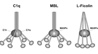
The lectin pathway. The lectin activa-tion pathway has been rather less in-tensely studied. Activation takes place when MBL binds mannose-containing surface proteins on pathogenic surfaces. In its ultra-structure, MBL closely resem-bles C1q, and along with the serine pro-teases MASP-1 and -2 (which themselves resemble C1r and C1s, respectively), forms a potent multi-enzyme complex (Figure 1).
Upon activation, MASP-2 catalyzes the cleavage of C2 and C4 in a similar man-ner to the classical pathway and forms a C3 convertase named C4b2a. MASP-1 is
capable of C2 and C3 cleavage, although to a much lesser extent (11,19). Subse-quently, C3 is cleaved into C3a and C3b, and by accretion of C3b to the C3 conver-tase, the C5 convertase is formed. A third serine protease MASP-3 has a distinct function compared with MASP-1 and -2, exerting inhibitory actions against MASP-2 (20).
In addition to the established activa-tion of the lectin pathway via MBL and MASPs, it was demonstrated that ficolins were also capable of initiating the lectin pathway by forming active complexes with MASPs. There are three distinct fi-colins named 1 (M-ficolin), ficolin-2 (L-ficolin) and ficolin-3 (H-ficolin or Hakata antigen). Structurally homolo-gous to the collectin MBL as well as C1q, ficolins are soluble collagen-like proteins that bind to sugar structures presented on microorganisms and dying host cells and consequently activate the innate im-mune system (8,10,21–23).
Surfactant protein A and D (A, SP-D), such as MBL, belong to the collectin family (24), but unlike MBL, they are not able to activate the complement directly.
The impact of the MBL pathway re-mains to be completely elucidated. It is suspected that its major role takes place during early childhood and in particular during the translational period from the passive immunity provided by the mother’s antibodies to the development of the body’s own mature immunity (25).
The lytic membrane attack complex.
The final stage of all three activation pathways is the formation of the lytic membrane attack complex (MAC). In contrast to the three different upstream
paths forming a C5 convertase, only the cleavage of C5 into the anaphylatoxin C5a and the active C5b represents an en-zymatic step, while the rest of the cas-cade is solely an accretion of stable pro-teins. In detail, C5b remains bound to the target cell followed by association of C6, resulting in a hydrophilic complex. By accretion of C7, a conformational change occurs—facilitating a stable linkage by exposure of lipophilic groups. Attach-ment of C8 with its binding component, C8b, induces the penetration of C8a-g into the lipid double layer of the target cell membrane. The final step toward formation of a stable transmembrane pore with a diameter of 10 angstrom is the binding of 10–15 C9 proteins, which generate a cylindrical structure (Figure 2). Assembly of such a pore may lead to osmotic imbalance through the constant flow of ions, small molecules and water along their concentration gradient, re-sulting in the lysis of the target cell (26).
It is noteworthy that the importance of these transmembrane pores should not be overrated, since, for instance, the blockage of the MAC only leads to a small increase in bacterial Neisseriainfection (25,27). The upstream effects of the complement sys-tem, such as the anaphylactoid reaction and the opsonization, appear to play a more important role (14,28).
Effects of the complement system.
The main effect of the complement sys-tem is the induction of a pathogen-asso-ciated and modulated enzymatic cascade that, once triggered, ends with the lysis of the target cell and protects the host from infection. In addition to this appar-ent effect, the complemappar-ent system also displays crucial additional activities that appear to be even more relevant.
One effect is the opsonization of the pathogen. Cleavage products such as C3b and C4b as well as C5b opsonize the surface of recognized pathogenic sub-stances and therefore facilitate phagocy-tosis. Additionally, opsonization is also important for the clearance of soluble, circulating antigen-antibody complexes. After the attachment of C3b and C4b to these complexes, they are bound to
com-plement receptor 1 (CR1) on erythrocytes and are subsequently transported to the spleen and liver, where the immune complexes are eliminated.
C3 cleavage products also bridge the innate and the adaptive immune systems. Opsonized antigens are bound to the complement receptor 2 (CR2) on B-cells via the C3-fragment C3d, initiating the production of specific antibodies as well as the differentiation of B-memory cells. Presumably, the most important func-tion is the inducfunc-tion of an anaphylactoid reaction. The small activation products C3a, C4a and particularly C5a are potent anaphylatoxins, capable of inducing the migration of phagocytes (29), smooth muscle relaxation, degranulation of mast cells and basophile granulocytes and therefore unleashing vasoactive sub-stances such as histamine, prostaglandins, kinins and serotonin. All can cause va-sodilation and capillary leakage (30) and induce the migrated cells to release eicosanoids, oxygen radicals and lysoso-mal enzymes, which cause damage to the pathogens (31–33). It is noteworthy, that complement acts far beyond “inflamma-tion,” as indicated by its close interaction with the coagulation cascade (34–36) and
its involvement in the regulation of apo-ptosis (37–40) and cellular growth (41). The anaphylactoid functions are medi-ated by the interaction of C3a and C5a with their corresponding transmembrane–spanning receptors C3aR and C5aR (CD88), respectively (see Figure 2). The role of the second C5a re-ceptor named C5L2 is not fully under-stood and is still controversially dis-cussed. However, there is increasing evidence that C5L2 represents a func-tional receptor acting as a negative regu-lator of the inflammatory response. For example, it was shown that inflammation in C5L2 knock-out mice was amplified (42) and that blockage of C5L2 increased serum interleukin (IL)-6 (43).
In summary, complement is highly ca-pable of inducing all classical signs of in-flammation, with the occurrence of pain, swelling, reddening, hyperthermia and impaired function.
New Activation Pathways
In addition to the established pathways, new pathways of complement activation were recently discovered (Figure 3).
During the last decade, more and more interaction sites between the two major
serine protease systems of the human body, namely the coagulation and the complement cascades, were found.
The potent serine protease thrombin is able to directly cleave C3 as well as C5 in a dose- and time-dependent manner, leading to biologically active C3a/C5a (44). In addition to thrombin, investiga-tions by our group indicated proteolytic cleavage of C3 and C5 also by FXa, FXIa and plasmin (35). Interestingly, FVIII and tissue factor failed to directly interact with C3 and C5 (35). Furthermore, FXIIa activates the classical complement path-way via C1q. Crosstalk between the lectin pathway and the coagulation cas-cade has only recently been ascertained by the observation that the complex of FVIII and von Willebrand factor pos-sesses lectin activity (45).
Vice versa, complement factors also interact with the coagulation system. C1 inhibitor not only blocks all three estab-lished complement pathways but also the endogenous coagulation path (46). Ikeda et al.(47) found evidence that C5a induces tissue factor activity on en-dothelial cells. Furthermore, crosstalk between the anaphylatoxin receptor C5aR and tissue factor was recently found (36,48).
Another possibility for complement ac-tivation exists via direct cellular interac-tions. Huber-Lang et al.demonstrated that phagocytic cells (macrophages, poly-morphonuclear leucocytes [PMNs]) are able to cleave C5 into biologically active C5a. This cleavage was conducted by a cell-bound serine protease that was in-ducible for alveolar macrophages, being more constitutively active on PMNs (49).
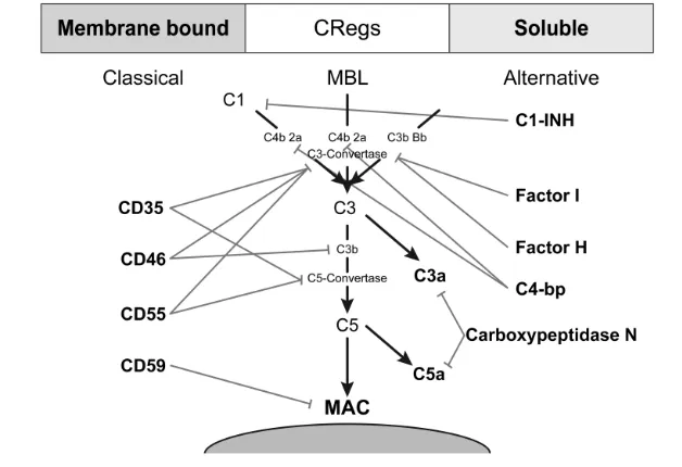
REGULATION OF THE COMPLEMENT SYSTEM
Soluble complement regulators The complement system can exert manifold detrimental effects not only on pathogenic or damaged tissue but also on healthy host tissue. To protect against a complement attack, the human body has developed various strategies. Princi-pally, there are both membrane-bound
and fluid phase complement regulators that are briefly covered (Figure 4).
The best known regulatory protein is the C1 inhibitor (C1-INH). C1-INH con-trols the activity of the classical pathway by binding to the C1 complex and initiat-ing the diffusion of the fragments C1r and C1s. This process leads to an irre-versible inactivation of the initiating ser-ine protease. As the classical and the
lectin pathway resemble each other in many ways, the C1-INH also inactivates MASP-1 and -2, thereby also inhibiting the lectin pathway. As well as inactivat-ing complement components, C1-INH also blocks certain parts of the kinin, fib-rinolytic and coagulation systems, such as coagulation factors XII and IX.
Factor I is a serine protease catalyzing the cleavage of the α-chain of C3b and
Figure 3.The established pathways of complement activation associated with the new activation pathway and crosstalk between cells and the coagulation cascade with the complement system.
C4b, leading to their permanent inactiva-tion. Cofactors for this enzymatic activity are factor H, C4-binding protein (C4-bp), CD35 and CD46.
Factor H is a protein that hinders the formation of the C3 convertase by com-peting for the binding site with factor B. Additionally, it facilitates the dissociation of already active C3 convertases and supports the proteolytic cleavage of C3b by factor I.
C4-bp also facilitates the proteolytic cleavage of the α-chain of C4b by factor I in a complex together with protein S.
Serumprotein S (Vitronectin) and clus-terin (Sp-40, 40) hinder the formation of the lytic membrane attack complex by adhesion to the lipophilic groups of C7, therefore leading to impaired anchorage in the cell membrane.
Carboxypeptidase N inactivates ana-phylatoxins of the complement system as well as other factors such as kinins and creatinine kinase MM through cleavage of terminal arginine and lysine residues of the peptides (17,50).
Membrane-Bound Complement Regulators
CD35 (complement receptor 1 [CR1]) is found on the surface of erythrocytes as well as on leukocytes and on podocytes in the glomerula of the kidney. CD35 facili-tates the decay of the C3/C5 convertase and also acts as a cofactor for factor I. CD46 (membrane cofactor protein [MCP]) also acts as a cofactor for factor I–mediated cleavage of C3b and is broadly expressed, except on erythrocytes.
CD55 (decay accelerating factor [DAF]) is widely expressed, except on natural killer cells and on a special subgroup of T-cells. The protein is phosphatidyl-inositol (GPI) anchored in the cell membrane and accelerates the decay of the classical as well as the alter-native C3 convertases by replacing C2a/Bb in these complexes.
CD59 (protectin) is expressed ubiqui-tously and is similarly integrated into the cell membrane by GPI anchors. It regu-lates the formation of the terminal lytic membrane attack complex by inhibiting
the interaction of the C8α-subchain and the first molecule of C9 so that integra-tion into the cell membrane and the cre-ation of a transmembrane pore is pre-vented (50–53).
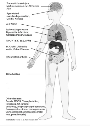
CLINICAL RELEVANCE AND COMPLEMENT-MEDIATED DISEASES
Sepsis and Complement
In contrast to all the beneficial effects for the host organism, the complement
system can also be detrimental for the host tissue (Figure 5).
Many distinct pathogenetic mecha-nisms may lead to the expression of an excessive and uncontrolled immune re-sponse. Depending on the individual’s immune status, this immune response leads to a proinflammatory systemic im-mune response syndrome or to compen-sated antiinflammatory response syn-drome. Clinical complications of these reactions can be progressive sepsis and
the development of multiple organ dys-function syndrome, with enhanced sus-ceptibility to infections.
Sepsis is defined as a systemic im-mune response syndrome with signs of infection. Excessive inflammation is in-duced by the recognition of pathogen-as-sociated molecular patterns on invading microorganisms or danger-associated molecular patterns of damaged tissue by the cellular “first line of defense” and the complement system. This result conse-quently leads to the robust release of cy-tokines from phagocytes (“cytokine storm”) to fight the infection. Although of benefit to the organism when acting locally, this pattern leads to a dramatic life-challenging event when occurring systemically (54–58).
Various studies proposed excessive complement activation during sepsis in
humans (59). Because of its potent in-flammatory profile, C5a appears to be the most detrimental molecule and has been described as “too much of a good thing” (54). When activated, C5a may lead to immune paralysis, multiorgan dysfunction and thymocyte apoptosis (37,40) as well as disturbance of the coag-ulation and fibrinolytic cascades (34).
In accordance, some protection of sep-tic mice has been shown by the applica-tion of a C5a receptor antagonist (60). Furthermore, C5aR antagonism during sepsis led to a changed cytokine profile, such as decreased levels of tumor necro-sis factor-αand IL-6, suggesting a direct or indirect role in the synthesis of these factors (40). Czermak et al.(61) showed an enhanced survival rate of septic rats treated with a C5a antibody. Addition-ally, these authors found that C5a binds
to neutrophils, which led to inactivation of their functions.
In a study by Flierl et al.(62), the ef-fects of complement on sepsis using C3–/–and C5–/–deficient mice were ex-amined. In the absence of either C3 or C5, a reduced production of proinflam-matory mediators was found.
On a cellular level, it has been shown that C5a effectively interacts with cells and modulates their apoptosis rate. In-terestingly, the effects on programmed cell death seem to be cell dependent, with a higher rate of apoptosis in thymo-cytes (37,40) but decreased apoptosis in neutrophils (39,63,64). Overall, the C5a-induced changes point toward an en-hanced susceptibility toward infections, as well as to a prolonged presence of neutrophils resulting in an exaggerated inflammatory response and host damage.
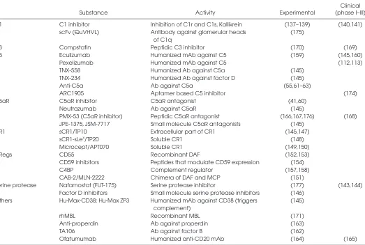
Table 1.Different complement-modulating drugs listed by activity level along with references of experimental/clinical application. Clinical
Substance Activity Experimental (phase I–III)
C1 C1 inhibitor Inhibition of C1r and C1s, Kallikrein (137–139) (140,141)
scFv (QuVHVL) Antibody against glomerular heads (175) of C1q
C3 Compstatin Peptidic C3 inhibitor (170) (169)
C5 Eculizumab Humanized mAb against C5 (159) (145,160)
Pexelizumab Humanized mAb against C5 (112,113)
TNX-558 Humanized Ab against C5a (145)
TNX-234 Humanized Ab against factor D (145)
Anti-C5a Ab against C5a (55,61–63)
ARC1905 Aptamer based C5 inhibitor (174)
C5aR C5aR inhibitor C5aR antagonist (41,60)
Neutrazumab Ab against C5aR (145)
PMX-53 (C5aR inhibitor) Peptidic C5aR antagonist (166,167,176) (168) JPE-1375, JSM-7717 Small molecule C5aR antagonists (145)
CR1 sCR1/TP10 Extracellular part of CR1 (145,147)
sCR1-sLex/TP20 Soluble CR1 (148)
Microcept/APT070 Soluble CR1 (149,150)
CRegs CD55 Recombinant DAF (152,153)
CD59 inhibitors Peptides that modulate CD59 expression (154)
C4BP Complement regulator (157,158)
CAB-2/MLN-2222 Chimera of DAF and MCP (151)
Serine protease Nafamostat (FUT-175) Serine protease inhibitor (177) (143,144)
Factor D inhibitors Small molecule serine protease inhibitors (146) Others Hu-Max-CD38; Hu-Max ZP3 Humanized mAb against CD38 (triggers (145)
complement)
rhMBL Recombinant MBL (171)
Anti-properdin Ab against properdin (163)
TA106 Ab against factor B (162)
Complement and Bone Biology In the emerging field of osteoim-munology, the role of complement in bone biology in general and fracture healing in particular has started to raise interest. Although some direct interac-tions between the immune system and bone cells have been found (65,66), few studies demonstrated the presence of complement components in bone cells. In particular, the expression of various complement factors might depend on the cell differentiation state. Murine os-teoblastic cells were shown to produce C3 in response to vitamin D3 (67,68). During osteoblastic differentiation in murine osteoblasts and in human cell lines, the complement components C1q, C4, C1 inhibitor, C3a receptor (C3aR), properdin and the complement factor H were upregulated (69), whereas the ex-pression of the subcomponents C1r and C1s together with factor H was de-creased (70). The expression of function-ally active C5a receptor (C5aR; CD88) that modulated IL-6 production was de-scribed in a human osteoblastic cell line (71,72). Recently, mRNA and protein ex-pression of C3aR and C5aR in human MSC were reported (73).
Immunohistochemical studies clearly indicated that complement was activated during enchondral ossification, possibly for the modulation of apoptosis (74,75). These studies indicated that some inter-action of the complement system with bone cells exists. However, the resulting function to date has been minimally in-vestigated. With the exception of our un-published data and the immunohisto-chemical studies in enchondral ossification, in vivodata on the expres-sion of complement components in bone are lacking, particularly with regard to fracture healing.
Recently, a major role of an excessively stimulated complement system in the posttraumatic inflammatory response was postulated (16,76,77). Prospective studies with polytraumatized patients showed a consumption and massive acti-vation of complement products, posi-tively correlating with the mortality rate
(78–80). Fracture healing is delayed in particular when additional severe in-juries stress the organism (81). The mas-sive trauma-induced inflammation corre-lates with consequent organ dysfunction (79) and may possibly be involved in the delayed fracture healing.
Studies with experimental blunt tho-racic trauma found that the complement system contributed to the inflammatory reaction, and additionally successful inhi-bition of various inflammatory mediators occurred upon application of an antibody against C5a (76). Ganter et al.reported an earlier complement activation after major trauma, for which magnitude correlated with the mortality rate (16). Unpublished data from our group underscore the role of complement in fracture healing, re-flected by the enhanced C5aR immunos-taining of bone sections.
Traumatic Brain Injury
The anaphylatoxins C3a and C5a have been reported to exert both protective and detrimental effects in the central nervous system (82,83). It is noteworthy that many cells of the central nervous system are more susceptible to a comple-ment attack, since they lack important complement regulators such as CD59 (84). Complement products were demon-strated to be dramatically upregulated in models of cerebral ischemia in rats (85), and evidence for deposition of C3d and C9 after experimental cerebral contusion was found (86). In agreement with these experimental findings, systemic comple-ment activation has been shown in stroke patients (87). A negative role for comple-ment has been proposed in traumatic brain injury, since systemic depletion of complement by infusion of the cobra venom factor improved blood flow and neurological function and reduced cere-bral edema during experimental intrac-erebral hemorrhage (88,89). Moreover, C1 deficiency (90) and complement inhibi-tion by infusion of the C1 inhibitor re-vealed some neuroprotective effects (91,92). In contradiction to these results for traumatic/ischemic injuries, C1q has a strong neuroprotective effect in
neu-rodegenerative diseases (93). Traumatic brain injury induces C5aR upregulation in an experimental model in rats with en-hanced C5a serum levels for >1 week. The functional consequences of traumatic brain injury–induced systemic C5a gen-eration have still to be elucidated (94).
Ischemia/Reperfusion Injuries and Cardiovascular Diseases
After ischemia/reperfusion injury, complement is activated via all three es-tablished pathways (classical, alternative and MBL) (95–97). Endothelium dam-aged by hypoxic stress activates comple-ment, leading to elevated vascular per-meability, release of anaphylatoxins and the accumulation of neutrophils (98). Ex-perimental studies indicated comple-ment activation after ischemia/reperfu-sion in several organs, including lung, liver, gut, kidney, myocardium and skeletal muscle (99–105).
Cardiac surgery and cardiopulmonary bypass surgery are known to cause a considerable immunologic impact to the body, resulting in a systemic inflamma-tory reaction (106,107). Various factors contribute to the extent of the immuno-logical impact. In addition to general surgical trauma, hypothermia and blood loss, the main mechanisms are exposure of blood to foreign surfaces of the car-diopulmonary bypass, the
ischemia/reperfusion injury by aortic cross-clamping and splanchnic hypoper-fusion leading to mucosal damage and consequently endotoxemia (108).
An important pathogenetic role for complement in ischemia/reperfusion of the myocardium was indicated experi-mentally, where inhibition of the comple-ment cascade greatly reduced myocar-dial damage after myocarmyocar-dial infarction (109–111).
im-plantation of drug-eluting stents in pa-tients with coronary heart disease (114).
It was recently found that high MBL levels and low plasma levels of sC5b-9 were associated with an increased risk of dysfunctional cardiac performance after myocardial infarction, possibly owing to the increased complement ac-tivity during ischemia/reperfusion, which generally triggers an inflamma-tory response (115).
However, activation of the comple-ment cascade after ischemia/reperfusion injury or exposure to foreign surfaces is not the only cause of harmful effects of the complement system. Several studies alluded to a role for complement in car-diovascular diseases such as atheroscle-rosis or vasculitis.
Kawasaki disease, a systemic vasculi-tis in childhood causing coronary artery aneurysmata, appears to be related to MBL deficiency according to genetic family studies by Biezeveld et al.(116). Rugonfalvi-Kiss et al.(117) found signif-icantly lower restenosis rates in females with low MBL levels undergoing throm-boendarteryectomy of the carotid artery because of atherosclerotical stenosis. Additionally, patients with MBL defi-ciency had a greater risk of myocardial infarction than individuals with normal MBL serum levels (118,119). The pro-posed protective effect of complement in the pathogenesis of atherosclerosis was experimentally underlined by C3–/– mice exhibiting accelerated develop-ment of atherosclerosis (120). Clinical analysis of C2-deficient humans re-vealed a significant increase in cardio-vascular diseases (121). In particular, classical pathway involvement was demonstrated by Bhatia et al.(122) when C1q protected against the devel-opment of atherosclerosis in combined C1q and LDLr (low-density-lipoprotein receptor) knockout mice.
In agreement with these results, defi-ciency of complement inhibitors such as CD59 promoted atherogenesis (123). However, it is noteworthy that both downstream complement defects and ac-tivation of the terminal pathway were
as-sociated with inherited proatherogenic effects and an increased risk of cardio-vascular disease (124).
In conclusion, tightly regulated com-plement activation seems to protect against atherogenesis, possibly through the clearance of apoptotic cells and other debris, whereas inhibition of the terminal pathway by complement regulators ap-pears to hinder proinflammatory effects (123,125). Once an atherogenic plaque has been formed, increasingly leading to ischemia and reperfusion injury, comple-ment is excessively activated and may lead to harmful tissue damage.
Infections
Generally, a major complication of complement insufficiency is an enhanced susceptibility toward infection. Several studies have shown low MBL serum lev-els (genetically derived) are correlated with enhanced development of systemic immune response syndrome/sepsis in pa-tients admitted to the intensive care unit (126,127). Low MBL serum concentrations were also associated with the develop-ment of pneumonia after surgery in colo-rectal cancer patients (128). Similarly, MASP-2 deficiency caused increased in-fection rates with Streptococcus pneumoniae (129). Furthermore, there is evidence for a higher rate of chorioamnionitis in patients with low MBL levels and preterm birth (130). Low MBL serum levels appear to be associated with recurrent spontaneous abortion based on in uteroinfection. This was proposed by Kruse et al.(131), who demonstrated that patients with MBL lev-els <100 ng/mL had a higher abortion rate. Some urogenital infections such as vulvovaginitis, herpes simplex virus-2 in-fection, vulvovaginal candidiasis and vestibulitis appear to be linked to low MBL levels in comparison to healthy con-trols (132–134). Downstream complement deficiencies are typically associated with recurrent invasive infections of Neisseria meningitidis and gonorrhea (135).
In defects further upstream, such as factor D or properdin, N. meningitidis also appear to be involved in most infec-tions (136). Deficiencies of the classical
pathway may also cause enhanced infec-tion rates, since invasive infecinfec-tion was the predominant clinical manifestation in patients with C2 deficiency, especially with S. pneumoniae(121).
IMMUNOMODULATION OF COMPLEMENT
Many efforts have been undertaken to effectively modulate the activity of com-plement, thereby trying to ameliorate or completely abolish the symptoms of complement-mediated diseases. It is tempting to speculate that the effective modulation of the broad spectrum of in-ternal diseases (as listed below) may also reflect an effective future target of vari-ous complement-dependent surgical diseases.
C1 inhibitor provided protective ef-fects in myocardial cell injury (137), transplantation (138) and various other diseases (139). C1-INH was effective in a randomized, double-blind clinical study in septic intensive care unit patients. Whereas the renal dysfunction was re-duced, the mortality rate remained un-changed in both groups (140). The only clinical application for C1 inhibitor so far is for C1 inhibitor deficiency leading to hereditary angioedema (141).
Nafamostat/FUT-175 is an unspecific serine protease inhibitor that, besides complement activation, also blocks abun-dant serine protease of the coagulation system (142). It is currently used clini-cally for the treatment of acute pancreati-tis and for the prevention of thrombosis in disseminated intravascular coagulation and extracorporal circulation (143,144), underscoring the crosstalk of the comple-ment and coagulation cascades (35).
Because of a lack of specificity and short half-life, other serine protease in-hibitors, such as factor D, were not trans-ferred to clinical trials, and further devel-opment was stopped (145,146).
protective effects in male patients under-going coronary artery bypass grafting but not in females (145,147). Therefore, clinical trials have been stopped (145). Similar substances such as sCR1-sLex/ TP20 with an improved structure (148) and Microcept/APT070 (149) are cur-rently under investigation for the treat-ment of diseases such as acute myocar-dial infarction, stroke and inflammatory diseases but have not entered clinical evaluation (150).
A hybrid form of the complement reg-ulators DAF and MCP was designed, named complement activity blocker 2 (CAB2), and entered clinical trials under the name MLN-2222 for the treatment of coronary artery bypass grafting (151).
The complement regulator CD55 (DAF) was recently shown to ameliorate the hepatic inflammation in experimental chronic hepatitis C (152) and autoim-mune posterior uveitis (153).
The complement regulator CD59 was investigated in the context of cancer re-search. Neuroblastoma cells are known to escape cell lysis by abundantly ex-pressing CD59. Therefore, a peptide was generated that was capable of sup-pressing CD59 expression that resulted in complement-mediated killing of the neuroblastoma cells (154). Experimental studies for the treatment of paroxysmal nocturnal hemoglobinuria with the sub-stitution of CD59 have recently been conducted (155), but definitive results are pending.
C4BP as well as factor H have both been experimentally used to successfully abrogate complement activation on artifi-cial surfaces and to inhibit the develop-ment of arthritis. However, these sub-stances have not yet been clinically evaluated (156–158).
Because of their specificity, antibodies against various complement factors ap-pear to be a more reliable treatment strategy.
Eculizumab is a monoclonal antibody against C5 investigated for the treatment of paroxysmal nocturnal hemoglobinuria (159) and is currently in a phase I clinical trial for the treatment of systemic lupus
erythematosus (160). The first clinical studies in patients suffering from atypi-cal hemolytic uremic syndrome were conducted and were partly promising, with the optimal dose and timing still to be determined (161).
Pexelizumab is also a monoclonal anti-body against C5 and has revealed benefi-cial effects after cardiopulmonary bypass surgery, but not after myocardial infarc-tion in clinical studies (112,113). Various other antibodies are currently in devel-opmental progress. Neutrazumab and TNX-558 are both directed against C5aR. TNX-234 is an antibody against factor D. TA106 blocks factor B from targeting, for example, in age-dependent macular dys-trophia and asthma (162). At least one antibody directed against properdin is currently undergoing testing (145). In ex-perimental studies, this antiproperdin monoclonal antibody was shown to be beneficial during coronary artery bypass grafting, reducing the activation of platelets and neutrophils (163).
Ofatumumab is a monoclonal anti-body against CD20 and exerts its effects not by inhibition, like most other com-plement therapeutics, but by stimula-tion of complement-dependent cytotox-icity. It is currently in clinical trials for the treatment of rheumatoid arthritis, B-cell chronic lymphocytic leukemia and follicular lymphoma (164,165). A similar approach that has not yet been clinically tested is the efficacy of the re-lated substances HuMax-CD38 for mul-tiple myeloma and HuMax-ZP3 for the treatment of colon, pancreatic and prostate cancer.
PMX-53 is a peptidic C5aR antagonist and has proven to be advantageous in experimental animal studies for neurode-generative diseases, rheumatoid arthritis, ischemia/reperfusion and inflammatory bowel disease (102,166,167). It is cur-rently being evaluated in clinical trials, but a recent study in humans failed to show significant effects on synovial in-flammation (168).
Compstatin, a peptidic C3 inhibitor, is considered a promising drug and is cur-rently in clinical trials for the treatment
of age-dependent macular dystrophia (169). Since compstatin has proven to be effective in various animal models for other conditions (170), its application does not appear to be limited to dependent macular dystrophia.
It is estimated that 30% of the human population present decreased plasma levels of MBL owing to genetic muta-tions. Therefore, a recombinant human form of MBL as a substitution therapy for MBL-deficient people was developed and found to be safe (171). The substance is currently under clinical investigation for people suffering from multiple mye-loma and undergoing high-dose chemo-therapy (145,172,173).
ARC1905 is an aptamer-based C5 in-hibitor that inhibits the cleavage of C5 into C5a and C5b. It is intended for in-travitreal application in age-dependent macular dystrophia and is currently un-dergoing phase I clinical trials (174). With JPE-1375 and JSM-7717, there now exists even more C5aR antagonists that are in preclinical evaluation for the treat-ment of inflammatory, renal and ocular diseases.
CONCLUSION
Although complement research has been in the center of interest for many years, our understanding and insight into the cascade mechanisms and their complex interaction with other protein cascades such as the coagulation cascade is still in its nascent phase. Currently, complement activation is not divisable into three canonical pathways with sepa-rate activation patterns. The exact role of complement in many diseases needs to be further clarified because successful complement interventions need to be matched to the individual patient and be as specific as possible.
DISCLOSURE
REFERENCES
1. Bordet J. (1895) Les leukocytes et les proprietes actives du serum chez les vaccines [in French].
Ann. Inst. Pasteur. 9:462–506.
2. Bordet J. (1898) Sur l’agglutination et la dissolution des globules rouge par le sérum d’animaux in-jecteies de sang defibiné [in French]. Ann. Inst. Pasteur. 12:688.
3. Ehrlich P, Morgenroth J. (1899) Zur Theorie der Lysenwirkung [in German]. Berlin Klin. Wchsr.
36:6.
4. Nuttall G. (1888) Experimente über die bacterien-feindliche Einflüsse des tierischen Körpers [in Ger-man]. Z. Hyg. Infec.ionskir.4:353.
5. Pillemer L, et al.(1954) The properdin system and immunity. I. Demonstration and isolation of a new serum protein, properdin, and its role in immune phenomena. Science120:279–85. 6. Kawasaki T, Etoh R, Yamashina I. (1978) Isolation
and characterization of a mannan-binding pro-tein from rabbit liver. Biochem. Biophys. Res. Com-mun.81:1018–24.
7. Super M, Thiel S, Lu J, Levinsky RJ, Turner MW. (1989) Association of low levels of mannan- binding protein with a common defect of opsonisation.
Lancet2:1236–9.
8. Matsushita M, Endo Y, Fujita T. (2000) Cutting edge: complement-activating complex of ficolin and mannose-binding lectin-associated serine protease. J. Immunol.164:2281–4.
9. Matsushita M, Fujita T. (1992) Activation of the classical complement pathway by mannose-bind-ing protein in association with a novel C1s-like serine protease. J. Exp. Med.176:1497–502. 10. Matsushita M, et al.(2002) Activation of the lectin
complement pathway by H-ficolin (Hakata anti-gen). J. Immunol.168:3502–6.
11. Matsushita M, Thiel S, Jensenius JC, Terai I, Fu-jita T. (2000) Proteolytic activities of two types of mannose-binding lectin-associated serine pro-tease. J. Immunol.165:2637–42.
12. Gewurz H, Ying SC, Jiang H, Lint TF. (1993) Nonimmune activation of the classical comple-ment pathway. Behring Inst. Mitt. 138–47. 13. Barrington R, Zhang M, Fischer M, Carroll MC.
(2001) The role of complement in inflammation and adaptive immunity. Immunol. Rev.180:5–15. 14. Gasque P. (2004) Complement: a unique innate
immune sensor for danger signals. Mol. Immunol.
41:1089–98.
15. Thurman JM, Holers VM. (2006) The central role of the alternative complement pathway in human disease. J. Immunol.176:1305–10. 16. Ganter MT, et al.(2007) Role of the alternative
pathway in the early complement activation fol-lowing major trauma. Shock28:29–34.
17. Liszewski MK, Farries TC, Lublin DM, Rooney IA, Atkinson JP. (1996) Control of the comple-ment system. Adv. Immunol. 61:201–83. 18. Harboe M, Mollnes TE. (2008) The alternative
complement pathway revisited. J. Cell. Mol. Med.
12:1074–84.
19. Hajela K, et al.(2002) The biological functions of
MBL-associated serine proteases (MASPs). Im-munobiology205:467–75.
20. Dahl MR, et al.(2001) MASP-3 and its association with distinct complexes of the mannan-binding lectin complement activation pathway. Immunity
15:127–35.
21. Liu Y, et al.(2005) Human M-ficolin is a secretory protein that activates the lectin complement pathway. J. Immunol.175:3150–6.
22. Matsushita M, Endo Y, Hamasaki N, Fujita T. (2001) Activation of the lectin complement path-way by ficolins. Int. Immunopharmacol. 1:359–63. 23. Matsushita M, Fujita T. (2001) Ficolins and the
lectin complement pathway. Immunol. Rev.
180:78–85.
24. Day AJ. (1994) The C-type carbohydrate recogni-tion domain (CRD) superfamily. Biochem. Soc. Trans. 22:83–8.
25. Walport MJ. (2001) Complement. First of two parts. N. Engl. J. Med.344:1058–66.
26. Dalmasso AP, Falk RJ, Raij L. (1989) The pathobi-ology of the terminal complement complexes.
Complement Inflamm. 6:36–48.
27. Walport MJ. (2001) Complement. Second of two parts. N. Engl. J. Med.344:1140–4.
28. Morgan BP. (1989) Mechanisms of tissue damage by the membrane attack complex of complement.
Complement Inflamm. 6:104–11.
29. Marder SR, Chenoweth DE, Goldstein IM, Perez HD. (1985) Chemotactic responses of human pe-ripheral blood monocytes to the derived peptides C5a and C5a des Arg. J. Im-munol.134:3325–31.
30. Schumacher WA, Fantone JC, Kunkel SE, Webb RC, Lucchesi BR. (1991) The anaphylatoxins C3a and C5a are vasodilators in the canine coronary vasculature in vitro and in vivo. Agents Actions
34:345–49.
31. Goldstein IM, Weissmann G. (1974) Generation of C5-derived lysosomal enzyme-releasing activ-ity (C5a) by lysates of leukocyte lysosomes. J. Im-munol.113:1583–88.
32. Mollnes TE, et al.(2002) Essential role of the C5a receptor in E coli-induced oxidative burst and phagocytosis revealed by a novel lepirudin-based human whole blood model of inflamma-tion. Blood100:1869–77.
33. Sacks T, Moldow CF, Craddock PR, Bowers TK, Jacob HS. (1978) Oxygen radicals mediate en-dothelial cell damage by complement-stimulated granulocytes. An in vitro model of immune vas-cular damage. J. Clin. Invest.61:1161–67. 34. Laudes IJ, et al.(2002) Anti-c5a ameliorates
coag-ulation/fibrinolytic protein changes in a rat model of sepsis. Am. J. Pathol.160:1867–75. 35. Amara U, et al.(2008) Interaction between the
co-agulation and complement system. Adv. Exp. Med. Biol. 632:71–9.
36. Markiewski MM, Nilsson B, Ekdahl KN, Mollnes TE, Lambris JD. (2007) Complement and coagu-lation: strangers or partners in crime? Trends Im-munol.28:184–92.
37. Guo RF, et al.(2000) Protective effects of anti-C5a
in sepsis-induced thymocyte apoptosis. J. Clin. Invest.106:1271–80.
38. Markiewski MM, et al.(2009) The regulation of liver cell survival by complement. J. Immunol.
182:5412–18.
39. Perianayagam MC, Balakrishnan VS, King AJ, Pereira BJ, Jaber BL. (2002) C5a delays apoptosis of human neutrophils by a phosphatidylinositol 3-kinase-signaling pathway. Kidney Int.61:456–63. 40. Riedemann NC, et al.(2002) C5a receptor and
thymocyte apoptosis in sepsis. FASEB J. 16:887–8. 41. Markiewski MM, et al.(2008) Modulation of the
antitumor immune response by complement.
Nat. Immunol. 9:1225–35.
42. Gerard NP, et al.(2005) An anti-inflammatory function for the complement anaphylatoxin C5a-binding protein, C5L2. J. Biol. Chem.280:39677–80. 43. Gao H, et al.(2005) Evidence for a functional role of the second C5a receptor C5L2. FASEB J. 19:1003–5. 44. Huber-Lang M, et al.(2006) Generation of C5a in
the absence of C3: a new complement activation pathway. Nat. Med.12:682–7.
45. Santizo F, et al.(2009) Lectin activity of the coag-ulation factor VIII/von Willebrand complex. To-hoku J. Exp. Med.217:209–15.
46. Davis AE 3rd. (2004) Biological effects of C1 in-hibitor. Drug News Perspect. 17:439–46.
47. Ikeda K, et al.(1997) C5a induces tissue factor activ-ity on endothelial cells. Thromb. Haemost. 77:394–8. 48. Ritis K, et al.(2006) A novel C5a receptor-tissue
factor cross-talk in neutrophils links innate im-munity to coagulation pathways. J. Immunol.
177:4794–802.
49. Huber-Lang M, et al.(2002) Generation of C5a by phagocytic cells. Am. J. Pathol.161:1849–59. 50. Meri S, Jarva H. (1998) Complement regulation.
Vox Sang. 74 Suppl 2:291–302.
51. Morgan BP. (1995) Complement regulatory mole-cules: application to therapy and transplantation.
Immunol. Today16:257–9.
52. Miwa T, Song WC. (2001) Membrane comple-ment regulatory proteins: insight from animal studies and relevance to human diseases. Int. Im-munopharmacol. 1:445–59.
53. Kim DD, Song WC. (2006) Membrane complement regulatory proteins. Clin. Immunol. 118:127–36. 54. Gerard C. (2003) Complement C5a in the sepsis
syndrome: too much of a good thing? N. Engl. J. Med.348:167–9.
55. Guo RF, Ward PA. (2006) C5a, a therapeutic tar-get in sepsis. Recent Pat. Antiinfect. Drug Discov. 1:57–65.
56. Rittirsch D, Flierl MA, Ward PA. (2008) Harmful molecular mechanisms in sepsis. Nat. Rev. Im-munol. 8:776–87.
57. Ward PA. (2004) The dark side of C5a in sepsis.
Nat. Rev. Immunol. 4:133–42.
58. Ward PA. (2008) Sepsis, apoptosis and comple-ment. Biochem. Pharmacol. 76:1383–8.
59. Riedemann NC, Guo RF, Ward PA. (2003) Novel strategies for the treatment of sepsis. Nat. Med.
9:517–24.
immunity by C5aR antagonist in septic mice.
FASEB J. 16:1567–74.
61. Czermak BJ, et al.(1999) Protective effects of C5a blockade in sepsis. Nat. Med.5:788–92. 62. Flierl MA, et al.(2008) Functions of the
comple-ment components C3 and C5 during sepsis.
FASEB J. 22:3483–90.
63. Guo RF, et al.(2006) In vivo regulation of neu-trophil apoptosis by C5a during sepsis. J. Leukoc. Biol.80:1575–83.
64. Perianayagam MC, Balakrishnan VS, Pereira BJ, Jaber BL. (2004) C5a delays apoptosis of human neutrophils via an extracellular signal-regulated kinase and Bad-mediated signalling pathway.
Eur. J. Clin. Invest.34:50–6.
65. Arron JR, Choi Y. (2000) Bone versus immune system. Nature408:535–36.
66. Takayanagi H. (2007) Osteoimmunology: shared mechanisms and crosstalk between the immune and bone systems. Nat. Rev. Immunol. 7:292–304. 67. Hong MH, et al.(1991) Transcriptional regulation of
the production of the third component of comple-ment (C3) by 1 alpha,25-dihydroxyvitamin D3 in mouse marrow-derived stromal cells (ST2) and pri-mary osteoblastic cells. Endocrinology129:2774–79. 68. Sato T, et al.(1991) The specific production of the
third component of complement by osteoblastic cells treated with 1 alpha,25-dihydroxyvitamin D3. FEBS Lett. 285:21–24.
69. Roman-Roman S, et al.(2003) Identification of genes regulated during osteoblastic differentiation by genome-wide expression analysis of mouse cal-varia primary osteoblasts in vitro. Bone32:474–82. 70. Billiard J, et al.(2003) Transcriptional profiling of
human osteoblast differentiation. J. Cell. Biochem.
89:389–400.
71. Jiang T, Gao H. (2008) Expression of C5a receptor in osteoblasts [abstract]. FASEB J. 22:1121.12. 72. Pobanz JM, Reinhardt RA, Koka S, Sanderson
SD. (2000) C5a modulation of interleukin-1 beta-induced interleukin-6 production by human osteoblast-like cells. J. Periodontal Res. 35:137–45. 73. Schraufstatter IU, Discipio RG, Zhao M,
Khal-doyanidi SK. (2009) C3a and C5a are chemotactic factors for human mesenchymal stem cells, which cause prolonged ERK1/2 phosphoryla-tion. J. Immunol.182:3827–36.
74. Andrades JA, et al.(1996) Complement proteins are present in developing endochondral bone and may mediate cartilage cell death and vascu-larization. Exp. Cell. Res. 227:208–13.
75. Sakiyama H, et al.(1997) Complement Cls, a classi-cal enzyme with novel functions at the endochon-dral ossification center: immunohistochemical staining of activated Cls with a neoantigen-specific antibody. Cell Tissue Res. 288:557–65.
76. Flierl MA, et al.(2008) The role of C5a in the in-nate immune response after experimental blunt chest trauma. Shock29:25–31.
77. Galvan MD, et al.(2008) Deficiency in complement C1q improves histological and functional locomo-tor outcome after spinal cord injury. J. Neurosci.
28:13876–88.
78. Donnelly TJ, et al.(1994) Cytokine, complement, and endotoxin profiles associated with the devel-opment of the adult respiratory distress syndrome after severe injury. Crit. Care Med.22:768–76. 79. Keel M, Trentz O. (2005) Pathophysiology of
polytrauma. Injury36:691–709. 80. Zilow G, Joka T, Obertacke U, Rother U,
Kirschfink M. (1992) Generation of anaphylatoxin C3a in plasma and bronchoalveolar lavage fluid in trauma patients at risk for the adult respiratory distress syndrome. Crit. Care Med.20:468–73. 81. Bhandari M, et al.(2003) Predictors of reoperation
following operative management of fractures of the tibial shaft. J. Orthop. Trauma17:353–61. 82. van Beek J, et al.(2001) Complement
anaphyla-toxin C3a is selectively protective against NMDA-induced neuronal cell death. Neuroreport
12:289–93.
83. Osaka H, Mukherjee P, Aisen PS, Pasinetti GM. (1999) Complement-derived anaphylatoxin C5a protects against glutamate-mediated neurotoxic-ity. J. Cell. Biochem.73:303–11.
84. Bradt BM, Kolb WP, Cooper NR. (1998) Comple-ment-dependent proinflammatory properties of the Alzheimer’s disease beta-peptide. J. Exp. Med.188:431–8.
85. Schafer MK, et al.(2000) Complement C1q is dra-matically up-regulated in brain microglia in re-sponse to transient global cerebral ischemia. J. Im-munol.164:5446–52.
86. Pasinetti GM, et al.(1992) Complement C1qB and C4 mRNAs responses to lesioning in rat brain.
Exp. Neurol. 118:117–25.
87. Pedersen ED, Waje-Andreassen U, Vedeler CA, Aamodt G, Mollnes TE. (2004) Systemic comple-ment activation following human acute is-chaemic stroke. Clin. Exp. Immunol. 137:117–22. 88. Vasthare US, et al.(1998) Complement depletion
improves neurological function in cerebral ische-mia. Brain Res. Bull. 45:413–9.
89. Xi G, Hua Y, Keep RF, Younger JG, Hoff JT. (2001) Systemic complement depletion diminishes peri-hematomal brain edema in rats. Stroke32:162–7. 90. Ten VS, et al.(2005) C1q-deficiency is neuropro-tective against hypoxic-ischemic brain injury in neonatal mice. Stroke36:2244–50.
91. De Simoni MG, et al.(2004) The powerful neuro-protective action of C1-inhibitor on brain ische-mia-reperfusion injury does not require C1q. Am. J. Pathol.164:1857–63.
92. Longhi L, et al.(2009) C1-inhibitor attenuates neurobehavioral deficits and reduces contusion volume after controlled cortical impact brain in-jury in mice. Crit. Care Med.37:659–65. 93. Benoit M, Tenner AJ. (2010) Regulation of
neu-ronal gene and miRNA expression by the com-plement protein C1q associated with neuropro-tection. Mol. Immunol.47:2250–51.
94. Stahel PF, Kossmann T, Morganti-Kossmann MC, Hans VH, Barnum SR. (1997) Experimental dif-fuse axonal injury induces enhanced neuronal C5a receptor mRNA expression in rats. Brain Res. Mol. Brain Res.50:205–12.
95. Arumugam TV, Shiels IA, Woodruff TM, Granger DN, Taylor SM. (2004) The role of the complement system in ischemia-reperfusion in-jury. Shock21:401–9.
96. Link C, et al.(1999) Selection of phage-dis-played anti-guinea pig C5 or C5a antibodies and their application in xenotransplantation.
Mol. Immunol.36:1235–47.
97. Stahl GL, et al.(2003) Role for the alternative complement pathway in ischemia/reperfusion injury. Am. J. Pathol.162:449–55.
98. Arumugam TV, et al.(2006) Complement medi-ators in ischemia-reperfusion injury. Clin. Chim. Acta. 374:33–45.
99. Buerke M, Murohara T, Lefer AM. (1995) Car-dioprotective effects of a C1 esterase inhibitor in myocardial ischemia and reperfusion. Circula-tion91:393–402.
100. Jaeschke H, Farhood A, Bautista AP, Spolarics Z, Spitzer JJ. (1993) Complement activates Kupffer cells and neutrophils during reperfu-sion after hepatic ischemia. Am. J. Physiol.
264:G801–9.
101. Proctor LM, et al.(2004) Comparative inflammatory activities of antagonists to C3a and C5a receptors in a rat model of intestinal is-chaemia/reperfusion injury. Br. J. Pharmacol.
142:756–64.
102. Arumugam TV, et al.(2003) A small molecule C5a receptor antagonist protects kidneys from ischemia/reperfusion injury in rats. Kidney Int.
63:134–42.
103. De Vries B, et al.(2003) Inhibition of comple-ment factor C5 protects against renal ischemia-reperfusion injury: inhibition of late apoptosis and inflammation. Transplantation75:375–82. 104. Kyriakides C, et al.(1999) Skeletal muscle
reper-fusion injury is mediated by neutrophils and the complement membrane attack complex.
Am. J. Physiol.277:C1263–8.
105. Kyriakides C, et al.(2000) Neutrophil mediated remote organ injury after lower torso ischemia and reperfusion is selectin and complement de-pendent. J. Trauma. 48:32–8.
106. Ascione R, et al.(2000) Inflammatory response after coronary revascularization with or with-out cardiopulmonary bypass. Ann. Thorac. Surg. 69:1198–204.
107. Butler J, Rocker GM, Westaby S. (1993) Inflam-matory response to cardiopulmonary bypass.
Ann. Thorac. Surg. 55:552–9.
108. Laffey JG, Boylan JF, Cheng DC. (2002) The sys-temic inflammatory response to cardiac sur-gery: implications for the anesthesiologist.
Anesthesiology97:215–52.
109. Langlois PF, Gawryl MS. (1988) Detection of the terminal complement complex in patient plasma following acute myocardial infarction.
Atherosclerosis70:95–105.
111. Vakeva A, et al.(1994) Time course of comple-ment activation and inhibitor expression after ischemic injury of rat myocardium. Am. J. Pathol.144:1357–68.
112. Mahaffey KW, et al.(2006) Effect of pex-elizumab on mortality in patients with acute myocardial infarction or undergoing coronary artery bypass surgery: a systematic overview.
Am. Heart J. 152:291–6.
113. Testa L, et al.(2008) Pexelizumab in ischemic heart disease: a systematic review and meta-analysis on 15,196 patients. J. Thorac. Cardiovasc. Surg. 136:884–93.
114. Speidl WS, et al.(2010) Coronary late lumen loss of drug eluting stents is associated with in-creased serum levels of the complement compo-nents C3a and C5a. Atherosclerosis208:285–9. 115. Haahr-Pedersen S, et al.(2009) Level of
comple-ment activity predicts cardiac dysfunction after acute myocardial infarction treated with pri-mary percutaneous coronary intervention. J. In-vasive Cardiol. 21:13–9.
116. Biezeveld MH, et al.(2003) Association of man-nose-binding lectin genotype with cardiovascu-lar abnormalities in Kawasaki disease. Lancet
361:1268–70.
117. Rugonfalvi-Kiss S, et al.(2005) High rate of early restenosis after carotid eversion en-darterectomy in homozygous carriers of the normal mannose-binding lectin genotype. Stroke
36:944–8.
118. Madsen HO, Videm V, Svejgaard A, Svennevig JL, Garred P. (1998) Association of mannose-binding-lectin deficiency with severe athero-sclerosis. Lancet352:959–60.
119. Saevarsdottir S, et al.(2005) Mannan binding lectin as an adjunct to risk assessment for myo-cardial infarction in individuals with enhanced risk. J. Exp. Med.201:117–25.
120. Buono C, et al.(2002) Influence of C3 deficiency on atherosclerosis. Circulation105:3025–31. 121. Jönnson G, et al.(2005) Homozygous C2
defi-ciency in Sweden: frequent occurrence of inva-sive infection, atherosclerosis and rheumatic disease. Medicine (Baltimore)84:23–34. 122. Bhatia VK, et al.(2007) Complement C1q
re-duces early atherosclerosis in low-density lipoprotein receptor-deficient mice. Am. J. Pathol.170:416–26.
123. Yun S, Leung VW, Botto M, Boyle JJ, Haskard DO. (2008) Brief report: accelerated atheroscle-rosis in low-density lipoprotein receptor-defi-cient mice lacking the membrane-bound com-plement regulator CD59. Arterioscler. Thromb. Vasc. Biol. 28:1714–6.
124. Schmiedt W, et al.(1998) Complement C6 defi-ciency protects against diet-induced atheroscle-rosis in rabbits. Arterioscler. Thromb. Vasc. Biol. 18:1790–5.
125. Oksjoki R, et al.(2007) Complement regulation in human atherosclerotic coronary lesions: im-munohistochemical evidence that C4b-binding protein negatively regulates the classical
com-plement pathway, and that C5b-9 is formed via the alternative complement pathway. Atheroscle-rosis192:40–8.
126. Fidler KJ, et al.(2004) Increased incidence and severity of the systemic inflammatory response syndrome in patients deficient in mannose-binding lectin. Intensive Care Med.30:1438–45. 127. Garred P, Strom J, Quist L, Taaning E, Madsen HO. (2003) Association of mannose-binding lectin polymorphisms with sepsis and fatal out-come, in patients with systemic inflammatory response syndrome. J. Infect. Dis. 188:1394–403. 128. Ytting H, Christensen IJ, Jensenius JC, Thiel S,
Nielsen HJ. (2005) Preoperative mannan-bind-ing lectin pathway and prognosis in colorectal cancer. Cancer Immunol. Immunother. 54:265–72. 129. Stengaard-Pedersen K, et al.(2003) Inherited
de-ficiency of mannan-binding lectin-associated serine protease 2. N. Engl. J. Med.349:554–60. 130. Annells MF, et al.(2005) Polymorphisms in
im-munoregulatory genes and the risk of histologic chorioamnionitis in Caucasoid women: a case control study. BMC Pregnancy Childbirth5:4. 131. Kruse C, et al.(2002) Low serum level of
man-nan-binding lectin is a determinant for preg-nancy outcome in women with recurrent spon-taneous abortion. Am. J. Obstet. Gynecol.
187:1313–20.
132. Babula O, Danielsson I, Sjoberg I, Ledger WJ, Witkin SS. (2004) Altered distribution of man-nose-binding lectin alleles at exon I codon 54 in women with vulvar vestibulitis syndrome. Am. J. Obstet. Gynecol.191:762–6.
133. Babula O, Lazdane G, Kroica J, Ledger WJ, Witkin SS. (2003) Relation between recurrent vulvovaginal candidiasis, vaginal concentra-tions of mannose-binding lectin, and a man-nose-binding lectin gene polymorphism in Lat-vian women. Clin. Infect. Dis. 37:733–7. 134. Gadjeva M, et al.(2004) Mannan-binding lectin
modulates the response to HSV-2 infection.
Clin. Exp. Immunol. 138:304–11.
135. Figueroa JE, Densen P. (1991) Infectious dis-eases associated with complement deficiencies.
Clin. Microbiol. Rev. 4:359–95.
136. Sjöholm AG. (2002) Deficiencies of mannose-binding lectin, the alternative pathway, and the late complement components. In: Rose NR, Hamilton RG, Detrick B (eds.) Manual of Clinical Laboratory Immunology.ASM Press, Washington, DC, pp. 847–54.
137. Fu J, et al.(2006) Anti-ischemia/reperfusion of C1 inhibitor in myocardial cell injury via regu-lation of local myocardial C3 activity. Biochem. Biophys. Res. Commun.350:162–8.
138. Kirschfink M. (2002) C1-inhibitor and trans-plantation. Immunobiology205:534–41. 139. Kirschfink M. (2001) Targeting complement in
therapy. Immunol. Rev.180:177–89.
140. Caliezi C, et al.(2002) C1-inhibitor in patients with severe sepsis and septic shock: beneficial effect on renal dysfunction. Crit. Care Med.
30:1722–8.
141. Davis AE 3rd. (2006) Mechanism of angioedema in first complement component inhibitor defi-ciency. Immunol. Allergy Clin. North Am. 26:633–51. 142. Fujii S, Hitomi Y. (1981) New synthetic
in-hibitors of C1r, C1 esterase, thrombin, plasmin, kallikrein and trypsin. Biochim. Biophys. Acta. 661:342–5.
143. Kobayashi T, Terao T, Maki M, Ikenoue T. (2001) Diagnosis and management of acute obstetrical DIC. Semin. Thromb. Hemost. 27:161–7. 144. Takahashi H, et al.(2003) Combined treatment
with nafamostat mesilate and aspirin prevents heparin-induced thrombocytopenia in a he-modialysis patient. Clin. Nephrol. 59:458–62. 145. Ricklin D, Lambris JD. (2007)
targeted therapeutics. Nat. Biotechnol.
25:1265–75.
146. Szalai AJ, et al.(2000) The Arthus reaction in ro-dents: species-specific requirement of comple-ment. J. Immunol.164:463–8.
147. Li JS, Jaggers J, Anderson PA. (2006) The use of TP10, soluble complement receptor 1, in car-diopulmonary bypass. Expert Rev. Cardiovasc. Ther. 4:649–54.
148. Rittershaus CW, et al.(1999) Recombinant glyco-proteins that inhibit complement activation and also bind the selectin adhesion molecules. J. Biol. Chem.274:11237–44.
149. Smith RA. (2002) Targeting anticomplement agents. Biochem. Soc. Trans. 30:1037–41. 150. De Silva RJ, et al.(2006) APT070 inhibits
comple-ment activation during in vitro cardiopulmonary bypass. Eur. J. Cardiothorac. Surg. 30:72–6. 151. Sahu A, Lambris JD. (2000) Complement
in-hibitors: a resurgent concept in anti-inflammatory therapeutics. Immunopharmacology49:133–48. 152. Chang ML, et al.(2009) Hepatic inflammation
mediated by hepatitis C virus core protein is ameliorated by blocking complement activa-tion. BMC Med. Genomics2:51.
153. An F, et al.(2009) Role of DAF in protecting against T-cell autoreactivity that leads to experi-mental autoimmune uveitis. Invest. Ophthalmol. Vis. Sci.50:3778–82.
154. Donev RM, et al.(2008) Modulation of CD59 ex-pression by restrictive silencer factor-derived peptides in cancer immunotherapy for neurob-lastoma. Cancer Res.68:5979–87.
155. Hill A, et al.(2006) Protection of erythrocytes from human complement-mediated lysis by membrane-targeted recombinant soluble CD59: a new approach to PNH therapy. Blood
107:2131–7.
156. Andersson J, Larsson R, Richter R, Ekdahl KN, Nilsson B. (2001) Binding of a model regulator of complement activation (RCA) to a biomater-ial surface: surface-bound factor H inhibits complement activation. Biomaterials22:2435–43. 157. Mikata S, et al.(1998) Regulation of
comple-ment-mediated swine endothelial cell lysis by a surface-bound form of human C4b binding pro-tein. Transplantation65:363–8.
(2009) C4b-binding protein (C4BP) inhibits de-velopment of experimental arthritis in mice.
Ann. Rheum. Dis. 68:136–42.
159. Hillmen P, et al.(2006) The complement in-hibitor eculizumab in paroxysmal nocturnal he-moglobinuria. N. Engl. J. Med.355:1233–43. 160. Robak E, Robak T. (2009) Monoclonal
antibod-ies in the treatment of systemic lupus erythe-matosus. Curr. Drug Targets10:26–37. 161. Mache CJ, et al.(2009) Complement inhibitor
eculizumab in atypical hemolytic uremic syn-drome. Clin. J. Am. Soc. Nephrol.4:1312–6. 162. Taube C, et al.(2006) Factor B of the alternative
complement pathway regulates development of airway hyperresponsiveness and inflammation.
Proc. Natl. Acad. Sci. U. S. A.103:8084–9. 163. Gupta-Bansal R, Parent JB, Brunden KR. (2000)
Inhibition of complement alternative pathway function with properdin monoclonal anti-bodies. Mol. Immunol.37:191–201.
164. Pawluczkowycz AW, et al.(2009) Binding of submaximal C1q promotes dependent cytotoxicity (CDC) of B cells opsonized with anti-CD20 mAbs ofatumumab (OFA) or rituximab (RTX): considerably higher levels of CDC are induced by OFA than by RTX. J. Im-munol.183:749–58.
165. Castillo J, Milani C, Mendez-Allwood D. (2009) Ofatumumab, a second-generation anti-CD20 monoclonal antibody, for the treatment of lym-phoproliferative and autoimmune disorders.
Expert Opin. Investig. Drugs18:491–500. 166. Woodruff TM, et al.(2006) Therapeutic activity
of C5a receptor antagonists in a rat model of neurodegeneration. FASEB J. 20:1407–17. 167. Woodruff TM, et al.(2005) Increased potency of
a novel complement factor 5a receptor antago-nist in a rat model of inflammatory bowel dis-ease. J. Pharmacol. Exp. Ther. 314:811–7. 168. Vergunst CE, et al.(2007) Blocking the receptor
for C5a in patients with rheumatoid arthritis does not reduce synovial inflammation.
Rheumatology (Oxford)46:1773–8.
169. Ricklin D, Lambris JD. (2008) Compstatin: a complement inhibitor on its way to clinical ap-plication. Adv. Exp. Med. Biol. 632:273–92. 170. Holland MC, Morikis D, Lambris JD. (2004)
Synthetic small-molecule complement in-hibitors. Curr. Opin. Investig. Drugs5:1164–73. 171. Valdimarsson H, et al.(2004) Human
plasma-derived mannose-binding lectin: a phase I safety and pharmacokinetic study. Scand. J. Im-munol.59:97–102.
172. Gupta K, Gupta RK, Hajela K. (2008) Disease associations of mannose-binding lectin and po-tential of replacement therapy. Indian J. Med. Res. 127:431–40.
173. Mollnes TE, Kirschfink M. (2006) Strategies of therapeutic complement inhibition. Mol. Im-munol.43:107–21.
174. Ni Z, Hui P. (2009) Emerging pharmacologic therapies for wet age-related macular degenera-tion. Ophthalmologica223:401–10.
175. Duvall MR, Hwang HY, Boackle RJ. (2010) Spe-cific inhibition of the classical complement path-way with an engineered single-chain Fv to C1q globular heads decreases complement activation by apoptotic cells. Immunobiology. 215:395–405. 176. Arumugam TV, et al. (2004) Protective effect of a
human C5a receptor antagonist against hepatic ischaemia-reperfusion injury in rats. J. Hepatol. 40:934–41.