Characteristics and Application Analysis of Nano-hydroxyapatite and its
Composite Biomaterials Based on Folic Acid Targeting
Naveenkumar Poonia
Department of Biotechnology and Microbiology, Karnatak University, Pavate Nagar, Dharwad, Karnataka, 580003, India
Keywords: Folic acid; Nano-hydroxyapatite;
Abstract. The research progress of nano-hydroxyapatite and its composite biomaterials, which are widely used, is summarized. The preparation techniques and applications of nano-hydroxyapatite and its composites in tissue engineering scaffolds, sustained and controlled release drug carriers, targeting drug delivery and environmental functional materials show that nano-hydroxyapatite and its composites will have good prospects in the field of biomedicine. The nano-hydroxyapatite composite biomaterial is a first phase or multi-phase material added to the nano-hydroxyapatite to obtain a favorable histological reaction, satisfactory strength and rigidity, and to synthesize a scaffold material for tissue regeneration. As a scaffold for guiding bone tissue regeneration, nano-hydroxyapatite composite material can also be used as a targeted drug release system for drug delivery system. It has good biocompatibility and degradability and has become a research hotspot in bone tissue engineering. The complex is expected to be applied to medical diagnostic fields such as tumor cell detection and nano biological probes.
Introduction
The chemical formula of hydroxyapatite is Ca10(PO4)6(OH)2, abbreviated as HAP, which is the
main inorganic component of the bones and teeth of organisms. In recent years, HAP has quickly become one of the alternative materials for hard tissue in human body. Hydroxyapatite has almost the same chemical composition and crystal structure as the main inorganic mineral components of human bone tissue. It does not cause rejection after introduction into human body. Therefore, it has been used as a substitute for bone repair in clinical application at home and abroad. Ten years [1]. Therefore, the use of high-tech to repair bone defects caused by trauma, inflammation, tumors and congenital malformations, is undoubtedly of great significance in relieving the suffering of millions of patients and restoring the hard tissue function of patients [2]. In recent years, functional nano-biomaterials have attracted more and more attention of researchers all over the world because of their diversity and particularity. Surface-modified metal nanoparticles can couple with specific cells, proteins, DNA, etc. [3]. Folic acid is a small molecule vitamin necessary for cells (especially proliferating cells), which participates in carbon transfer through various metabolic pathways. It can specifically bind to folate receptors. Nano-hydroxyapatite is more similar to human tissue composition and has better biological properties [4]. Because nanoparticles have surface effect, small size effect and quantum effect, nano-HAP has stronger biological activity, mechanical properties and solubility than ordinary HAP. Among many bone repairing materials, trans-apatite (HA) / polymer composite biomaterials have attracted much attention due to their combination of HA bioactivity and toughness of macromolecule components.
Definition of Nano-light-based Apatite Composite Biomaterials
In recent years, clinical application studies have found that there are many cases of artificial prostheses prepared from metal materials such as drill-base alloys and alloys. In addition to the friction and wear factors caused by the corrosion and brittleness of the material itself, the fundamental reason is that the rigidity of bone tissue does not match the rigidity of metal. The mechanical properties of the main metal bone repair materials and bone are shown in Table 1.
Table 1 Main metal bone repair materials and bone mechanical properties Material type Elongation at break(%) Tensile strength(MPa)
Stainless steel 9 1000
Co-Cr-Mo 13 1200
Ti-A1-V 10 1100
Nano-hydroxyapatite composite biomaterials mainly refer to the addition of second-phase or multi-phase materials in nano-hydroxyapatite to obtain favorable histological reaction, satisfactory strength and rigidity, and to synthesize scaffolds for tissue regeneration [5]. Although HAP is close to the composition of human skeleton and teeth, its trace elements, void fraction and mechanical strength are still different from those of natural bone tissue. Folic acid has the advantages of small molecular weight, low price, and high affinity with cell surface receptors for proteins, DNA and other substances. Therefore, it is often preferred to prepare various functional nanomaterials as corresponding coupling agents [6]. The organic matrix provides the desired shape for the bone repair material hydroxyapatite. The nano-hydroxyapatite composite biomaterial fully exerts the biocompatibility of the nano-hydroxyapatite, and also fully exerts the superior processing performance and biodegradability of the organic polymer. Nano-HAP inhibits the growth of certain cancer cells and inhibits HIV. These biological properties have increased the interest of scientists in their in-depth research.
Bio-composite of Nano-hydroxyapatite and Natural Macromolecule Materials Compound of Nano-hydroxyapatite and Collagen
Natural bone is a complex of inorganic (nanocrystalline hydroxyapatite) and biomacromolecules (collagen and a small amount of polysaccharide). Therefore, the preparation of collagen, nano-hydroxyapatite composite biomaterials, which are the composition, structure and properties of natural bone, is one of the hotspots of biomaterial research [7]. Although nano-hydroxyapatite has good biocompatibility and inorganic chemical composition is close to the chemical composition of bone, its disadvantages are poor mechanical properties, brittleness, insufficient compressive strength and bending strength. In human bone, the AH nanocrystal size is 5-20x60nn, and the bone apatite crystal is also needle-like. In the enamel, the apatite crystal length can reach 120nm, and the n-HA crystal prepared under normal pressure is in It has bionics in morphological dimensions [8]. It is an excellent biomaterial for guiding tissue regeneration. It has good antigenicity and biocompatibility, and can participate in the process of tissue healing. It has been widely used in hemostasis, promoting wound healing, as burn wound dressing, bone graft substitute material and tissue regeneration inducer. The results show that when the ratio of collagen to nHA is 3:6, the implant material has the best effect and can solve the defects of poor mechanical properties of collagen and low absorbability of nHA [9]. Nano-hydroxyapatite deposits evenly on collagen clusters; it can be seen by transmission electron microscopy that nano-hydroxyapatite is embedded in collagen in a preferred orientation; through infrared spectroscopy analysis of the complex, it is known that nano-hydroxyapatite and collagen form a close bond.
Compound of Nano-hydroxyapatite and Bone Morphogenetic Protein
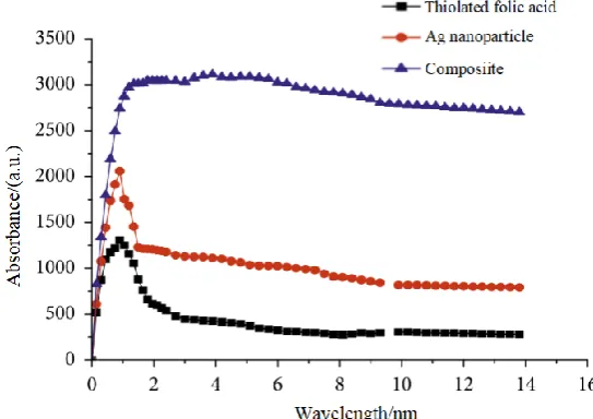
Fig. 1 UV-visible spectra of different substances
Bone morphogenetic protein (BMP) is a kind of bioactive substance existing in bone matrix. It is a small molecule acidic polypeptide substance. It has high bone induction effect and shows the biological characteristics of non-species-specific induction of bone formation. The raw materials used in chemical precipitation are new ecological calcium oxide and phosphoric acid, and calcium hydroxide can react with phosphoric acid, etc. The particle size distribution of the product is wide, the powder uniformity is poor, and agglomeration may occur. In natural bone tissues, apatite crystals are also acicular, which is related to the hexagonal symmetry of bone apatite crystals and the compression of six surfaces into equivalence state [10]. However, the growth process of apatite crystals in bone tissue involves the participation of biomacromolecules. After centrifugation, the molecules were self-assembled with thiolated folic acid in a ratio of 2:3, left to stand for about 8 h, and then centrifuged 5 times to remove excess thiolated folic acid to obtain a thiolated folate-modified silver nanoparticle composite. Nano-hydroxyapatite/chitosan-bone morphogenetic protein composite artificial bone was prepared by nano-hydroxyapatite/chitosan composite bone morphogenetic protein, and the composite artificial bone was implanted into animal proximal humeral bone defect model. Observe. It has been found to functionally promote the expression of osteogenic related genes, improve the activity of alkaline phosphatase and mineral deposition, significantly enhance the cell adhesion and proliferation of osteoblasts, and have good biocompatibility and bone regeneration. ability.
Composite of Nano-hydroxyapatite with Chitin and Its Derivative Chitosan
Chitin, also known as chitin, is a natural non-toxic polymer compound with a molecular structure similar to cellulose, which has good biocompatibility and can be degraded by organisms. In the electrolyte containing calcium and phosphorus ions at a low temperature, a coating is obtained on the metal substrate by an applied electric field, and the coating is processed to form HAP. If the biomacromolecule interacts with the apatite crystal to distort the apatite crystal structure, the characteristic peak of the apatite in the bone tissue spectrum may change [11]. Chitosan is a biosynthetic natural polysaccharide. It is the only alkaline polysaccharide in the known natural polysaccharide. It is obtained by deacetylation of Chitin. Because of its unique one-dimensional structure, high specific surface area and excellent intrinsic physical and mechanical properties, hydroxyapatite has been used to enhance its mechanical strength. Carbon nanotubes/hydroxyapatite composites showed bactericidal effect without antibiotics. Recognition of target tumor cells by functional groups on the surface of HA particles can achieve targeted drug release, reduce its toxicity and reduce side effects. It can promote the growth of vascular endothelial cells and tissue regeneration and repair, and has good biodegradability, biocompatibility, non-toxicity and biological function.
The polyamide is usually a white to pale yellow opaque solid, has a structure similar to collagen, has excellent toughness, and has good biocompatibility. The preparation of nano-hydroxyapatite by sol-gel method has the characteristics of good powder dispersion and uniform particle size. However, the use of organic raw materials and organic solvents makes the production cost high, which hinders the implementation of the method. It is generally believed that this substitution does not substantially destroy the crystal structure of the light-based apatite, and it is non-polluting to the light-based apatite crystal and can be regarded as a normal substitution [12-20]. The nano-hydroxyapatite has good biocompatibility, rigidity, stability of nano-crystal size and toughness of polyamide 66. Its morphology, structure and composition are very similar to human bone apatite nano-needle crystal. Good biocompatibility. This is particularly important in the formation of AH nanoneedle crystals. According to relevant experiments, if the AH sediment is laid down for 3 days before hydrothermal treatment, only very small crystals will be formed [21-26]. The fluorescence intensity of folic acid, mercaptofolic acid and mercaptofolic acid modified silver nanoparticles was measured by fluorescence spectrophotometer. In the reconstruction of vertebral structure, the nano-hydroxyapatite/polyamide 66 composite bioactive support material can effectively restore the height and structure of the vertebral body, heal with the vertebral body and reconstruct the vertebral structure [27 -30]. Therefore, it is necessary to use organic-inorganic method to prepare composite materials in order to obtain bone tissue scaffolds with high strength and better biocompatibility to meet the needs of biomedicine.
Composition of Nano-hydroxyapatite and Polyester
Polylactic acid (PLA) has good biocompatibility, biodegradability and moderate mechanical properties. Bone tissue engineering scaffolds were prepared by suitable preparation methods of PLA and nano-hydroxyapatite. It has no foreign body reaction, non-toxic, degradable, anticoagulant and protease activation, so its composite material has better biocompatibility and biological activity. In the pressurized method, the transport capacity of water as the crystal growth medium is larger than that of organic solution, so the crystal size is larger than that of the formation crystal prepared under normal pressure. Folic acid derivatives containing disulfide bond were synthesized by esterification of disulfide with folic acid, and thiolated folic acid containing free thiol group was prepared by cleavage of disulfide bond with reductant. Nano-hydroxyapatite was prepared by room temperature chemical coprecipitation method, and then modified nano-hydroxyapatite was obtained by polymerizing grafted nano-hydroxyapatite with L-lactide in xylene solution [31 – 36]. There are two contradictory tendencies in crystal growth. One tendency is to release the most energy and minimize its surface energy. Another tendency is to make this process as fast as possible. The addition of nano-hydroxyapatite to the polylactic acid matrix can increase the tensile strain and the area shrinkage of polylactic acid and improve its plasticity. The modified nano-hydroxyapatite and polylactic acid have better mechanical properties. In addition, the composite material also showed good effects in physiological and chemical activities, osteoblast adhesion, drug controlled release and the like.
Composite of Nano-hydroxyapatite and Polyvinyl Alcohol
superparamagnetic properties, enabling targeted gene therapy. This is actually manifested by changing the other parameters to enhance solubility and mass transfer. When the mass distribution coefficient of nano-hydroxyapatite is less than 18%, the tensile strength increases with the increase of nano-hydroxyapatite. However, when it reaches 24%, the tensile strength decreases remarkably, and the compressive strength increases as the nano-hydroxyapatite increases.
Conclusion
In recent years, research on nano-hydroxyapatite and its composite materials and its application in tissue engineering have made gratifying progress, but there are still many problems. If the strength and toughness of the composite material are not fundamentally improved to meet the clinical requirements, the problem of the degradation rate of the composite material in the body and the bone growth rate cannot be completely solved. In human bone, HA nanocrystal size is 5-20x60nn, and the bone apatite crystal is also needle-like. In the enamel, the apatite crystal length can reach 100lun, which is bionic. When the temperature is raised rapidly, the aspect ratio of the n-HA crystal is close to the aspect ratio of the HA crystal in the human dental axis, and some crystal sizes reach the apatite crystal length in the enamel. Mercaptofolic acid was successfully synthesized by mercaptoylation reaction to modify silver nanoparticles. The intermediates and final products were verified by infrared characterization. The complexity of new bone formation mechanism leads to tremendous challenges for guided bone regeneration engineering. Composite hydroxyapatite nanomaterials with better physical and chemical properties, biological activity and osteoinductive ability bring new breakthroughs for bone tissue regeneration. Despite many problems, nano-hydroxyapatite and its composite biomaterials are still the important development direction of tissue engineering materials.
References
[1] A. Akinc, A. Zumbuehl, M. Goldberg, E.S. Leshchiner, V. Busini, N. Hossain, S.A. Bacallado, D.N. Nguyen, J. Fuller, R. Alvarez, A combinatorial library of lipid-like materials for delivery of RNAi therapeutics, Nature biotechnology, 26 (2008) 561.
[2] R.L. Ball, K.A. Hajj, J. Vizelman, P. Bajaj, K.A. Whitehead, Lipid nanoparticle formulations for enhanced co-delivery of siRNA and mRNA, Nano letters, (2018).
[3] T. Bettinger, R.C. Carlisle, M.L. Read, M. Ogris, L.W. Seymour, Peptide-mediated RNA delivery: a novel approach for enhanced transfection of primary and post-mitotic cells, Nucleic acids research, 29 (2001) 3882-3891.
[4] R. Zhang, B.D. Ulery, Synthetic vaccine characterization and design, Journal of Bionanoscience, 12 (2018) 1-11.
[5] K.A. Whitehead, J.R. Dorkin, A.J. Vegas, P.H. Chang, O. Veiseh, J. Matthews, O.S. Fenton, Y. Zhang, K.T. Olejnik, V. Yesilyurt, Degradable lipid nanoparticles with predictable in vivo siRNA delivery activity, Nature communications, 5 (2014) 4277.
[6] K.A. Whitehead, R. Langer, D.G. Anderson, Knocking down barriers: advances in siRNA delivery, Nature reviews Drug discovery, 8 (2009) 129.
[7] S. Wilhelm, A.J. Tavares, Q. Dai, S. Ohta, J. Audet, H.F. Dvorak, W.C. Chan, Analysis of nanoparticle delivery to tumours, Nature reviews materials, 1 (2016) 16014.
[9] A. Wroblewska, M. Dhainaut, B. Ben-Zvi, S.A. Rose, E.S. Park, E.-A.D. Amir, A. Bektesevic, A. Baccarini, M. Merad, A.H. Rahman, Protein Barcodes Enable High-Dimensional Single-Cell CRISPR Screens, Cell, 175 (2018) 1141-1155. e1116.
[10] H. Wu, H. Fan, Z. Shou, M. Xu, Q. Chen, C. Ai, Y. Dong, Y. Liu, Z. Nan, Y. Wang, Extracellular vesicles containing miR-146a attenuate experimental colitis by targeting TRAF6 and IRAK1, International immunopharmacology, 68 (2019) 204-212.
[11] Q. Wu, M. Chen, M. Buchwald, R.A. Phillips, A simple, rapid method for isolation of high quality genomic DNA from animal tissues, Nucleic acids research, 23 (1995) 5087.
[12] R. Xavier, D. Podolsky, Unravelling the pathogenesis of inflammatory bowel disease, Nature, 448 (2007) 427.
[13] S.D. Xiang, C. Selomulya, J. Ho, V. Apostolopoulos, M. Plebanski, Delivery of DNA vaccines: an overview on the use of biodegradable polymeric and magnetic nanoparticles, Wiley Interdisciplinary Reviews: Nanomedicine and Nanobiotechnology, 2 (2010) 205-218.
[14] Z. Yaari, D. Da Silva, A. Zinger, E. Goldman, A. Kajal, R. Tshuva, E. Barak, N. Dahan, D. Hershkovitz, M. Goldfeder, Theranostic barcoded nanoparticles for personalized cancer medicine, Nature communications, 7 (2016) 13325.
[15] R. Zhang, C.N. Leeper, X. Wang, T.A. White, B.D. Ulery, Immunomodulatory vasoactive intestinal peptide amphiphile micelles, Biomaterials science, 6 (2018) 1717-1722.
[16] P.W. Laird, A. Zijderveld, K. Linders, M.A. Rudnicki, R. Jaenisch, A. Berns, Simplified mammalian DNA isolation procedure, Nucleic acids research, 19 (1991) 4293.
[17] R. Zhang, J.D. Smith, B.N. Allen, J.S. Kramer, M. Schauflinger, B.D. Ulery, Peptide Amphiphile Micelle Vaccine Size and Charge Influence the Host Antibody Response, ACS Biomaterials Science & Engineering, 4 (2018) 2463-2472.
[18] A.J. Mukalel, R.S. Riley, R. Zhang, M.J. Mitchell, Nanoparticles for Nucleic Acid Delivery: Applications in Cancer Immunotherapy, Cancer letters, (2019).
[19] J.D. Smith, L.N. Cardwell, D. Porciani, J.A. Nguyen, R. Zhang, F. Gallazzi, R.R. Tata, D.H. Burke, M.A. Daniels, B.D. Ulery, Aptamer-displaying peptide amphiphile micelles as a cell-targeted delivery vehicle of peptide cargoes, Physical biology, 15 (2018) 065006.
[20] X. Luo, B. Li, X. Zhang, W. Zhao, A. Bratasz, B. Deng, D. McComb, Y. Dong, Dual-functional lipid-like nanoparticles for delivery of mRNA and MRI contrast agents, Nanoscale, 9 (2017) 1575-1579.
[21] R. Zhang, J.S. Kramer, J.D. Smith, B.N. Allen, C.N. Leeper, X. Li, L.D. Morton, F. Gallazzi, B.D. Ulery, Vaccine Adjuvant Incorporation Strategy Dictates Peptide Amphiphile Micelle Immunostimulatory Capacity, The AAPS journal, 20 (2018) 73.
[22] G. Kumar, D. Rambhau, S. Apte, Potential Oral Protective Effects of SNEDDS of Diclofenac Sodium on Experimental Gastric Ulcers in Rats, Biochem Pharmacol (Los Angel), 7 (2018) 2167-0501.1000254.
[23] R. Zhang, L.D. Morton, J.D. Smith, F. Gallazzi, T.A. White, B.D. Ulery, Instructive Design of Triblock Peptide Amphiphiles for Structurally Complex Micelle Fabrication, ACS Biomaterials Science & Engineering, (2018).
[24] C. Leonhardt, G. Schwake, T.R. Stögbauer, S. Rappl, J.-T. Kuhr, T.S. Ligon, J.O. Rädler, Single-cell mRNA transfection studies: delivery, kinetics and statistics by numbers, Nanomedicine: Nanotechnology, Biology and Medicine, 10 (2014) 679-688.
by microfluidic mixing exhibit an electron-dense nanostructured core, The Journal of Physical Chemistry C, 116 (2012) 18440-18450.
[26] B. Li, Y. Dong, Preparation and optimization of lipid-like nanoparticles for mRNA delivery, in: RNA Nanostructures, Springer, 2017, pp. 207-217.
[27] B. Li, X. Luo, B. Deng, J.B. Giancola, D.W. McComb, T.D. Schmittgen, Y. Dong, Effects of local structural transformation of lipid-like compounds on delivery of messenger RNA, Scientific reports, 6 (2016) 22137.
[28] B. Li, X. Zhang, Y. Dong, Nanoscale platforms for messenger RNA delivery, Wiley Interdisciplinary Reviews: Nanomedicine and Nanobiotechnology, (2018) e1530.
[29] M.P. Lokugamage, C.D. Sago, J.E. Dahlman, Testing thousands of nanoparticles in vivo using DNA barcodes, Current Opinion in Biomedical Engineering, (2018).
[30] K.T. Love, K.P. Mahon, C.G. Levins, K.A. Whitehead, W. Querbes, J.R. Dorkin, J. Qin, W. Cantley, L.L. Qin, T. Racie, Lipid-like materials for low-dose, in vivo gene silencing, Proceedings of the National Academy of Sciences, 107 (2010) 1864-1869.
[31] M. Lueckheide, J.R. Vieregg, A.J. Bologna, L. Leon, M.V. Tirrell, Structure-property relationships of oligonucleotide polyelectrolyte complex micelles, Nano letters, (2018).
[32] R. Zhang, M.M. Billingsley, M.J. Mitchell, Biomaterials for vaccine-based cancer immunotherapy, Journal of Controlled Release, (2018).
[33] C.A. Machado, I.R. Smith, D.A. Savin, Self-Assembly of Oligo-and Polypeptide-Based Amphiphiles: Recent Advances and Future Possibilities, Macromolecules, (2019).
[34] A. Marabelle, H. Kohrt, C. Caux, R. Levy, Intratumoral immunization: a new paradigm for cancer therapy, Clinical Cancer Research, 20 (2014) 1747-1756.
[35] C.J. McKinlay, N.L. Benner, O.A. Haabeth, R.M. Waymouth, P.A. Wender, Enhanced mRNA delivery into lymphocytes enabled by lipid-varied libraries of charge-altering releasable transporters, Proceedings of the National Academy of Sciences, 115 (2018) E5859-E5866.
[36] J.M. McLendon, S.R. Joshi, J. Sparks, M. Matar, J.G. Fewell, K. Abe, M. Oka, I.F. McMurtry, W.T. Gerthoffer, Lipid nanoparticle delivery of a microRNA-145 inhibitor improves experimental pulmonary hypertension, Journal of Controlled Release, 210 (2015) 67-75.
[37] P. Midoux, C. Pichon, Lipid-based mRNA vaccine delivery systems, Expert review of vaccines, 14 (2015) 221-234.
[38] J.B. Miller, S. Zhang, P. Kos, H. Xiong, K. Zhou, S.S. Perelman, H. Zhu, D.J. Siegwart, Non‐ Viral CRISPR/Cas Gene Editing In Vitro and In Vivo Enabled by Synthetic Nanoparticle Co‐ Delivery of Cas9 mRNA and sgRNA, Angewandte Chemie, 129 (2017) 1079-1083.
[39] J.B. Miller, S. Zhang, P. Kos, H. Xiong, K. Zhou, S.S. Perelman, H. Zhu, D.J. Siegwart, Non‐ viral CRISPR/Cas gene editing in vitro and in vivo enabled by synthetic nanoparticle co‐delivery of Cas9 mRNA and sgRNA, Angewandte Chemie International Edition, 56 (2017) 1059-1063.
[40] G. Minigo, A. Scholzen, C.K. Tang, J.C. Hanley, M. Kalkanidis, G.A. Pietersz, V. Apostolopoulos, M. Plebanski, Poly-L-lysine-coated nanoparticles: a potent delivery system to enhance DNA vaccine efficacy, Vaccine, 25 (2007) 1316-1327.
[41] M.J. Mitchell, R.K. Jain, R. Langer, Engineering and physical sciences in oncology: challenges and opportunities, Nature Reviews Cancer, 17 (2017) 659.