Journal of Engineering and Applied Sciences 14 (Special Issue 9): 10659-10664, 2019 ISSN: 1816-949X
© Medwell Journals, 2019
Preparation and Characterization of Bioceramic Composite Based on
Hydroxyapatite and Silver Nanoparticles
Mohsin Abbas Aswad, Hayder Kraidi Rashed and Azhar abd Selman
Department of Engineering of Ceramic and Building Materials, College of Materials Engineering,
University of Babylon, Hillah, Iraq
Abstract: In this study, composite material consistsof silver (Ag) nanoparticles added to hydroxyapatite as matrixderived from bovine bone was prepared to investigate the possibility of using hydroxyapatite as bone substitute in high load bearing locations. The phase composition and structure of the sintered bio-ceramics composite were identified by X-Ray Diffraction (XRD) and Scanning Electron Microscopy (SEM-EDX), respectively. Densification rate of HA/Ag was assessed by measuring the density and apparent porosity. A mechanical properties of the sintered HA/Ag was carried out by technique of vickers indentations and compressive strength determinations. The results showed that the bioceramic composite present higher densification, better mechanical properties compared to pure hydroxyapatite.
Key words:Hydroxyapatite, silver nanoparticles, biocomposites, bovine hydroxyapatite and mechanical properties, HA/Ag, measuring
INTRODUCTION
Bone can regenerate themselves and is one of few human tissues. Activity osteogenesis causes the fracture of bone but ultimately the fracture heals without any scar creation. The bones have the ability to reappear but this ability slow down with age and will affect with diseases and other sources and this will happens will small bone flaws. The grafts are important to help bone repair after loss of bone is too high such as sarcoma of bone in cases of excision of bone tumour or other disease of bones in some accidents will cause bone loss and in fracture of complicated bone that cannot repair themselves. Many grafts can choice in clinics polymers, metal, bioglasses and calcium phosphate biomaterials have studied for repairing of the bone (Kokubo et al., 2003; Ducheyne and Qiu, 1999; Jones et al., 2006). Calcium phosphates are preferred as a materials for bone grafts inengineering of hard tissue because of their bioactivity and superior biocompatibility (Kalita et al., 2007).
The normal HA ceramics have low mechanical strength that will restrict its uses for low load bearing applications, the fracture toughness and other mechanical properties for HA will improve with increasing its surface area that will lead to improve sinterability and enhanced densification (Zhou and Lee, 2011).
The current trend in biomedical ceramics is focused on using and developing biocomposites ceramic (Pattanayak et al., 2011). The fracture toughness and other mechanical properties limiting of bioceramics materials for load bearing applications (Best et al., 2008).
In current research silver nanoparticles (Ag) which used for reinforcing hydroxyapatite in orthopedic implant considerations. Precisely, the effect of silver nanoparticle on the mechanical properties of crystalline HA will be studied in this research. Silver in the recent research will take attention because its unique important properties for example resistance of corrosion and antimicrobial activities (Zhang et al., 2017). The infections of the implanted part was protected by its antimicrobial activities (Victor and Roberto, 2015).
MATERIALS AND METHODS
Experimental procedures
Preparation of hydroxyapatite from bovine bone and HA/Ag bioceramicscomposite processing: Preparation of hydroxyapatite from bovine bonethe procedure discussed by Khoo et al. (2015) commercial silver powder was 80 nm particle size. (Hongwu International Group Ltd, China). Hydroxyapatite was mixed with Ag nanoparticles in a ball mill under variation of the Ag nanoparticles contents. The percentage of Ag nanoparticles was 5, 10 and 15 Vol. % from total amount of hydroxyapatite. By this method three types of ceramic samples containing different percentages of Ag nanoparticles were prepared. HA/Ag powder was uniaxially pressed at 152 MPa into green bodies using a 10 mm for cylindrical and 60×5 mm for rectangular dies and sintered at various sintering temperatures of 1200 and 1250°C. There are wide variations in the reported mechanical property of HA depend on its sources and manufactures.
Fig. 1: XRD results of HA specimen at 1000, 1200 and 1250°C
Fig. 2: XRD results of HA/5, 10, 15 vol.% Ag specimens at 1200°C
RESULTS AND DISCUSSION
X-ray diffraction: Figure 1 shows the result of x-ray diffraction analysis of HA powder calcined at 1000°C with a heating rate of 10°C/min and soaking time of (2 h). The result shows that all peaks corresponding with hydroxyapatite phase and matching with card number (JCPDS, card NO. 09-0432) but the XRD result of HA powder sample when increasing the sintering temperature to 1250°C shows appear minor phase of β-tricalcium phosphate and matched with card number (JCPDS, card NO. 09-0432) for hydroxyapatite (JCPDS, card NO. 09-0169) for β-tricalcium phosphate The temperature was used for sintering HA is 1200°C due to no decomposition of hydroxyapatite (Al-Dujaili et al., 2017).
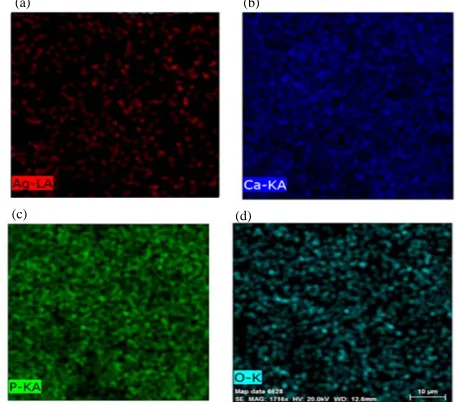
Fig. 3(a-d): SEM-EDX map for HA-5 Vol.% Ag
Fig. 4(a-d): SEM-EDX map for HA-0 Vol.% Ag Figure 2 shows the XRD of HA -5, 10 and 15 Vol. % Ag, respectively. The results show that sliver did not disrupt the apatite structure (Bharti et al., 2014; Nirmala et al., 2011). XRD data reveals that the sample does not lose its crystallinity after the incorporation of AgNPs which makesa good Ag-HA composite material (Maidaniuc et al., 2017).
Figure 3-5 show SEM-EDX map for HA/Ag composite with the increased concentration of Ag particles display a slight agglomeration tendency (Maidaniuc et al., 2017; Zhuk et al., 2015). The actual rate of penetration of Ag might vary largely depending on the actual wettability and the porous structure (Bracco and Holst, 2013).
HA pure 1000°C HA pure 1200°C HA pure 1250°C
10 20 30 40 50
2 (degree)
HA
-TCP
HA/15 Vol.% Ag HA/10 Vol. % Ag HA/5 Vol. % Ag
Ag pure
0 10 20 30 40 50 60 70 80 90 0 10 20 30 40 50 60 70 80 90
2 (degree)
(a) (b)
(c) (d)
(a) (b)
[image:2.612.112.290.102.290.2] [image:2.612.321.549.327.528.2] [image:2.612.86.282.332.503.2]J. Eng. Applied Sci., 14 (Special Issue 9): 10659-10664, 2019
[image:3.612.161.467.98.345.2]Fig. 5(a-d): SEM-EDX map for HAP- 15 Vol.% Ag
Fig. 6: FTIR of HA powder calcination at 1000°C Fourier Transform Infrared Spectrometer (FTIR): Infrared characterization was carried out for the sample to study the spectral characteristics indicative of the chemical bonding in the synthesized HAP powder calcination at 1000°C. The spectrum Figure 6 can be divided into four regions with peaks having wave numbers around 3571, 1075, 1036 and 602 cmG1
(Mondal et al., 2012).
After 6 h of milling in a planetary ball mill of the prepared hydroxyapatite. It can be observed that the prepared powder have multimodal distribution, the
particles size distribute on the 0.290 and 5.132 μm with a mean size of 0.886 µm as shown in Fig. 7.
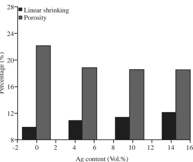
Relative density, porosity and shrinkage: The result of relative density was measured before sintering (B. Sintering) and after sintering (A. Sintering) but the other properties such as porosity and linear shrinkage were measured for HA and HA/Ag composite as sintering at 1200°C are shown in Fig. 8 and 9, respectively.
1076 P-O
1036 P-O
651 O-H
602 O-H
581 P-O
3600 3000 2400 1950 1650 1350 1050 900 750 600 450 1/cm 14
12
10
8
6
4
2
-0
11 cm
T (%) 3571
O-H
(a) (b)
[image:3.612.153.469.379.568.2]J. Eng. Applied Sci., 14 (Special Issue 9): 10659-10664, 2019
-2 0 2 4 6 8 10 12 14 16 Ag content (Vol.%)
100 95 90 85 80 75 70 65 60 55 50 R el a ti ve s de n si ty ( % ) A. sintering B. sintering 28 24 20 16 12 8 Pe rc en ta g e (% )
-2 0 2 4 6 8 10 12 14 16
Ag content (Vol.%) Linear shrinking
[image:4.612.137.477.102.287.2]Porosity
[image:4.612.298.516.108.485.2]Fig. 7: particle size distribution of hydroxyapatite powder
Fig. 8: Relation between relative density and Ag content before and after sintering
From the result in Fig. 10 shows that when adding Ag lead to increase in the relative density which is due to aneffect of adding Ag which act aid sintering and enhancing densification by Ag caused stagnation of grain growth by pinning the grain boundaries (Maidaniuc et al., 2016).
The higher relative green densities of the composites are due to better packing of the mixture of HA/Ag particles and due to plastic deformation of Ag pa rticles under compaction (Maidaniuc et al., 2017). The sintering temperature had a significant influence on the densification of HA-Ag composites. The densities of both pure HA and HA-Ag compositesincreased rapidly with the sintering temperature to 1200°C (Chaki and Wang, 1994).
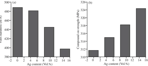
Compression strength and vickers hardness: The result of compression strength and vickers hardness for HA
Fig. 9: Relation porosity and shrinkage versus Ag content and HA-Ag composite as sintering at 1200°C shown in Fig. 10. This result calculated according to ASTM standard C-773 and ASTM standard C1327-90, respectively.
The maximum compressive strength was obtained at temperature 1200°C due to complete sintering process of HA/Ag specimen with high density value. However, the compressive strength of hydroxyapatite was increase with increasing Ag content because the porosity of the sample decrease at high volume fraction of silver. The compressive strength of HA/Ag is a property depends on the crystal defects and porosities. The Vickers hardness results are shown in Fig. 10, not the similar trend of compression strength and the lowest hardness was obtained at 15 Vol.% from Ag at the same temperature as shown above because the high content of silver leads to increase the ductility phase.
100.0 90.0 80.0 70.0 60.0 50.0 40.0 30.0 20.0 10.0 0.0 Cum u (%)
0.0 0.1 1.0 10.0 100.0 1000.0 8.1 7.2 6.3 5.4 4.5 3.6 2.7 1.8 0.9 0.0 Size (um) D10 = 0.290
D50 = 0.886 D90 = 5.132
Cum
u
[image:4.612.321.518.322.488.2] [image:4.612.86.287.325.475.2]J. Eng. Applied Sci., 14 (Special Issue 9): 10659-10664, 2019 500 480 460 440 420 400 380 Vd es h a rd n ess (H/ N )
-2 0 2 4 6 8 10 12 14 16 Ag content (Vol.%)
326 324 320 318 316 314 312 310 Co m p ou n d on s tre n g th ( M Pa )
-2 0 2 4 6 8 10 12 14 16 Ag content (Vol.%)
(a) (b)
Fig. 10(a, b): Relation compression strength and hardness versus AG content CONCLUSION
According to the results of the experimental work can be concluded. Extraction method from bovine bone considers the suitable, economical and safe method to produce pure hydroxyapatite. Silver is an appropriate metal to addition in traditional sintering. Addition of Ag to hydroxyapatite leads to increase the density, compression strength and decrease the vickers hardness of hydroxyapatite.
REFERENCES
Al-Dujaili, M.A.A., S. Jaheel and H.N. Abbas, 2017. Preparation of HA/ß-TCP scaffold and mechanical strength optimization using a genetic algorithm method. J. Aust. Ceram. Soc., 53: 41-48.
Best, S.M., A.E. Porter, E.S. Thian and J. Huang, 2008. Bioceramics: Past, present and for the future. J. Eur. Ceram. Soc., 28: 1319-1327.
Bharti, A., S. Singh, V.K. Meena and N. Goyal, 2014. Synthesis of novel multiple shaped silver nanoparticles incorporated hydroxyapatite anocomposite for orthopaedic body implants. Adv. Sci. Lett., 20: 1297-1302.
Bracco, G. and B. Holst, 2013. Surface Science Techniques. 1st Edn., Springer, Berlin, Germany, ISBN: 978-3-642-34242-4, Pages: 663.
Chaki, T.K. and P.E. Wang, 1994. Densification and s t r e n g t h e n i n g o f s i l v e r - r e i n f o r c e d hydroxyapatite-matrix composite prepared by sintering. J. Mater. Sci. Mater. Med., 5: 533-542. Ducheyne, P. and Q. Qiu, 1999. Bioactive ceramics:
The effect of surface reactivity on bone formation and bone cell function. Biomaterials, 20: 2287-2303.
Jones, J.R., L.M. Ehrenfried and L.L. Hench, 2006. Optimising bioactive glass scaffolds for bone tissue engineering. Biomaterials, 27: 964-973.
Kalita, S.J., A. Bhardwaj and H.A. Bhatt, 2007. Nanocrystalline Calcium Phosphate ceramics in biomedical engineering. Mater. Sci. Eng. C., 27: 441-449.
Khoo, W., F.M. Nor, H. Ardhyananta and D. Kurniawan, 2015. Preparation of natural hydroxyapatite from bovine femur bones using calcination at various temperatures. Procedia Manuf., 2: 196-201.
Kokubo, T., H.M. Kim and M. Kawashita, 2003. Novel bioactive materials with different mechanical properties Biomaterials, 24: 2161-2175.
Maidaniuc, A., F. Miculescu, A.C. Mocanu, S.I. Voicu, M. Miculescu, A. Purcaru and M.E. Rada, 2017. Sinterability study of bovine-derived hydroxyapatite and silver microcomposites. Univ. Politehnica Bucharest Sci. Bull. Ser. B. Chem. Mater. Sci., 79: 145-154.
Maidaniuc, A., M. Miculescu, S.I. Voicu, L.T. Ciocan, M. Niculescu, M. C. Corobea and F. Miculescu, 2016. Effect of micron sized silver particles concentration on the adhesion induced by sintering and antibacterial properties of hydroxyapatite microcomposites. J. Adhes. Sci. Technol., 30: 1829-1841.
Mondal, S., B. Mondal, A. Dey and S.S. Mukhopadhyay, 2012. Studies on processing and characterization of Hydroxyapatite biomaterials from different bio wastes. J. Miner. Mater. Charact. Eng., 11: 55-67.
Nirmala, R., F.A. Sheikh, M.A. Kanjwal, J.H. Lee, S.J. Park, R. Navamathavan and H.Y. Kim, 2011. Synthesis and characterization of bovine femur bone hydroxyapatite containing silver nanoparticles for the biomedical applications. J. Nanopart Res., 13: 1917-1927.
[image:5.612.140.454.101.241.2]Victor, S.U. and V.B.J. Roberto, 2015. Gold and silver nanotechology on medicine. J. Chem. Biochem., 3: 21-33.
Zhang, X., W. Chaimayo, C. Yang, J. Yao, B.L. Miller and M.Z. Yates, 2017. Silver-hydroxyapatite composite coatings with enhanced antimicrobial activities through heat treatment. Surf. Coat. Technol., 325: 39-45.
Zhou, H. and J. Lee, 2011. Nanoscale hydroxyapatite particles for bone tissue engineering. Acta Biomater., 7: 2769-2781.