Metabolic Modeling of
Streptococcus mutans
Reveals Complex
Nutrient Requirements of an Oral Pathogen
Kenan Jijakli,a,b Paul A. Jensena,b,c
aDepartment of Bioengineering, University of Illinois at Urbana-Champaign, Urbana, Illinois, USA
bCarl R. Woese Institute for Genomic Biology, University of Illinois at Urbana-Champaign, Urbana, Illinois, USA cDepartment of Microbiology, University of Illinois at Urbana-Champaign, Urbana, Illinois, USA
ABSTRACT Streptococcus mutans is a Gram-positive bacterium that thrives under
acidic conditions and is a primary cause of tooth decay (dental caries). To better un-derstand the metabolism of S. mutans on a systematic level, we manually con-structed a genome-scale metabolic model of the S. mutans type strain UA159. The model, called iSMU, contains 675 reactions involving 429 metabolites and the prod-ucts of 493 genes. We validated iSMU by comparing simulations with growth experi-ments in defined medium. The model simulations matched experimental results for 17 of 18 carbon source utilization assays and 47 of 49 nutrient depletion assays. We also simulated the effects of single gene deletions. The model’s predictions agreed with 78.1% and 84.4% of the gene essentiality predictions from two experimental data sets. Our manually curated model is more accurate thanS. mutansmodels gen-erated from automated reconstruction pipelines and more complete than other manually curated models. We used iSMU to generate hypotheses about the S. mu-tans metabolic network. Subsequent genetic experiments confirmed that (i) S. mu-tanscatabolizes sorbitol via a sorbitol-6-phosphate 2-dehydrogenase (SMU_308) and (ii) the Leloir pathway is required for growth on complex carbohydrates such as raffinose. We believe the iSMU model is an important resource for understanding the metabolism ofS. mutansand guiding future experiments.
IMPORTANCE Tooth decay is the most prevalent chronic disease in the United
States. Decay is caused by the bacterium Streptococcus mutans, an oral pathogen that ferments sugars into tooth-destroying lactic acid. We constructed a complete metabolic model of S. mutans to systematically investigate how the bacterium grows. The model provides a valuable resource for understanding and targeting S. mutans’ ability to outcompete other species in the oral microbiome.
KEYWORDS Streptococcus mutans, dental caries, flux balance analysis, metabolic
modeling
S
treptococcus mutansis one of over 600 species of bacteria in the oral microbiome (1). This Gram-positive, lactic acid bacterium thrives in the oral environment in part due to its metabolic flexibility.S. mutanscan feed on several carbohydrates (2) and has complex, interdependent amino acid auxotrophies (3).S. mutansis the primary cause of tooth decay (dental caries). By fermenting a wide array of dietary sugars into lactic acid,S. mutanscreates a highly acidic microenvironment near the tooth surface (as low as pH 3.0) (4). The lactic acid demineralizes the tooth structure, resulting in decay.Understanding the acidogenic capabilities of S. mutans requires an unbiased, systems-level approach. Previous studies have shown that acid production and toler-ance inS. mutansrequire large changes in gene expression and metabolic pathway utilization (5). For example, decreasing pH increases glycolytic activity and branched-chain amino acid synthesis without increasing cell growth (6). A drop in pH is also
CitationJijakli K, Jensen PA. 2019. Metabolic modeling ofStreptococcus mutansreveals complex nutrient requirements of an oral pathogen. mSystems 4:e00529-19.https://doi .org/10.1128/mSystems.00529-19.
EditorKatrine L. Whiteson, University of California, Irvine
Copyright© 2019 Jijakli and Jensen. This is an open-access article distributed under the terms of theCreative Commons Attribution 4.0 International license.
Address correspondence to Paul A. Jensen, [email protected].
A genome-scale metabolic model predicts nutrient requirements and gene essentiality of the oral pathogen Streptococcus mutans.
Received25 August 2019 Accepted6 October 2019 Published
Molecular Biology and Physiology
29 October 2019
on September 8, 2020 by guest
http://msystems.asm.org/
accompanied by an increased expression of F-ATPases to maintain a higher intracellular pH (7).
Mathematical models aid in our understanding of how an organism’s genes collec-tively give rise to a phenotype. Models translate bioinformatic features (differential expression, presence/absence of genes) into biological function (flux distributions, uptake and secretion rates, and fitness). Constraint-based reconstruction and analysis (COBRA) of genome-scale models is widely used to integrate genetic and metabolic data to produce phenotypic predictions (8, 9). Models of microbial metabolism and transcriptional regulation predict responses to gene deletions (10, 11), mutation (12, 13), metabolic shifts (14, 15), and long-term evolution (16, 17). The models identify emergent properties of a metabolic network, including links between pathways and interdependencies among genes (18, 19).
We present iSMU v1.0, a genome-scale metabolic model for theS. mutanstype strain UA159. Our model is manually curated using multiple databases, literature evidence, and phenotyping experiments. Model predictions were tested using growth and mu-tagenesis experiments, demonstrating the utility of the iSMU model in guiding predic-tions and experiments. Our investigation ofS. mutansfocused on metabolism for two reasons: (i) the primary metabolic products ofS. mutans, lactic acid and biofilm matrix, are responsible for the pathogen’s cariogenicity, and (ii) metabolic networks are among the best-characterized intracellular networks with established computational tech-niques. Given metabolism’s central role in cariogenesis, we believe the iSMU model will improve our understanding ofS. mutans’ role in oral health.
RESULTS
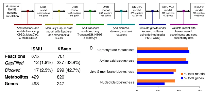
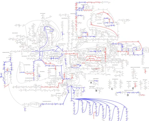
Manual curation produces an annotated metabolic model for S. mutans.We manually reconstructed anin silicometabolic model forS. mutanstype strain UA159 (Fig. 1A). Our model, named iSMU for “in silico S. mutans,” includes major metabolic pathways for carbohydrate metabolism and synthesis of amino acids, nucleotides, lipids, vitamins, and cofactors. The model includes 675 reactions transforming 429 metabolites. The reactions are catalyzed by the products of 493 genes (Fig. 1B). Figure 2 shows all reactions in the iSMU model.
Our assembly of iSMU began with reaction databases and an annotated genome. Draft models assembled from genome annotations are often incomplete because of gaps in the genome annotation or spontaneous reactions that lack an associated
FIG 1 (A) Reconstruction of theS. mutansmetabolic network began with an annotated UA159 genome. The draft model was refined with bioinformatics databases and experimental results. (B) The manually reconstructed model (iSMU) has fewer GapFilled (non-gene-associated) and blocked reactions than a model generated automatically by the KBase system. Metabolite counts do not reflect compartmentalization, e.g., “glucose[c]” and “glucose[e]” are counted as one metabolite. (C) The reactions and genes in the iSMU model are distributed across a range of KEGG subsystems.
on September 8, 2020 by guest
http://msystems.asm.org/
enzyme. Several computational methods attempt to identify and add these missing reactions in a process called GapFilling (20). Rather than rely on automated GapFilling algorithms, we manually GapFilled iSMU by examining the reactions in each pathway and testing for growth in chemically defined medium (CDM) with glucose as the sole carbon source. We attempted to close gaps in any pathway that (i) was complete except for a small number of reactions or (ii) was blocked (unable to carry flux) due to metabolites that could not be produced or consumed. We also attempted to find unannotated or misannotated genes that could catalyze the GapFilled reactions. Com-pared to other metabolic reconstructions, our manually GapFilled model contains fewer incomplete pathways (Fig. 1B; see also Table S3 in the supplemental material). On average, published metabolic models of well-studied organisms lack gene annotations for 53% of the models’ reactions (Table S3). These models also average 32.7% blocked reactions, i.e., reactions that cannot carry a steady-state flux because they lack upstream or downstream pathways. Our iSMU model has only 23% reactions without an associ-ated enzyme and 2.5% blocked reactions.
Metabolic models simulate growth by collecting cellular building blocks into a biomass reaction. The biomass reaction is used as the objective function for metabolic
FIG 2 A custom pathway map shows all reactions in the iSMU model. (A high-resolution image is available as Data Set S4 in the supplemental material.)S.
mutansUA159 appears to lack complete pathways for synthesizing thiamine (A), riboflavin (B), pyridoxine (C), pantothenate (D), and biotin (E). Reactions are
colored by agreement between the essentiality predictions of the associated genes and Tn-seq data from the work of Shields et al. (31). Blue reactions agree with the Tn-seq essentiality results; red reactions disagree. Overall agreement between the data sets is 84.8% (Fig. 4).
on September 8, 2020 by guest
http://msystems.asm.org/
simulations. A nonzero flux through the biomass reaction indicates growth in the metabolic environment specified by the model’s inputs (called exchange reactions). We modified the biomass reaction from a model ofEnterococcus faecalisV583 (21) to create a biomass reaction forS. mutansUA159. BothE. faecalisandS. mutansare lactic acid bacteria, with similar metabolic capabilities. To tailor the biomass reaction toS. mutans, we changed the relative ratios of nucleotides and amino acids. We also changed the cell membrane composition to reflect membrane sugar polysaccharides specific to S. mutans. We replaced UDP-N-acetyl-D-galactosamine, which, based on genetic evidence, is not produced byS. mutans, with UDP-N-acetyl-D-mannosamine and UDP-N -acetyl-D-glucosamine (22). We also adjusted the cell wall fatty acids to their measured propor-tions at pH 7.0 (23). The final biomass reaction consumes 56 metabolites to produce a unit of biomass.
Manual curation improves model consistency and accuracy. Several software systems can generate draft metabolic models using reaction databases and annotated genomes, a process called automatic reconstruction. Typically, metabolic models are reconstructed by first using a software package to generate a draft model and then manually curating that draft model to increase its consistency and reconcile it with other biological data. Our first attempt at reconstructing the metabolism ofS. mutans
used a draft model from the KBase system (24). Unfortunately, the draft model lacked many of the metabolic features of lactic acid bacteria and included several subsystems known to be inactive in homofermentative anaerobes. We therefore abandoned the KBase model and began a manual reconstruction process. We compared the final iSMU model to the KBase model to quantify the disagreement between the manual and automated reconstruction pipelines.
Our manually curated reconstruction differs substantially from the reconstruction produced automatically by KBase. Our iSMU model has 3.7% fewer reactions than the KBase model (675 versus 701, respectively) but 99.5% more genes (493 versus 247, respectively) (Fig. 1B). Thus, the manually curated model has a larger proportion of gene-associated reactions than the automated reconstruction. The dearth of gene associations in the KBase model is due in part to the 237 reactions added during GapFilling, since GapFilled reactions are added without genomic evidence for the reaction. By comparison, our iSMU model required only 12 GapFilled reactions to enable growth on defined medium.
The KBase model contains 41% more metabolites than our iSMU model. Unfortu-nately, 22 of the metabolites in the KBase model are “dead-end” metabolites that lack either a producing or a consuming reaction. The dead-end metabolites block flux through 299 (43%) of the KBase model’s reactions. At steady state, these blocked reactions cannot carry flux or be analyzed using flux balance analysis (FBA). About 2.5% of the reactions in our iSMU model are blocked, indicating more complete reaction pathways than in the model created by automated reconstruction.
A comparison of iSMU to other microbial genome-scale models. Existing genome-scale metabolic models play an important role in the reconstruction of models for new species. It is common for the new models to be built by comparison to the older ones, just as our iSMU model builds on the biomass reaction from anE. faecalis
model. Automated reconstruction pipelines in particular depend on existing models (24–26). The earliest reconstruction of a microbial genome-scale metabolic model was for the Gram-negative Escherichia coli K-12 MG1655 (27), and this model has had periodic updates, culminating in the model iML1515 (28). Among the first Gram-positive reconstructions was a model ofBacillus subtilis168 (29). The latestB. subtilis168 model is iBSU1144 (30). Both models represent a well-studied microorganism with a wealth of existing data.
Relative to the total number of genes in each organism, iSMU contains 24.2% of all
S. mutansgenes, while iML1515 contains 35% of allE. coligenes and iBSU1144 contains 16.5% of allB. subtilisgenes. The high fraction of modeled genes in theE. colimodel may reflect its 2-decade history of curation and updates. The number of blocked
on September 8, 2020 by guest
http://msystems.asm.org/
reactions in iSMU is significantly lower than in both other models (2.5% compared to 35.6% in iML1515 and 67.9% in iBSU1144). Finally, the number of components that are required for the production of biomass in iML1515 and iBSU1144 is higher than in iSMU. The biomass reaction of iML1515 includes 101 components, nearly twice the number of components found in iSMU’s biomass reaction (Table S4).
Primary metabolism (metabolism of carbohydrates, amino acids, nucleotides, lipids, fatty acids, and cell membrane components) includes 308 genes in iSMU, or 62.5% of all genes in the model. Primary metabolism in iML1515 includes 607 genes (40%) and in iBSU1144 includes 398 genes (55.4%). The largest proportion of genes in iSMU are involved in carbohydrate metabolism (120 genes, 24.3%). This is followed by amino acid metabolism (118 genes, 23.9%) and then metabolism of lipids, fatty acids, and cell membrane components (84 genes, 17%), and then, finally, nucleotide metabolism (63 genes, 12.7%) (Fig. 1C). In contrast, the plurality of genes in iML1515 and iBSU1144 were involved in amino acid metabolism. In iML1515, amino acid metabolism involved 229 genes (15.1%), followed by carbohydrate metabolism (205 genes, 13.5%); metabolism of lipids, fatty acids, and cell membrane components (179 genes, 11.8%); and finally nucleotide metabolism (131 genes, 8.6%). There were 195 genes involved in amino acid metabolism in iBSU1144 (27.1%), followed by 122 genes involved in carbohydrate metabolism (17%). Metabolism of lipids, fatty acids, and cell membrane components and metabolism of nucleotides were tied at 100 genes each (13.9%). Compared toE. coli or B. subtilis, a larger fraction of the genome of S. mutans is associated with carbohydrate metabolism. The focus on carbohydrates is consistent with S. mutans’ need to scavenge dietary sugars while living solely in the human mouth.
Only 54 genes in iSMU are involved in the biosynthesis and metabolism of vitamins and cofactors, compared to 178 genes in iML1515 and 117 genes in iBSU1144. The low number of biosynthetic genes is consistent withS. mutans’ dependence on its host for these nutrients. S. mutans is auxotrophic for several vitamins and cofactors (Fig. 3), whereas the free-living E. coli and B. subtilis can synthesize their own secondary metabolites.
Gene deletion simulations match experimental data.Metabolic models contain chemical reactions and gene associations that link reactions to their corresponding enzymes. Gene associations are expressed as logical statements describing the required gene products for a reaction to carry flux. An enzymatic complex of two proteins is expressed using “and” (subunit 1 and subunit 2). A pair of isozymes that could each independently catalyze a reaction would be written with an “or” (isozyme 1 or isozyme 2). Flux balance analysis and the model’s gene associates can be combined to simulate the effects of gene deletions on growth. The logical rules in the gene associations are evaluated to identify reactions that cannot carry flux in a deletion strain. Reactions that cannot carry flux are removed from the model before calculating the maximum biomass flux. Deletion of an essential gene will prevent any nonzero biomass flux. Comparing experimentally determined essential genes with the model’s predictions validates the model’s gene associations.
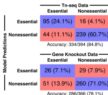
We simulated the effects of all single deletions for the 493 genes in the iSMU model (Data Set S5). We compared thein silicodeletions to two experimental gene deletion studies inS. mutansUA159: a transposon mutagenesis sequencing (Tn-seq) experiment (31) and a screen of an ordered array of single-gene deletion strains (32). We checked every disagreement between the model simulations and the experimental data to correct errors in gene associations. Changes were validated by literature searches whenever possible.
The Tn-seq study used a mariner-family transposon to generate random insertions across the UA159 genome (31). The transposon-genomic DNA junctions were amplified and sequenced to quantify fitness after growth in a defined medium (FMC). Genes lacking transposon insertions sites are predicted to be essential in FMC. Overall, 84.8% of the essentiality predictions from the iSMU model were consistent with the Tn-seq
on September 8, 2020 by guest
http://msystems.asm.org/
data (Fig. 4). A comparison between iSMU’s essential gene predictions and the Tn-seq data is shown in the iSMU map in Fig. 2.
The gene deletion strains in the work of Quivey et al. (32) were constructed individually using homologous recombination with a selective marker. The deletion library contains strains for 1,112 of the 1,956 genes inS. mutansUA159, including 366 of the 488 genes in the iSMU metabolic model. The remaining 122 genes are hypoth-esized to be essential or could not be deleted due to technical limitations. Deletion strains were grown in brain heart infusion medium (BHI), a rich and undefined medium. We simulated BHI by opening all exchange reactions in the iSMU model. As shown in Fig. 4, 78.1% of the experimental essentiality results agreed with the model predictions. Agreement between iSMU’s essential gene predictions and the data from the work of Quivey et al. (32) is highlighted in the iSMU map in Data Set S4.
S. mutanshas complex nutrient requirements.Several studies have investigated the minimal requirements forS. mutansgrowthin vitro(3, 33–36). Like many obligate
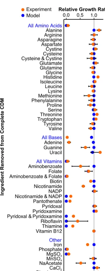
FIG 3 Model predictions (blue) match growth experiments forS. mutansUA159 in defined medium (orange). Growth rate was measured for CDM lacking the specified component(s). Blue labels indicate removal of all components listed below. Experimental data are means from three independent trials with error bars representing the standard deviation. Growth rates are normalized toS. mutansUA159 grown in complete CDM.
on September 8, 2020 by guest
http://msystems.asm.org/
human pathogens,S. mutansrequires a combination of carbon, nitrogen, sulfur, and phosphorus sources; inorganic minerals; nucleotides; and vitamins and cofactors. We used our iSMU model and phenotypic assays to systematically explore auxotrophies in
S. mutans.
TheS. mutansUA159 genome encodes complete biosynthetic pathways for all 20 amino acids (22). S. mutans can grow without any exogenous amino acids using ammonium as the sole nitrogen source (33). The iSMU model can similarly produce biomass with ammonium and no amino acids. Using a series of leave-one-out exper-iments, we confirmed that the removal of individual amino acids from a defined medium does not affectS. mutansgrowth in vitro (Fig. 3). Simultaneous removal of cysteine and cystine does not significantly reduce growth, indicating thatS. mutanscan catabolize another sulfur source, possibly sulfate or methionine.
S. mutans can theoretically synthesize nucleotides (adenine, guanine, cytosine, uracil, and thymine) but only through the nonoxidative branch of the pentose phos-phate pathway (37). S. mutansUA159 apparently lacks the more efficient oxidative branch of the pentose phosphate pathway (38). The nonoxidative pentose phosphate pathway is bidirectional and can produce or recycle ribose 5-phosphate and other pentose sugars. These sugars are necessary precursors for nucleotide biosynthesis. The model iSMU requires no nucleotides in the medium for growth. However, we found that removing all nucleotides from CDM prevents growth ofS. mutans(Fig. 3). We know that cytosine and thymine are not required forS. mutansgrowth since they are not present in CDM (Table 1). Consistent with model predictions, uracil and guanine can also be removed from CDM. Removing uracil does not significantly alter growth, but removing guanine causes a 30% decrease in growth rate (Fig. 3). Only the removal of adenine completely abolished growth in CDM, which does not agree with our model predictions (Fig. 3).
S. mutansis unable to synthesize thiamine, riboflavin, pyridoxal 5-phosphate, NAD⫹/ NADP⫹, pantothenate, and folate. Anabolic pathways for these vitamins and cofactors are incomplete, and key enzymes are not encoded in theS. mutansUA159 genome (Fig. 2). All of these nutrients (or their metabolic precursors) are ingredients in two chemically defined media used to culture streptococci (CDM [39] and FMC [40]). Our model predicts that aminobenzoate (an ingredient in CDM) can substitute for folate, but at least one of these nutrients is required for growth. Indeed, we found that S. mutansUA159 can grow in CDM without either folate or aminobenzoate but is unable to grow in medium lacking both (Fig. 3).
S. mutanscannot synthesize NAD⫹/NADP⫹de novo. The iSMU model predicts that both NAD⫹and NADP⫹can be produced from any of NAD⫹, NADP⫹, nicotinamide, or
FIG 4 iSMU essentiality predictions align with two experimental studies. Shields et al. (31) (top) used transposon mutagenesis sequencing (Tn-seq) to identify essential genes in the defined medium FMC. Quivey et al. (32) (bottom) screened a library of single-gene knockout strains for growth in rich medium. Blue boxes indicate the number (percentage) of genes in the model and data set that are both essential or both nonessential. Red boxes indicate disagreements between the model and experiments.
on September 8, 2020 by guest
http://msystems.asm.org/
nicotinate alone. CDM includes two of these four metabolites (NADP⫹and nicotin-amide). Consistent with our model,S. mutanscan grow in CDM missing either NADP⫹ or nicotinamide, but not both (Fig. 3).
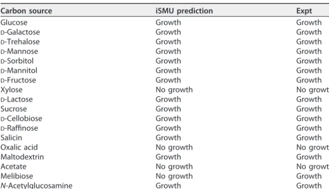
The iSMU model predicts carbon source utilization ofS. mutans.S. mutanscan metabolize a wide range of carbon sources (41, 42), allowing the organism to thrive in the oral cavity of humans with varied diets.S. mutans’ robustness to changing carbon sources may be essential for its survival and cariogenicity. The iSMU model can grow in CDM with 24 carbon sources: glucose, fructose, sucrose, lactose, trehalose, ascorbate, arbutin, maltose, cellobiose, salicin, sorbitol, mannitol, mannose,N-acetylglucosamine, fructan, galactose, galactinol, epimelibiose, melibiitol, melibiose, raffinose, maltodex-trin, stachyose, and malate. Growth on glucose, sucrose, andN-acetylglucosamine is consistent with previous experimental studies (41, 42). To validate our model, we tested iSMU’s growth predictions for 18 carbon sources (Table 2). The model predictions and experimental results agreed for 17 of the 18 carbon sources. The sole disagreement was growth on melibiose. The iSMU model can grow on melibiose, butS. mutansUA159 did TABLE 1CDM includes 22 amino acids, 11 vitamins, 3 nucleobases, 8 inorganic salts, and glucose
Component Concn (g/liter)
Deionized H2O
Iron 0.006
Phosphate 18.3
MgSO4·7H2O 0.7
MnSO4·H2O 0.005
NaC2H3O2·3H2O 4.5
DL-Alanine 0.1
L-Arginine 0.1
L-Aspartic acid 0.1
L-Asparagine 0.1
L-Cysteine HCl 0.65
L-Cystine 0.05
L-Glutamic acid 0.1
L-Glutamine 0.2
Glycine 0.1
L-Histidine 0.1
L-Isoleucine 0.1
L-Leucine 0.1
L-Lysine 0.1
L-Methionine 0.1
L-Phenylalanine 0.1
L-Proline 0.1
Hydroxy-L-proline 0.1
L-Serine 0.1
L-Threonine 0.2
L-Tryptophan 0.1
L-Tyrosine 0.1
L-Valine 0.1
p-Aminobenzoic acid 0.0002
Biotin 0.0002
Folic acid 0.0008
Nicotinamide 0.001
B-NADP 0.0025
Pantothenate Ca salt 0.002
Pyridoxal 0.001
Pyridoxamine diHCl 0.001
Riboflavin 0.002
Thiamine hydrochloride 0.001
Vitamin B12 0.0001
Adenine 0.02
Guanine hydrochloride 0.02
Uracil 0.02
CaCl2 0.00676
NaHCO3 2.5
Glucose 10.0
on September 8, 2020 by guest
http://msystems.asm.org/
not grow on this carbon source. Other S. mutans strains can catabolize melibiose (43–46). The iSMU model imports melibiose using a multiple sugar transporter (Msm-EFGK) that reportedly supports melibiose catabolism under anaerobic conditions with a semidefined medium (47). It is possible thatS. mutansUA159 requires an ingredient not found in CDM to utilize melibiose via MsmEFGK or that the strain’s melibiose pathway is incomplete or inactive.
iSMU predictions guide experiments. Metabolic models like iSMU link growth phenotypes to the organism’s genotype. These predictions can guide experiments to validate gene annotations for metabolic enzymes. The iSMU model predicts—and our experiments confirm—thatS. mutanscan grow on sorbitol (Table 2). There are several enzymatic pathways for sorbitol utilization, including conversion of sorbitol into fruc-tose by a sorbitol dehydrogenase (EC 1.1.1.14 or 1.1.1.15), the conversion of sorbitol to sorbose (EC 1.1.99.21 or 1.1.1.289) and the subsequent utilization of sorbose, or the conversion of sorbitol into glucose by an aldehyde reductase (EC 1.1.1.21). In the iSMU model, sorbitol is phosphorylated and converted into fructose-6-phosphate by a pu-tative sorbitol-6-phosphate 2-dehydrogenase encoded by the gene SMU_308 (Fig. 5A). If the model is correct, SMU_308 should be essential for growth on sorbitol.
We tested the predicted essentiality of SMU_308 by deleting the gene from S. mutansUA159. While the wild-type UA159 strain was able to grow on sorbitol, the ΔSMU_308 strain was unable to grow (Fig. 5B). Growth on sorbitol was restored when the SMU_308 gene was added back to the ΔSMU_308 strain on a plasmid (Fig. 5B). These experiments confirm the essentiality of gene SMU_308 for growth on sorbitol, supporting the model’s mechanism for sorbitol catabolism.
We used iSMU to predict more indirect links between genotype and phenotype. The model predicts that galactose is produced as a by-product during the utilization of oligosaccharides and sugar alcohols (raffinose, stachyose, epimelibiose, melibiose, ga-lactinol, and melibiitol). These complex carbohydrates are broken down into simpler sugars like galactose that must be exported or catabolized by the cell. Thus, disrupting galactose catabolism can indirectly affectS. mutans’ ability to grow on oligosaccharides and sugar alcohols.
Flux balance analysis assumes a steady state with no net accumulation or depletion of metabolites. Our manual curation process for iSMU provided no genomic evidence for a galactose exporter, so intracellular galactose must be converted to another metabolite to avoid accumulation. Our simulations with iSMU agree— blocking galac-tose catabolism prevents growth on galacgalac-tose or any complex carbohydrate that produces galactose as an intermediate metabolite.
TABLE 2Model predictions and measured growth forS. mutansUA159 on 18 carbon sources
Carbon source iSMU prediction Expt
Glucose Growth Growth
D-Galactose Growth Growth
D-Trehalose Growth Growth
D-Mannose Growth Growth
D-Sorbitol Growth Growth
D-Mannitol Growth Growth
D-Fructose Growth Growth
Xylose No growth No growth
D-Lactose Growth Growth
Sucrose Growth Growth
D-Cellobiose Growth Growth
D-Raffinose Growth Growth
Salicin Growth Growth
Oxalic acid No growth No growth
Maltodextrin Growth Growth
Acetate No growth No growth
Melibiose No growth Growth
N-Acetylglucosamine Growth Growth
on September 8, 2020 by guest
http://msystems.asm.org/
Galactose utilization in S. mutansfollows the Leloir pathway (Fig. 5C), including phosphorylation by galactokinase (GalK, EC 2.7.1.6) and subsequent conversion into UDP-galactose by a uridylyltransferase (GalT, EC 2.7.7.12) (48). Our model predicts that deletion ofgalKorgalT(or both) would abolish galactose catabolism. We deleted both
WT
ΔgalKTΔgalKT +pGalKT 0.0 0.5 1.0 1.5 2.0 2.5 OD 600 afte r 2 4 h * ns
CDM+galactose
UDP-raffinose (extracellular) raffinose sucrose D-fructose melibiose D-glucose galactose galactose-1-phosphate glucose-1-phosphate UDP-GalK GalT GalT galactose glucoseD
C
0 4 8 12 16 20 24
0.00 0.25 0.50 0.75 1.00 1.25 time [h] OD 600
CDM+raffinose
WT 'galKT 'galKT +pGalKT WT ΔgalKTΔgalKT +pGalKT 0.00 0.05 0.10 0.15 gr owth r ate [h -1]CDM+raffinose
nsE
F
SMU_308 D-fructose-6-phosphate sorbitol-6-phosphate sorbitol (extracellular)B
A
WT ΔSMU_308ΔSMU_308 +pSMU_308 0.0 0.2 0.4 0.6 0.8 1.0 OD 600 afte r 2 4 hCDM+sorbitol
* * *FIG 5 iSMU guides experiments withS. mutans. (A) The model predicts that sorbitol catabolism requires the product of SMU_308. (B) Wild-type (WT)S. mutansUA159 grows on sorbitol as a carbon source, but a strain with a scarless deletion of SMU_308 does not. Complementing the deletion strain with a plasmid carrying SMU_308 restores growth. (C) The Leloir pathway breaks down raffinose into galactose. Enzymes GalK and GalT are required to utilize galactose. There is no predicted exporter for galactose inS. mutansUA159. (D) The genesgalKandgalTare required for growth on galactose. (E) The ΔgalKTstrain has a growth defect on raffinose, potentially due to intracellular accumulation of galactose. (F) Adding thegalKTgenes back to the ΔgalKTstrain restores the growth rate on raffinose to wild-type levels. For panels B, D, and F, bar height indicates the mean from three biological replicates.*indicatesP⬍0.001 byttest. ns, not significant. The representative growth curves in panel E correspond to the biological replicate in panel F with the median growth rate.
on September 8, 2020 by guest
http://msystems.asm.org/
genes fromS. mutansUA159 and confirmed that the ΔgalKTstrain is unable to grow on galactose (Fig. 5D). Adding the genes back to the ΔgalKT strain via a plasmid is sufficient to restore growth on galactose (Fig. 5D). These results agree with previous mutagenesis studies of the Leloir pathway inS. mutans(49).
Our model predicts that the complex sugar raffinose is broken down into sucrose, fructose, and melibiose. The melibiose is further broken down into galactose and glucose. Growth on raffinose should indirectly depend on galactose catabolism to avoid a toxic buildup of galactose in the cell. Wild-typeS. mutans UA159 grows on raffinose (Fig. 5E), but the ΔgalKTmutant has a growth defect when raffinose is the sole carbon source (Fig. 5E and F). Adding thegalKTgenes back to the ΔgalKTstrain via a plasmid relieved the growth defect (Fig. 5E and F). These results demonstrate that the Leloir pathway is required for efficient growth on complex sugars like raffinose and suggest that accumulation of intermediate galactose may be toxic to the bacterium.
DISCUSSION
iSMU is the first whole-genome metabolic model of the cariogenic pathogen S. mutansUA159. The model captures the entire metabolism of the organism and was validated by comparing model predictions to experimental evidence. Metabolism plays a dual role in the pathogenicity ofS. mutans. First, fermenting sugars creates caries-causing lactic acid. A significant portion of the S. mutans genome is dedicated to carbohydrate metabolism, reflecting the plasticity of S. mutans’ metabolism. Second, multiple metabolic subsystems are required forS. mutansto tolerate acid and outcom-pete noncariogenic streptococci. A mathematical model allows us to investigate con-nections among metabolic pathways during pathogenesis.
iSMU’s predictions agree with most of the nutrient depletion experiments, but some of S. mutans’ auxotrophies are unexplained by the model. For example, the UA159 genome encodes a complete pathway for adenine synthesis, but exogenous adenine is required for growth in vitro. The adenine synthesis pathway in iSMU may not be expressed or functional in UA159 when grown aerobically in CDM. Other experiments agree qualitatively but not quantitatively with the model. When guanine, aminoben-zoate, nicotinamide, or sodium acetate is removed from CDM,S. mutansgrows slower, but that defect is not predicted by the model. The model also underpredicts growth rates when pyridoxal and pyridoxamine, riboflavin, and thiamine are removed. Differ-ences like these are expected with constraint-based models that lack kinetic details for nutrient uptake and enzymatic turnover.
S. mutans UA159 can grow on minimal medium with ammonium as the sole
nitrogen source (33), and the iSMU model can produce biomass under these conditions. Experimentally, growth on ammonium requires an anaerobic environment, but the model can produce biomass with or without oxygen. Oxygen may repress expression of enzymes required for scavenging nitrogen from ammonium, and the lack of regu-lation in our model would explain why iSMU can grow aerobically using ammonium. Several factors could explain the disagreements between the model’s essentiality prediction and experimental results. First, we note that both methods for identifying essential genes have technical strengths and limitations. In the ordered gene deletion library, any gene for which a deletion mutant cannot be constructed is labeled essential. A nonessential gene located in a region of the chromosome refractory to homologous recombination would be incorrectly labeled as essential. The Tn-seq libraries in the work of Shields et al. (31) were constructed usingin vitrotransposon mutagenesis followed by homologous recombination, so the same limitation applies. The Tn-seq libraries were grown for⬃30 generations before sequencing. Such a large expansion can bias the library against mutants with large fitness defects. Although these mutants may be viable, they appear at such low frequency in the final pool that they are missed during sequencing. The corresponding genes would be incorrectly labeled as essential. Another factor that can explain the disagreement is that the wrongly predicted genes encode enzymes that may have secondary functions essential for the cell. A significant number of genes that were predicted to be nonessential
on September 8, 2020 by guest
http://msystems.asm.org/
encode kinases and phosphatases that may be involved in cell signaling. The disagree-ment between the Tn-seq and defined deletion library suggest that the “essential genome” ofS. mutansUA159 has not been fully elucidated.
Inaccuracies in the iSMU model also contribute to disagreements over essential genes. After decades of curation, metabolic models for the model organismsE. coliand
S. cerevisiae still miss some essential gene predictions (50, 51). Unannotated genes could catalyze redundant routes to synthesize essential metabolites in vivo, creating false-positive essentiality predictions in iSMU. Regulation, loss-of-function mutations, and missing cofactors can also restrict the metabolic capabilities ofS. mutans, making the pathogen less metabolically flexible than the iSMU model.
Overall, we believe the model’s predictions could be improved by either (i) incor-porating regulatory rules during simulations or (ii) using gene expression or other high-throughput data to tailor the model to anaerobic, aerobic, acidic, or other conditions. S. mutans’ metabolic requirements change as the bacterium encounters different niches in the mouth. Before the organism forms thick biofilms and deep dental caries, growth conditions are likely aerobic with abundant nutrients from saliva and food consumed by the host. Deep dental caries may create anaerobic conditions with limited nutrient availability. In this environment,S. mutans would need to synthesize many biomass componentsde novo.
The steady-state (or quasi-steady-state) assumption of FBA is often perceived as a limitation of the method. However, the steady-state assumption allows us to predict toxic accumulation of intermediates, as evidenced by our experiments with galactose and the ΔgalKTstrains. It is important to note that the buildup of galactose was due to the irreversibility of the galactose importer. If we assumed that the transporter was bidirectional, excess galactose could be exported and the model predictions would not have matched the organism’s phenotype. Careful curation of reversibility is just as important to model fidelity as correct genome annotations.
S. mutansis a model organism in oral microbiology (52). Our iSMU model draws from hundreds of studies to form an accurate, genome-wide picture ofS. mutans metabo-lism. The model also highlights the value added by manual curation. The metabolism ofS. mutansis well characterized on the molecular and pathway levels. Incorporating manually curated models ofS. mutansand other lactic acid bacteria may improve the accuracy of automatic reconstruction pipelines.
MATERIALS AND METHODS
Model construction.The metabolic network ofS. mutansUA159 was reconstructed according to best practices in the COBRA modeling community (53). As summarized in Fig. 1A, reconstruction began with the annotated UA159 genome (RefSeq GCA_000007465.2). Metabolic enzymes and the associated reactions were initially collected from KEGG (54) and Uniprot (55). The Metacyc (56), RHEA (57), ModelSEED (25), BiGG (58), and ChEBI (59) databases were used as secondary sources for metabolic reactions. Transport reactions were verified with TransportDB (60). KEGG identifiers were used for metabolites and reactions for consistency with other databases. Reactions without gene associations were added only when supported by experimental or literature evidence. These 12 reactions are explained in Table S1 in the supplemental material. All chemical species and formulas were converted to their protonation state at pH 7.0 using the ModelSEED database. A custom map of the iSMU model was constructed using Escher version 1.6.0 (Fig. 2) (61).
Model simulations using flux balance analysis. All simulations were performed with Matlab (version R2016b; MathWorks, Natick, MA) using the COBRA toolbox (62). Mathematical programs were solved with Gurobi Optimizer (version 7.5; Gurobi Optimization, Beaverton, OR). Gene set enrichment for KEGG pathways was performed using KEGG Mapper (54). Details on simulations using flux balance analysis are available in Text S1 in the supplemental material.
Model availability.The final model is available as an SBML file and a spreadsheet compatible with the COBRA toolbox (Data Sets S1 and S2). The model map is available as a JSON file (Data Set S3) and SVG image (Data Set S4). Future versions of the model and map will be available on the authors’ website (http://jensenlab.net).
Strains and reagents.Strains and plasmids are listed in Table 3.S. mutansUA159 (ATCC 700610) was cultured in Todd-Hewitt broth (63) with 0.3% yeast extract (THY) liquid medium or agar plates. Strains were grown overnight in 5% CO2at 37°C unless specified. Growth experiments were performed in a chemically defined medium (CDM) (39) with 22 amino acids, 11 vitamins, 3 nucleobases, 8 inorganic salts, and a carbon source (Table 1). Two sets of growth experiments were performed: a set involving leave-one-out CDM variants with glucose as a carbon source and a set involving complete CDM with
on September 8, 2020 by guest
http://msystems.asm.org/
alternative carbon sources. All CDM-based media were prepared fresh weekly from concentrated stocks (39). All components were purchased from Sigma-Aldrich USA or Fisher Scientific USA and were sterilized by autoclaving or filtration.
Oligonucleotides were synthesized by Integrated DNA Technologies (Coralville, IA, USA). Peptides were purchased from GenScript, Inc. (Piscataway, NJ, USA). All enzymes were manufactured by New England Biolabs (Ipswich, MA, USA).
Growth assays. Overnight cultures of S. mutans were washed three times in sterile water or phosphate-buffered saline (PBS). The overnight culture was concentrated 5⫻(from 5 ml to 1 ml). For the leave-one-out experiments, 1l of the concentrate was used to inoculate wells of a 96 well plate containing 200l of defined medium. Plates were incubated without agitation in 5% CO2at 37°C. Optical density was measured by absorbance at 600 nm every hour for 16 h using a Tecan Infinite 200 Pro plate reader (Tecan, Männedorf, Switzerland). Exponential growth rates were calculated using the R package CellGrowth (version 3.7; Ludwig Maximilian University of Munich [https://www.bioconductor.org/ packages/release/bioc/html/cellGrowth.html]) with default settings. Growth rates were normalized to the growth rate in complete CDM.
For the carbon source growth experiments, 25l of the concentrated overnight culture was used to inoculate 5 ml of defined medium in culture tubes. The tubes were also incubated without agitation in 5% CO2at 37°C. Optical density was measured by absorbance at 600 nm right after inoculation and at 24 h using a NanoDrop OneC (Thermo Fisher Scientific, Waltham, MA, USA).
S. mutanstransformation.S. mutanswas transformed by induced competence using a protocol adapted from reference 64.S. mutansUA159 was grown overnight in 5 ml THY or CDM plus glucose. The overnight culture was diluted 1:40 into 5 ml of fresh medium and grown to an optical density (OD) of
⬃0.15. A 500-l aliquot was placed in a 2-ml centrifuge tube, and 500 ng of competence-stimulating peptide (CSP-18; SGSLSTFFRLFNRSFTQA) was added. DNA (1g) was added after 20 to 30 min, followed by incubation for 2 h. Up to 250l of the mixture was plated on THY agar plates with 200g/ml spectinomycin (Sigma) or 500g/ml kanamycin (Sigma). Single colonies were picked after 24 to 36 h and grown in 5 ml THY under selection. For markerless mutants, the overnight culture was replated on THY agar plates with 4 mg/mlp-chlorophenylalanine (4-CP) (Sigma). All strains were verified by PCR ampli-fication and Sanger sequencing (ACGT, Inc., Wheeling, IL).
Markerless mutagenesis.Gene deletion strains ofS. mutansUA159 were constructed with direct repeat-mediated cloning-independent markerless mutagenesis (DR-CIMM) (65). DR-CIMM replaces the target gene with an antibiotic resistance marker (aph3conferring kanamycin resistance) and a negative selection marker (pheS*, a mutated phenylalanyl-tRNA synthetase conferring sensitivity to p-chlorophenylalanine [4-CP]). A small region of DNA directly upstream of the target gene is repeated downstream ofpheS*to allow homologous recombination that removes both theaph3andpheS*genes. The target gene is deleted using selection on kanamycin. Subsequent plating on 4-CP selects for homologous recombination that removes both marker genes, leaving a scarless deletion.
The primer sequences in Table S2 in the supplemental material follow the DR-CIMM nomenclature in reference 65. Homology regions upstream and downstream of the target gene were amplified with primers upF, upR, dnF, and dnR. The direct repeat region was amplified with primers DR-F and DR-R. The
aph3andpheS*cassette was amplified with primers IFDC3-F and IFDC3-R. All primers except upF and
dnR contained tails to allow assembly by overlap extension PCR (66) into two constructs: (i) the upstream homology region plus the selection cassette and (ii) the selection cassette, direct repeat, and down-stream homology region.S. mutansUA159 was transformed simultaneously with the two constructs.
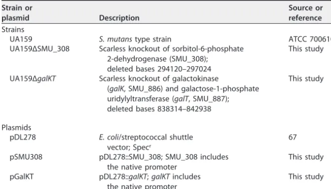
Plasmid construction.GenesgalKTand SMU_308 were cloned into the shuttle vector pDL278 (67) to complement the deletion strains. The genes were amplified by Q5 polymerase using the primers in TABLE 3Bacterial strains and plasmids used in this study
Strain or
plasmid Description
Source or reference Strains
UA159 S. mutanstype strain ATCC 700610
UA159ΔSMU_308 Scarless knockout of sorbitol-6-phosphate 2-dehydrogenase (SMU_308);
deleted bases 294120–297024
This study
UA159ΔgalKT Scarless knockout of galactokinase
(galK, SMU_886) and galactose-1-phosphate uridylyltransferase (galT, SMU_887); deleted bases 838314–842938
This study
Plasmids
pDL278 E. coli/streptococcal shuttle vector; Specr
67
pSMU308 pDL278::SMU_308; SMU_308 includes the native promoter
This study
pGalKT pDL278::galKT;galKTincludes the native promoter
This study
on September 8, 2020 by guest
http://msystems.asm.org/
Table S2 containing tails with EcoRI restriction sites. Purified pDL278 was digested with EcoRI and dephosphorylated with rSAP. The linearized plasmid was ligated with the EcoRI-digested amplicon using T4 DNA ligase. The deletion strains were transformed using 5l of the ligation mixture.
SUPPLEMENTAL MATERIAL
Supplemental material for this article may be found at https://doi.org/10.1128/ mSystems.00529-19.
TEXT S1, PDF file, 0.02 MB. TABLE S1, PDF file, 0.04 MB. TABLE S2, PDF file, 0.03 MB. TABLE S3, PDF file, 0.1 MB. TABLE S4, PDF file, 0.03 MB. DATA SET S1, TXT file, 1.1 MB. DATA SET S2, XLSX file, 0.1 MB. DATA SET S3, TXT file, 1.2 MB. DATA SET S4, PDF file, 1.3 MB. DATA SET S5, XLSX file, 0.03 MB.
ACKNOWLEDGMENTS
We thank Robert Shields for assistance with the Tn-seq data, John Gerlt for sharing equipment and reagents, and Walden Li for help with DR-CIMM. Plasmid pDL278 was provided by Garry Dunny. Constructs for DR-CIMM were provided by Justin Merritt.
This work was supported by the National Institutes of Health (grants DE026817 and EB027396 to P.A.J.) and the University of Illinois at Urbana-Champaign.
We declare no financial conflict of interest.
REFERENCES
1. Dewhirst FE, Chen T, Izard J, Paster BJ, Tanner ACR, Yu W-H, Lakshmanan A, Wade WG. 2010. The human oral microbiome. J Bacteriol 192: 5002–5017.https://doi.org/10.1128/JB.00542-10.
2. Daneo-Moore L, Terleckyj B, Shockman GD. 1975. Analysis of growth rate in sucrose-supplemented cultures of Streptococcus mutans. Infect Im-mun 12:1195–1205.
3. Terleckyj B, Shockman GD. 1975. Amino acid requirements of Strepto-coccus mutans and other oral streptococci. Infect Immun 11:656 – 664. 4. Quivey RG, Faustoferri RC, Clancy KA, Marquis RE. 1995. Acid adaptation in Streptococcus mutans UA159 alleviates sensitization to environmen-tal stress due to RecA deficiency. FEMS Microbiol Lett 126:257–261. https://doi.org/10.1111/j.1574-6968.1995.tb07427.x.
5. Baker JL, Abranches J, Faustoferri RC, Hubbard CJ, Lemos JA, Courtney MA, Quivey R. 2015. Transcriptional profile of glucose-shocked and acid-adapted strains of Streptococcus mutans. Mol Oral Microbiol 30: 496 –517.https://doi.org/10.1111/omi.12110.
6. Hamilton IR, Ellwood DC. 1978. Effects of fluoride on carbohydrate metabolism by washed cells of Streptococcus mutans grown at various pH values in a chemostat. Infect Immun 19:434 – 442.
7. Kuhnert WL, Zheng G, Faustoferri RC, Quivey RG. 2004. The F-ATPase operon promoter of Streptococcus mutans is transcriptionally regulated in response to external pH. J Bacteriol 186:8524 – 8528.https://doi.org/ 10.1128/JB.186.24.8524-8528.2004.
8. Oberhardt MA, Palsson BØ, Papin JA. 2009. Applications of genome-scale metabolic reconstructions. Mol Syst Biol 5:320.https://doi.org/10.1038/ msb.2009.77.
9. Hyduke DR, Lewis NE, Palsson BØ. 2013. Analysis of omics data with genome-scale models of metabolism. Mol Biosyst 9:167–174.https://doi .org/10.1039/c2mb25453k.
10. Suthers PF, Zomorrodi A, Maranas CD. 2009. Genome-scale gene/ reaction essentiality and synthetic lethality analysis. Mol Syst Biol 5:301. https://doi.org/10.1038/msb.2009.56.
11. Shlomi T, Berkman O, Ruppin E. 2005. Regulatory on/off minimization of metabolic flux changes after genetic perturbations. Proc Natl Acad Sci U S A 102:7695–7700.https://doi.org/10.1073/pnas.0406346102. 12. Lewis NE, Hixson KK, Conrad TM, Lerman JA, Charusanti P, Polpitiya AD,
Adkins JN, Schramm G, Purvine SO, Lopez᎑Ferrer D, Weitz KK, Eils R, König R, Smith RD, Palsson BØ. 2010. Omic data from evolved E. coli are
consistent with computed optimal growth from genome᎑scale models. Mol Syst Biol 6:390.https://doi.org/10.1038/msb.2010.47.
13. Applebee MK, Herrgård MJ, Palsson BØ. 2008. Impact of individual mutations on increased fitness in adaptively evolved strains of Esche-richia coli. J Bacteriol 190:5087–5094.https://doi.org/10.1128/JB.01976 -07.
14. Jensen PA, Papin JA. 2011. Functional integration of a metabolic net-work model and expression data without arbitrary thresholding. Bioin-formatics 27:541–547.https://doi.org/10.1093/bioinformatics/btq702. 15. Vu TT, Stolyar SM, Pinchuk GE, Hill EA, Kucek LA, Brown RN, Lipton MS,
Osterman A, Fredrickson JK, Konopka AE, Beliaev AS, Reed JL. 2012. Genome-scale modeling of light-driven reductant partitioning and car-bon fluxes in diazotrophic unicellular cyanobacterium Cyanothece sp. ATCC 51142. PLoS Comput Biol 8:e1002460.https://doi.org/10.1371/ journal.pcbi.1002460.
16. Fong SS, Palsson BØ. 2004. Metabolic gene– deletion strains of
Esche-richia colievolve to computationally predicted growth phenotypes. Nat
Genet 36:1056 –1058.https://doi.org/10.1038/ng1432.
17. Ibarra RU, Edwards JS, Palsson BO. 2002. Escherichia coli K-12 undergoes adaptive evolution to achievein silicopredicted optimal growth. Nature 420:186 –189.https://doi.org/10.1038/nature01149.
18. Bilu Y, Shlomi T, Barkai N, Ruppin E. 2006. Conservation of expression and sequence of metabolic genes is reflected by activity across meta-bolic states. PLoS Comput Biol 2:e106.https://doi.org/10.1371/journal .pcbi.0020106.
19. Burgard AP, Nikolaev EV, Schilling CH, Maranas CD. 2004. Flux coupling analysis of genome-scale metabolic network reconstructions. Genome Res 14:301–312.https://doi.org/10.1101/gr.1926504.
20. Pan S, Reed JL. 2018. Advances in gap-filling genome-scale metabolic models and model-driven experiments lead to novel metabolic discov-eries. Curr Opin Biotechnol 51:103–108.https://doi.org/10.1016/j.copbio .2017.12.012.
21. Veith N, Solheim M, van Grinsven KWA, Olivier BG, Levering J, Grosseholz R, Hugenholtz J, Holo H, Nes I, Teusink B, Kummer U. 2015. Using a genome-scale metabolic model of Enterococcus faecalis V583 to assess amino acid uptake and its impact on central metabolism. Appl Environ Microbiol 81:1622–1633.https://doi.org/10.1128/AEM.03279-14. 22. Ajdic´ D, McShan WM, McLaughlin RE, Savic´ G, Chang J, Carson MB,
on September 8, 2020 by guest
http://msystems.asm.org/
Primeaux C, Tian R, Kenton S, Jia H, Lin S, Qian Y, Li S, Zhu H, Najar F, Lai H, White J, Roe BA, Ferretti JJ. 2002. Genome sequence of Streptococcus mutans UA159, a cariogenic dental pathogen. Proc Natl Acad Sci U S A 99:14434 –14439.https://doi.org/10.1073/pnas.172501299.
23. Quivey RG, Faustoferri R, Monahan K, Marquis R. 2000. Shifts in mem-brane fatty acid profiles associated with acid adaptation of Streptococ-cus mutans. FEMS Microbiol Lett 189:89 –92.https://doi.org/10.1111/j .1574-6968.2000.tb09211.x.
24. Arkin AP, Cottingham RW, Henry CS, Harris NL, Stevens RL, Maslov S, Dehal P, Ware D, Perez F, Canon S, Sneddon MW, Henderson ML, Riehl WJ, Murphy-Olson D, Chan SY, Kamimura RT, Kumari S, Drake MM, Brettin TS, Glass EM, Chivian D, Gunter D, Weston DJ, Allen BH, Baumohl J, Best AA, Bowen B, Brenner SE, Bun CC, Chandonia J-M, Chia J-M, Colasanti R, Conrad N, Davis JJ, Davison BH, DeJongh M, Devoid S, Dietrich E, Dubchak I, Edirisinghe JN, Fang G, Faria JP, Frybarger PM, Gerlach W, Gerstein M, Greiner A, Gurtowski J, Haun HL, He F, Jain R, Joachimiak MP, Keegan KP, Kondo S, Kumar V, Land ML, Meyer F, Mills M, Novichkov PS, Oh T, Olsen GJ, Olson R, Parrello B, Pasternak S, Pearson E, Poon SS, Price GA, Ramakrishnan S, Ranjan P, Ronald PC, Schatz MC, Seaver SMD, Shukla M, Sutormin RA, Syed MH, Thomason J, Tintle NL, Wang D, Xia F, Yoo H, Yoo S, Yu D. 2018. KBase: the United States Department of Energy systems biology knowledgebase. Nat Bio-technol 36:566 –569.https://doi.org/10.1038/nbt.4163.
25. Henry CS, DeJongh M, Best AA, Frybarger PM, Linsay B, Stevens RL. 2010. High-throughput generation, optimization and analysis of genome-scale metabolic models. Nat Biotechnol 28:977–982.https://doi.org/10.1038/ nbt.1672.
26. Notebaart RA, van Enckevort FH, Francke C, Siezen RJ, Teusink B. 2006. Accelerating the reconstruction of genome-scale metabolic networks. BMC Bioinformatics 7:296.https://doi.org/10.1186/1471-2105-7-296. 27. Edwards JS, Palsson BO. 2000. The Escherichia coli MG1655 in silico
metabolic genotype: its definition, characteristics, and capabilities. Proc Natl Acad Sci U S A 97:5528 –5533.https://doi.org/10.1073/pnas.97.10 .5528.
28. Monk JM, Lloyd CJ, Brunk E, Mih N, Sastry A, King Z, Takeuchi R, Nomura W, Zhang Z, Mori H, Feist AM, Palsson BO. 2017. iML1515, a knowledge-base that computes Escherichia coli traits. Nat Biotechnol 35:904 –908. https://doi.org/10.1038/nbt.3956.
29. Oh Y-K, Palsson BO, Park SM, Schilling CH, Mahadevan R. 2007. Genome-scale reconstruction of metabolic network in Bacillus subtilis based on high-throughput phenotyping and gene essentiality data. J Biol Chem 282:28791–28799.https://doi.org/10.1074/jbc.M703759200.
30. Kocabas¸ P, Çalık P, Çalık G, Özdamar TH. 2017. Analyses of extracellular protein production in Bacillus subtilis—I: genome-scale metabolic model reconstruction based on updated gene-enzyme-reaction data. Biochem Eng J 127:229 –241.https://doi.org/10.1016/j.bej.2017.07.005. 31. Shields RC, Zeng L, Culp DJ, Burne RA. 2018. Genomewide identification
of essential genes and fitness determinants ofStreptococcus mutans UA159. mSphere 3:e00031-18. https://doi.org/10.1128/mSphere.00031 -18.
32. Quivey RG, Grayhack EJ, Faustoferri RC, Hubbard CJ, Baldeck JD, Wolf AS, MacGilvray ME, Rosalen PL, Scott᎑Anne K, Santiago B, Gopal S, Payne J, Marquis RE. 2015. Functional profiling in Streptococcus mutans: con-struction and examination of a genomic collection of gene deletion mutants. Mol Oral Microbiol 30:474 – 495.https://doi.org/10.1111/omi .12107.
33. Martin EJS, Wittenberger CL. 1980. Regulation and function of ammonia-assimilating enzymes in Streptococcus mutans. Infect Immun 28: 220 –224.
34. Cowman RA, Perrella MM, Fitzgerald RJ. 1974. Influence of incubation atmosphere on growth and amino acid requirements of Streptococcus mutans. Appl Environ Microbiol 27:86 –92.
35. Griffith CJ, Carlsson J. 1974. Mechanism of ammonia assimilation in streptococci. J Gen Microbiol 82:253–260. https://doi.org/10.1099/ 00221287-82-2-253.
36. Carlsson J. 1970. Nutritional requirements of Streptococcus mutans. Caries Res 4:305–320.https://doi.org/10.1159/000259653.
37. Bridges RB. 1977. Ribose biosynthesis in Streptococcus mutans. Arch Oral Biol 22:139 –145.https://doi.org/10.1016/0003-9969(77)90091-7. 38. Richardson AR, Somerville GA, Sonenshein AL. 2015. Regulating the
intersection of metabolism and pathogenesis in Gram-positive bacteria. Microbiol Spectr 3:MBP-0004-2014. https://doi.org/10.1128/microbiol spec.MBP-0004-2014.
39. Chang JC, LaSarre B, Jimenez JC, Aggarwal C, Federle MJ. 2011. Two
group A streptococcal peptide pheromones act through opposing Rgg regulators to control biofilm development. PLoS Pathog 7:e1002190. https://doi.org/10.1371/journal.ppat.1002190.
40. Terleckyj B, Willett NP, Shockman GD. 1975. Growth of several cariogenic strains of oral streptococci in a chemically defined medium. Infect Immun 11:649 – 655.
41. Ikeda T, Sandham HJ. 1972. A high-sucrose medium for the identification of Streptococcus mutans. Arch Oral Biol 17:781–783.https://doi.org/10 .1016/0003-9969(72)90204-x.
42. Moye ZD, Burne RA, Zeng L. 2014. Uptake and metabolism of N-acetylglucosamine and glucosamine by Streptococcus mutans. Appl Environ Microbiol 80:5053–5067.https://doi.org/10.1128/AEM.00820-14. 43. Arimoto T, Igarashi T. 2008. Role of prolipoprotein diacylglyceryl trans-ferase (Lgt) and lipoprotein-specific signal peptidase II (LspA) in local-ization and physiological function of lipoprotein MsmE in Streptococcus mutans. Oral Microbiol Immunol 23:515–519.https://doi.org/10.1111/j .1399-302X.2008.00455.x.
44. McLaughlin RE, Ferretti JJ. 1996. The multiple-sugar metabolism (msm) gene cluster of Streptococcus mutans is transcribed as a single operon. FEMS Microbiol Lett 140:261–264.https://doi.org/10.1016/ 0378-1097(96)00191-7.
45. Russell RR, Aduse-Opoku J, Sutcliffe IC, Tao L, Ferretti JJ. 1992. A binding protein-dependent transport system in Streptococcus mutans responsi-ble for multiple sugar metabolism. J Biol Chem 267:4631– 4637. 46. Tao L, Sutcliffe IC, Russell RR, Ferretti JJ. 1993. Cloning and expression of
the multiple sugar metabolism (msm) operon of Streptococcus mutans in heterologous streptococcal hosts. Infect Immun 61:1121–1125. 47. Webb AJ, Homer KA, Hosie A. 2008. Two closely related ABC
transport-ers in Streptococcus mutans are involved in disaccharide and/or oligosaccharide uptake. J Bacteriol 190:168 –178.https://doi.org/10 .1128/JB.01509-07.
48. Holden HM, Rayment I, Thoden JB. 2003. Structure and function of enzymes of the Leloir pathway for galactose metabolism. J Biol Chem 278:43885– 43888.https://doi.org/10.1074/jbc.R300025200.
49. Abranches J, Chen Y-Y, Burne RA. 2004. Galactose metabolism by Strep-tococcus mutans. Appl Environ Microbiol 70:6047– 6052.https://doi.org/ 10.1128/AEM.70.10.6047-6052.2004.
50. Orth JD, Conrad TM, Na J, Lerman JA, Nam H, Feist AM, Palsson BØ. 2011. A comprehensive genome᎑scale reconstruction of Escherichia coli me-tabolism—2011. Mol Syst Biol 7:535.https://doi.org/10.1038/msb.2011 .65.
51. Zomorrodi AR, Maranas CD. 2010. Improving the iMM904 S. cerevisiae metabolic model using essentiality and synthetic lethality data. BMC Syst Biol 4:178.https://doi.org/10.1186/1752-0509-4-178.
52. Lemos JA, Quivey RG, Koo H, Abranches J. 2013. Streptococcus mutans: a new Gram-positive paradigm? Microbiology 159:436 – 445.https://doi .org/10.1099/mic.0.066134-0.
53. Thiele I, Palsson BØ. 2010. A protocol for generating a high-quality genome-scale metabolic reconstruction. Nat Protoc 5:93–121.https:// doi.org/10.1038/nprot.2009.203.
54. Kanehisa M, Furumichi M, Tanabe M, Sato Y, Morishima K. 2017. KEGG: new perspectives on genomes, pathways, diseases and drugs. Nucleic Acids Res 45:D353–D361.https://doi.org/10.1093/nar/gkw1092. 55. The UniProt Consortium. 2017. UniProt: the universal protein
knowl-edgebase. Nucleic Acids Res 45:D158 –D169.https://doi.org/10.1093/ nar/gkw1099.
56. Caspi R, Billington R, Fulcher CA, Keseler IM, Kothari A, Krummenacker M, Latendresse M, Midford PE, Ong Q, Ong WK, Paley S, Subhraveti P, Karp PD. 2018. The MetaCyc database of metabolic pathways and enzymes. Nucleic Acids Res 46:D633–D639.https://doi.org/10.1093/nar/gkx935. 57. Morgat A, Lombardot T, Axelsen KB, Aimo L, Niknejad A, Hyka-Nouspikel
N, Coudert E, Pozzato M, Pagni M, Moretti S, Rosanoff S, Onwubiko J, Bougueleret L, Xenarios I, Redaschi N, Bridge A. 2017. Updates in Rhea—an expert curated resource of biochemical reactions. Nucleic Acids Res 45:D415–D418.https://doi.org/10.1093/nar/gkw990. 58. King ZA, Lu J, Dräger A, Miller P, Federowicz S, Lerman JA, Ebrahim A,
Palsson BO, Lewis NE. 2016. BiGG Models: a platform for integrating, standardizing and sharing genome-scale models. Nucleic Acids Res 44:D515–D522.https://doi.org/10.1093/nar/gkv1049.
59. Hastings J, Owen G, Dekker A, Ennis M, Kale N, Muthukrishnan V, Turner S, Swainston N, Mendes P, Steinbeck C. 2016. ChEBI in 2016: improved services and an expanding collection of metabolites. Nucleic Acids Res 44:D1214 –D1219.https://doi.org/10.1093/nar/gkv1031.
60. Elbourne LDH, Tetu SG, Hassan KA, Paulsen IT. 2017. TransportDB 2.0: a
on September 8, 2020 by guest
http://msystems.asm.org/
database for exploring membrane transporters in sequenced genomes from all domains of life. Nucleic Acids Res 45:D320 –D324.https://doi .org/10.1093/nar/gkw1068.
61. King ZA, Dräger A, Ebrahim A, Sonnenschein N, Lewis NE, Palsson BO. 2015. Escher: a web application for building, sharing, and embedding data-rich visualizations of biological pathways. PLoS Comput Biol 11: e1004321.https://doi.org/10.1371/journal.pcbi.1004321.
62. Heirendt L, Arreckx S, Pfau T, Mendoza SN, Richelle A, Heinken A, Haraldsdóttir HS, Wachowiak J, Keating SM, Vlasov V, Magnusdóttir S, Ng CY, Preciat G, Žagare A, Chan SHJ, Aurich MK, Clancy CM, Modamio J, Sauls JT, Noronha A, Bordbar A, Cousins B, Assal DCE, Valcarcel LV, Apaolaza I, Ghaderi S, Ahookhosh M, Guebila MB, Kostromins A, Som-pairac N, Le HM, Ma D, Sun Y, Wang L, Yurkovich JT, Oliveira MAP, Vuong PT, Assal LPE, Kuperstein I, Zinovyev A, Hinton HS, Bryant WA, Artacho FJA, Planes FJ, Stalidzans E, Maass A, Vempala S, Hucka M, Saunders MA, Maranas CD, Lewis NE, Sauter T, Palsson BØ, Thiele I, Fleming R. 2017. Creation and analysis of biochemical constraint-based models: the CO-BRA Toolbox v3.0. arXiv:1710.04038. [q-bio.QM]
63. Todd EW, Hewitt LF. 1932. A new culture medium for the production of antigenic streptococcal haemolysin. J Pathol 35:973–974. https://doi .org/10.1002/path.1700350614.
64. Khan R, Rukke HV, Høvik H, Åmdal HA, Chen T, Morrison DA, Petersen FC. 2016. Comprehensive transcriptome profiles of Streptococcus mutans UA159 map core streptococcal competence genes. mSystems 1:e00038 -15.https://doi.org/10.1128/mSystems.00038-15.
65. Zhang S, Zou Z, Kreth J, Merritt J. 2017. Recombineering in Streptococ-cus mutans using direct repeat-mediated cloning-independent marker-less mutagenesis (DR-CIMM). Front Cell Infect Microbiol 7:202.https:// doi.org/10.3389/fcimb.2017.00202.
66. Bryksin AV, Matsumura I. 2010. Overlap extension PCR cloning: a simple and reliable way to create recombinant plasmids. Biotechniques 48: 463– 465.https://doi.org/10.2144/000113418.
67. LeBlanc DJ, Lee LN, Abu-Al-Jaibat A. 1992. Molecular, genetic, and functional analysis of the basic replicon of pVA380-1, a plasmid of oral streptococcal origin. Plasmid 28:130 –145.https://doi.org/10.1016/0147 -619X(92)90044-B.