1
The present and future of serum diagnostic tests for testicular germ cell tumours
Matthew J. Murray1,2, Robert A. Huddart3,4 and Nicholas Coleman1,5
1
Department of Pathology, University of Cambridge, Tennis Court Road, Cambridge, CB2 1QP, UK.
2
Department of Paediatric Haematology and Oncology, Cambridge University Hospitals NHS Foundation Trust, Cambridge, CB2 0QQ, UK.
3
The Institute of Cancer Research, Downs Road, Sutton, Surrey, SM2 5PT, UK. 4
The Royal Marsden NHS Foundation Trust, Downs Road, Sutton, Surrey, SM2 5PT, UK.
5
Department of Histopathology, Cambridge University Hospitals NHS Foundation Trust, Cambridge, CB2 0QQ, UK.
Correspondence to M.J.M. and N.C.
2 human chorionic gonadotrophin (HCG) and lactate dehydrogenase (LDH) show some utility in the management of testicular malignant GCT. However, AFP and HCG display limited sensitivity and specificity, being indicative of yolk sac tumour (AFP) and choriocarcinoma or syncytiotrophoblast (HCG) subtypes. Furthermore, LDH is a very nonspecific biomarker. Consequently, seminomas and NSGCTs comprising a pure embryonal carcinoma subtype are generally negative for these conventional markers. As a result, novel universal biomarkers for testicular malignant GCTs are required. MicroRNAs are short, non-protein-coding RNAs that show much general promise as biomarkers. MicroRNAs from two ‘clusters’, miR-371–373 and miR-302–367, are overexpressed in all malignant GCTs, regardless of age (adult or paediatric), site (gonadal or extragonadal) and subtype (seminomas, yolk sac tumours or embryonal carcinomas). A panel of four circulating microRNAs from these two clusters (miR-371a-3p, miR-372-3p, miR-373-3p and miR-367-3p) is highly sensitive and specific for the diagnosis of malignant GCT, including seminoma and embryonal carcinoma. In the future, circulating microRNAs might be useful in diagnosis, disease monitoring and prognostication of testicular malignant GCTs, which might also reduce reliance on serial CT scanning. For translation into clinical practice, important practical considerations now need addressing.
Key points
3 AFP and HCG show limited sensitivity and specificity for all malignant GCTs, being representative of yolk sac tumour and choriocarcinoma or synctiotrophoblast subtypes, respectively; LDH is a very nonspecific biomarker Novel universal biomarkers for testicular malignant GCTs are required,
particularly for seminoma and embryonal carcinoma subtypes that are typically negative for conventional markers
MicroRNAs are short, non-protein-coding RNAs that show much promise as universal markers in malignant GCTs
Individual microRNAs from two microRNA clusters, 371–373 and miR-302–367, are overexpressed in all malignant GCTs, regardless of patient age, tumour site and subtype
A panel of four circulating microRNAs from these two clusters (miR-371a-3p, miR-372-3p, miR-373-3p and miR-367-3p) is highly sensitive and specific for the diagnosis of malignant GCT, including seminoma and embryonal carcinoma subtypes
Practical considerations need to be addressed to standardize the translation of circulating microRNA studies from a research tool to a routine clinical test
4 disease. Testicular GCTs are broadly classified into seminoma (a malignant subtype) and nonseminomatous GCTs (NSGCTs). The latter group include the malignant subtypes yolk sac tumour (YST), embryonal and choriocarcinoma (CHC), as well as teratoma. Teratomas in prepubertal patients are generally considered benign, but postpubertal cases might display malignant potential2. GCTs that contain more than one subtype are known as mixed malignant GCTs; those with both seminomatous and nonseminomatous components are classified as combined NSGCTs3.
The histological classification and clinical staging of testicular GCTs guide subsequent treatment decisions. Stage I testicular GCTs (that is, those confined to the testes) are initially treated with orchidectomy, although management after orchidectomy varies and can be dictated by clinician and/or national preference4 and/or known risk factors5-10. Chemotherapy is routinely administered for patients with stage II–IV testicular GCTs according to International Germ Cell Cancer Collaboration (IGCCC) prognostic criteria11.
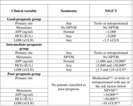
5 to be useful in diagnosis or monitoring3 (Table 2). Current priorities in GCT research include the identification of universal circulating biomarkers of malignant disease, which might assist timely diagnosis and reduce the need for repeated cross-sectional CT imaging in follow-up monitoring, along with the associated radiation burden and risk of second cancer development14. One recent study, for example, showed that only 3% of patients relapsing following a diagnosis of stage I seminoma were identified using conventional serum tumour markers (CTMs), but that figure was 87% for CT imaging9. Other important issues that remain to be fully addressed include the most appropriate management of stage I testicular GCTs, particularly NSGCTs (active surveillance versus adjuvant chemotherapy), and the early identification of patients who are destined to have poor clinical outcomes within the overall IGCCC poor-risk disease cohort 11. These issues are important, as many concerns exist regarding the substantial long-term sequelae of conventional chemotherapy treatment15-21, which include pulmonary fibrosis (with bleomycin), nephrotoxicity and/or ototoxicity (with cisplatin), neuropathy (with cisplatin and taxol agents) 20. Particular concerns also exist about the increased incidence of cardiovascular disease17, 18 and second cancer15, 21 associated with conventional chemotherapy. Minimizing these risks through improved patient stratification is important; novel circulating biomarkers might assist with this challenge.
non-6 protein-coding RNAs termed microRNAs in GCTs, and discusses potential clinical applications of these markers. Practical aspects that must be considered before such microRNA testing can be incorporated into future routine clinical practice are also highlighted. The ultimate aims of developing circulating microRNA testing alongside conventional tumour marker testing are threefold. Firstly, to rationally reduce or omit the use of CT scans in follow-up for patients whose disease is usually negative for conventional markers (predominantly seminoma subtype) at diagnosis. Secondly, to improve quality of patient survival, through the rational reduction and/or omission of chemotherapy, along with its concomitant late effects, for patients with stage I disease, who are predicted to have excellent outcomes. Thirdly, to improve overall survival for those patients with poor-risk disease, through identification of a robust prognostic signature.
[H1] AFP, HCG and LDH in testicular GCT
The serum protein markers AFP, HCG and LDH have all shown utility for the management of testicular GCTs and are currently widely used in clinical practice3.
[H2] Physiology, background and limitations
AFP is a glycoprotein comprising a carbohydrate moiety attached to an ~600 amino acid α-globulin molecule22
7 determined using a quantitative automated chemiluminescent sandwich enzyme immunoassay 24, and measured against known AFP standards according to WHO International Standard 72/225 22. The half-life of AFP in the bloodstream is usually ~5–7 days 3, 22, 25 and levels are measured as ng/ml or kU/l, with normal values being <12 ng/ml (<10 kU/l; conversion 1 ng/ml = 0.84 kU/l) 22. The features of AFP production during development are pertinent to the elevations observed in the serum of some patients with GCTs. Raised serum AFP levels are predominantly observed in GCTs containing YST components, although moderately elevated values can also occur in some embryonal carcinoma and immature teratoma lesions 22 (Table 1). In pure immature teratoma, serum AFP can be raised owing to the presence of immature liver and/or gastrointestinal tissues within the lesion22.
8 relevant drug history 22. In patients in whom potential liver disease is a concern, a full serum liver function test should be performed alongside AFP estimation 22.
HCG measurements can be of the free ß-subunit alone (monomer), the α–ß dimer (‘intact HCG’), or both (‘total HCG’, often referred to as ‘ß-HCG’) 3
. HCG is measured with a double antibody immunometric assay 3, typically using lanthanide fluorescence. Normal HCG levels are <2 IU/l, although levels can be measured in ng/ml (where 5 IU/l = 1 ng/ml). The half-life of HCG is much shorter than AFP, usually ~12–36 h 3, 29, and, therefore, levels should decay much more rapidly. HCG is produced primarily by CHC components, but low levels of HCG production can be observed in patients with pure seminoma containing syncytiotrophoblast (placental-like) cells (Table 1). However, even patients with advanced-stage pure seminoma only show raised HCG values in <20% of cases 3. HCG, like AFP, also shows limitations in sensitivity and specificity. Levels can also be elevated in other malignancies, including neuroendocrine, bladder, renal and lung carcinomas 3. The treating clinician needs to be aware of the limited sensitivity of both HCG and AFP to ensure appropriate patient management.
9 very variable. LDH is often routinely measured at the time of cancer diagnosis, providing a surrogate measure for tumour bulk, and high levels are associated with an increased risk of tumour lysis syndrome, which can occur in patients with malignant GCTs 30. Increased levels are also observed in lymphoma, lung cancer and bone tumours such as osteosarcoma and Ewing sarcoma, as well as in any non-malignant condition that results in cellular lysis or injury, such as such as myocardial infarction or liver or muscle damage or disease 3. These variations limit the use of LDH as a biomarker.
[H2] Diagnosis and early follow-up monitoring
[H3] Preorchidectomy. Measurement of CTM levels before orchidectomy is advised in all cases, as a substantially elevated preoperative AFP level precludes a diagnosis of pure seminoma, regardless of histological findings, and, in addition, this approach assists the interpretation of postorchidectomy estimations 3. Furthermore, in rare cases, such as testicular GCTs with considerable tumour burden where urgent treatment is required, or extragonadal cases (such as retroperitoneal or mediastinal primary), in which the risks of surgical biopsy are deemed excessive, a substantially raised AFP and/or HCG might be sufficient for diagnosis 3.
10 malignant GCTs will develop clinical relapse during follow-up monitoring, and therefore for some patients, postorchidectomy risk stratification is undertaken.
11 ~10% relapse risk; moderate, with a ~30–40% relapse risk; and high, with a ~70% relapse risk) 7, although this index remains to be prospectively validated in a trial setting.
Risk stratification of patients with seminoma has been more controversial than for those with NSGCTs. A review of 638 patients identified tumour size, RTI and LVI as being risk factors for relapse on univariate analysis with tumour size and RTI remaining as risk factors on multivariate analysis32. A risk of relapse of 12%, 15% and 31% was identified if zero, one or two risk factors were present, respectively 32. This prognostic index has never been fully validated and indeed, in an analysis of data from 685 patients with stage I seminoma, only tumour size was a significant risk factor for relapse33. Risk of relapse increased from 9% for a 1 cm diameter tumour to 26% for an 8 cm diameter tumour 33. Accurate prediction of relapse risk in this patient group remains elusive.
Novel circulating biomarkers could offer considerable additional value and refinement to such stratification systems in patients with stage I disease. For instance, a marker that shows increased sensitivity for residual microscopic disease might substantially reduce the need for adjuvant therapy.
13 resection of residual tumour masses - surgery should go ahead unless CTMs are increasing, in which case a change to alternative chemotherapy is likely to be indicated3.
15 AFP and/or HCG values that show an increase during therapy (aside from an initial surge or flare), usually indicate (YST-containing and/or CHC-containing) resistant or refractory disease and warrant a change in management3. Novel circulating biomarkers to better assist identification of all truly cisplatin-refractory cases upfront, either at the time of diagnosis or during early treatment (in both IGCCC intermediate-risk and poor-risk cohorts) would therefore be welcomed 20.
[H2] Detection of testicular GCT recurrence
16 sensitive markers, evidence for the clinical benefit of CTM estimation during follow-up visits is lacking in this population, and, therefore, not recommended 3. For example, a 2015 study showed that only 3% of stage I seminoma recurrences were detected by CTM evaluation, and even for stage I NSGCT cohorts, more than half of all relapses were negative by CTM estimation9. In the latter groups, CTM evaluation was only positive in 60% and 41% of stage I NSGCT patients with and without LVI, respectively 9, explaining the reliance on 3D imaging in follow-up assessment and the highlighting the clinical need for markers of increased sensitivity and specificity for malignant GCT disease.
low-17 stage or advanced-stage, NSGCT or seminoma), owing to its lack of specificity 47, 48, and, therefore, such evaluation is not advised 3.
[H1] Other potential markers and investigations
As AFP, HCG and LDH are not universal markers of malignant GCTs, researchers have attempted to identify other potential candidate blood-based biomarkers and investigations for GCTs.
[H2] Circulating XIST transcripts
Demethylated promoter regions of the long noncoding RNA X inactive specific transcript (XIST) gene at Xq13.2 have been reported in the plasma of men with testicular GCTs49. However, the 2004 study showing this finding was small, it only demonstrated an overall sensitivity of 64% for malignant testicular GCTs49 and the finding has not been confirmed by other reports. Thus, XIST promoter demethylation currently lacks the sensitivity and specificity required for a clinical test. Consequently, an urgent need remains to identify highly sensitive and specific serum markers for all malignant testicular GCTs, particularly for the detection of seminoma and embryonal carcinoma subtypes, which are predominantly marker-negative by CTM estimation.
[H2] Circulating mRNA transcripts
18
[H2] PET scans
18
F-fluorodeoxyglucose (FDG)–PET scans show better sensitivity and specificity for detecting active disease in seminoma masses after chemotherapy than does anatomical CT imaging, and consequently has become standard-of-care52, 53. 18FDG–PET might also have a role in detecting sites of relapse in patients with rising CTM levels after chemotherapy 54. However, despite encouraging pilot data, 18FDG–PET was insufficiently sensitive in patients with stage I high-risk LVI-positive NSGCT to identify a substantial proportion of patients who subsequently relapsed55. Likewise, this imaging modality has not been useful in predicting the histology of residual masses in patients with NSGCT treated with surgery. A full description of the role of PET scans in testicular GCT is outside the scope of this Review.
[H1] MicroRNAs in diagnosis and monitoring
19 the specific overexpression of the miR-371–373 cluster in testicular GCTs was confirmed in a study that also included some adult ovarian malignant GCT cases57. These findings were then extended in a report demonstrating that the miR-371–373 cluster and, in addition, the miR-302–367 cluster (miR-302a-d plus miR-367; at 4q25) were both overexpressed in all malignant GCTs, independent of patient age (paediatric or adult), tumour histological subtype (YST, seminoma or embryonal carcinoma) or anatomical site (gonadal or extragonadal) 58, representing the first universal molecular abnormality identified in this disease20. Across >100 clinical cases, the expression levels of the eight main microRNA members from the miR-371–373 and miR-302–367 clusters accurately separated malignant GCTs from nonmalignant samples, comprising normal gonadal control samples and teratomas (Fig 1)58 These findings suggest that microRNAs from the miR-371–373 and miR-302–367 clusters are potential highly sensitive and specific universal biomarkers of all malignant GCTs20. These observations have now been independently confirmed59, including the observation that these microRNA changes occur in germ cell neoplasia in situ60, the precursor lesion to testicular GCTs, implying that overexpression of these microRNAs represents an early molecular change that is likely to have a fundamental role in GCT pathogenesis. Importantly, these microRNAs have not been shown to be co-ordinately overexpressed in any other cancer or disease state, adding to their biomarker potential58.
20 substantial promise for cancer diagnosis and monitoring63. For malignant GCTs, a multiplexed quantitative reverse transcription (qRT)-PCR methodology approach was used to demonstrate that circulating levels of all eight main members of the miR-371–373 and miR-302–367 clusters were elevated in the serum of an index patient with extragonadal disease, compared with levels in pooled normal serum63. Levels of miR-372-3p fell to normal levels during treatment and during uneventful clinical follow-up monitoring63. This proof-of-principle was followed by a small study across the clinical spectra of age (paediatric and adult), anatomical site [gonadal (including testicular) and extragonadal] and histological subtype (YST, seminoma and embryonal carcinoma), confirming universal elevation at malignant GCT diagnosis of serum levels of miR-372-3p and miR-367-3p64. Importantly, most of the patients described in this study were marker-negative by CTM estimation (specifically the seminoma and embryonal carcinoma cases), demonstrating potential clinical utility in these cohorts.
These initial findings have since been replicated and extended, based on the qRT-PCR methodology described 12, 13, 64-70, predominantly in malignant testicular GCTs. Indeed, a panel of just four circulating microRNAs (namely 371a-3p, 372-3p, 373-3p and miR-367-3p) is highly sensitive and specific for diagnosis of malignant GCT 12, 66, 70. Circulating microRNA levels from the panel fall after definitive surgery and/or chemotherapy treatment12, 13, 63, 66, 70
, and are also highly sensitive for detecting relapse 12.
21 Circulating microRNAs are now starting to be be studied in prospective clinical trials in patients with GCTs, with the ultimate aim of embedding microRNA quantification in routine clinical practice. However, at present no agreed protocol exists for sample collection, RNA extraction, quality control assessment or actual PCR quantification, and indeed, the full panel of four microRNAs that might offer the greatest sensitivity and specificity for detecting all malignant testicular GCTs has not always been quantified (Table 3). These issues are all important and need to be overcome to ensure optimal reliability and reproducibility and to drive these research technologies towards future clinical use in testicular GCTs.
[H3] Sample collection.
22 be extracted, which is important as mutational profiles in testicular GCTs using whole-exome sequencing have now been reported 72. Mutations occurred in 43% of the tumours interrogated, with aberrations in the KIT gene (occurring in 14.3% of tumours) being the most common. Of particular interest, two treatment-refractory cases were identified, both of which harboured XRCC2 mutations, a gene implicated in cisplatin resistance72. Collection of plasma in prospective studies might, therefore, offer further opportunities for the molecular diagnosis and risk stratification of malignant GCTs. [H3] RNA extraction.
23 importance, as this step minimizes technical bias and maximizes detection of true biological variation between serum samples12. Early studies did not include such normalization steps 63, 64, as no stable housekeeping microRNAs had been established. In many subsequent reports, no clear rationale is given for the selection of different housekeeping genes for this purpose (Table 3). However, one MIQE-compliant 2016 report has identified miR-30b-5p as being the most stable microRNA in serum, based on a global microRNA study (n = 741) and subsequent validation in serum from patients with malignant GCTs12. Some researchers suggest that such relative quantification might be unnecessary for clinical testing and that raw PCR results provide adequate diagnostic discrimination69. However, such an approach may not be optimal when using current qRT-PCR methodology to quantify the full panel of four circulating microRNAs that offer the greatest accuracy for malignant GCT diagnosis12. The dynamic range for circulating 372-3p, 373-3p and 367-3p is lower than for circulating miR-371a-3p12. Consequently, differences in extraction efficiency will have a relatively high impact on overall PCR measurements and, therefore, careful control via relative quantification approaches is still recommended12.
[H3] Haemolysis.
24 differences in microscopic haemolysis between samples 12 (Table 3). MiR-23a-3p quantification is used as a housekeeping gene to enable a ΔCt value to be estimated, and miR-451a levels directly reflect the degree of haemolysis of the sample being interrogated, as this microRNA is one of the most abundant in red blood cells12. Other methods, such as spectrophotometry for quantification of free haemoglobin in the serum, can quantify the degree of haemolysis within a sample. The value of such assessments in circulating microRNA testing remains to be established for the diagnosis of malignant GCT. However, we recommend that a formal method of haemolysis assessment should continue to be performed at present, to inform how much contribution haemolysis makes to any potential false positive results in future trials of circulating microRNAs in malignant GCTs, and to enable appropriate adjustments to be made.
[H3] Using the full panel of four microRNAs.
25 locus (4q25) to the other three microRNAs in the panel (19q13)12. Such assessment is important, as although individual microRNAs from either the 371–373 or the miR-302–367 cluster have been shown to be increased in certain malignancies, coordinate overexpression of microRNAs from both clusters have not been demonstrated in any tumour other than malignant GCTs, or in any other disease state 57. We have proposed a pipeline for the quantification of circulating microRNAs in malignant GCTs that addresses these issues (Fig 2).
26 malignant GCT group and the control patients (ref 73 van Agthoven and Looijenga). Neither study used a formal haemolysis method to assess the samples (Table 3).
[H3] Cost and the future.
Currently, as a relatively low-throughput research method using the described pipeline (Fig 2)12, circulating microRNA analysis costs approximately UK£40 per sample. This figure compares favourably with the cost of a single CT scan of ~£200. In due course, when such a method is in routine clinical use with higher throughput, economies of scale are likely to reduce these costs further. In the future, robust absolute quantification PCR methods might overcome the need for relative quantification when measuring circulating microRNA levels, although the requirement to use nonhuman spike-ins or other appropriate standards will remain, to ensure the accuracy of RNA extraction steps and subsequent PCR quantification within and between samples. Such measures should improve sensitivity and specificity overall and provide greater confidence in borderline cases, ensuring that clinical decisions are not undermined by subtle technical effects 12.
[H1] Conclusions
27 improved targeted imaging and detection of relapse at an earlier stage. Their use might also enable new approaches to increase overall survival among IGCCC poor-risk and relapsed disease cohorts by modifying treatment and identifying patients who should receive intensified treatment.
28
Acknowledgements
The authors would like to acknowledge grant funding from CwCUK/GOSHCC (M.J.M. N.C. grant W1058), SPARKS (M.J.M. N.C. grant 11CAM01), CRUK (N.C. grant A13080) MRC (M.J.M. grant MC_EX_G0800464) and National Health Service funding to the Royal Marsden/Institute of Cancer Research National Institute for Health Research Biomedical Research Centre for Cancer (R.A.H.). The authors also thank the Max Williamson Fund, the Josh Carrick Foundation and The Perse Preparatory School, Cambridge for support.
Competing interests
M.J.M. and N.C. receive laboratory funding from AstraZeneca as part of a collaborative research programme investigating clinical applications of circulating microRNA quantification. R.A.H. declares no competing interests.
29
References
1. Trabert, B., Chen, J., Devesa, S.S., Bray, F. & McGlynn, K.A. International patterns and trends in testicular cancer incidence, overall and by histologic subtype, 1973-2007. Andrology3, 4-12 (2015).
2. Moch, H., Humphrey, P.A., Ulbright, T.M. & Reuter, V.E. in IARC WHO Classification of Tumours, Fourth Edition (ed. World Health Organization) (2016).
3. Gilligan, T.D. et al. American Society of Clinical Oncology Clinical Practice Guideline on uses of serum tumor markers in adult males with germ cell tumors. J Clin Oncol28, 3388-404 (2010).
4. de Wit, R. & Fizazi, K. Controversies in the management of clinical stage I testis cancer. J Clin Oncol24, 5482-92 (2006).
5. Cohn-Cedermark, G., Stahl, O., Tandstad, T. & Swenoteca. Surveillance vs. adjuvant therapy of clinical stage I testicular tumors - a review and the SWENOTECA experience. Andrology3, 102-10 (2015).
6. Daugaard, G. et al. Surveillance for stage I nonseminoma testicular cancer: outcomes and long-term follow-up in a population-based cohort. J Clin Oncol32, 3817-23 (2014).
30 8. Kobayashi, K. et al. Oncological outcomes in patients with stage I testicular seminoma and nonseminoma: pathological risk factors for relapse and feasibility of surveillance after orchiectomy. Diagn Pathol8, 57 (2013).
9. Kollmannsberger, C. et al. Patterns of relapse in patients with clinical stage I testicular cancer managed with active surveillance. J Clin Oncol33, 51-7 (2015). 10. Lago-Hernandez, C.A. et al. A refined risk stratification scheme for clinical stage
1 NSGCT based on evaluation of both embryonal predominance and lymphovascular invasion. Ann Oncol26, 1396-401 (2015).
11. International Germ Cell Consensus Classification: a prognostic factor-based staging system for metastatic germ cell cancers. International Germ Cell Cancer Collaborative Group. J Clin Oncol15, 594-603 (1997).
12. Murray, M.J. et al. A pipeline to quantify serum and cerebrospinal fluid microRNAs for diagnosis and detection of relapse in paediatric malignant germ-cell tumours. Br J Cancer114, 151-62 (2016).
13. Dieckmann, K.P. et al. MicroRNAs miR-371-3 in serum as diagnostic tools in the management of testicular germ cell tumours. Br J Cancer107, 1754-60 (2012). 14. Tarin, T.V., Sonn, G. & Shinghal, R. Estimating the risk of cancer associated with
imaging related radiation during surveillance for stage I testicular cancer using computerized tomography. J Urol181, 627-32; discussion 632-3 (2009).
31 16. Haugnes, H.S. et al. Long-term and late effects of germ cell testicular cancer
treatment and implications for follow-up. J Clin Oncol30, 3752-63 (2012).
17. Haugnes, H.S. et al. Cardiovascular risk factors and morbidity in long-term survivors of testicular cancer: a 20-year follow-up study. J Clin Oncol 28, 4649-57 (2010).
18. Huddart, R.A. et al. Cardiovascular disease as a long-term complication of treatment for testicular cancer. J Clin Oncol21, 1513-23 (2003).
19. Kollmannsberger, C. et al. Late toxicity following curative treatment of testicular cancer. Semin Surg Oncol17, 275-81 (1999).
20. Murray, M.J., Nicholson, J.C. & Coleman, N. Biology of childhood germ cell tumours, focussing on the significance of microRNAs. Andrology 3, 129-139 (2015).
21. Travis, L.B. et al. Second cancers among 40,576 testicular cancer patients: focus on long-term survivors. J Natl Cancer Inst97, 1354-65 (2005).
22. Murray, M.J. & Nicholson, J.C. alpha-Fetoprotein. Arch Dis Child Educ Pract Ed 96, 141-7 (2011).
23. Gitlin, D., Perricelli, A. & Gitlin, G.M. Synthesis of -fetoprotein by liver, yolk sac, and gastrointestinal tract of the human conceptus. Cancer Res 32, 979-82 (1972).
32 25. Blohm, M.E., Vesterling-Horner, D., Calaminus, G. & Gobel, U. Alpha
1-fetoprotein (AFP) reference values in infants up to 2 years of age. Pediatr
Hematol Oncol15, 135-42 (1998).
26. Johnson, P.J. Role of alpha-fetoprotein in the diagnosis and management of hepatocellular carcinoma. J Gastroenterol Hepatol14 Suppl, S32-6 (1999). 27. Schneider, D.T., Calaminus, G. & Gobel, U. Diagnostic value of alpha
1-fetoprotein and beta-human chorionic gonadotropin in infancy and childhood.
Pediatr Hematol Oncol18, 11-26 (2001).
28. Germa, J.R., Llanos, M., Tabernero, J.M. & Mora, J. False elevations of alpha-fetoprotein associated with liver dysfunction in germ cell tumors. Cancer 72, 2491-4 (1993).
29. Murray, M.J., Nicholson J.C. Germ Cell Tumours in Children and Adolescents.
Paediatrics and Child Health20, 109-116 (2010).
30. Murray, M.J. et al. Intra-abdominal metastasis of an intracranial germinoma via ventriculo-peritoneal shunt in a 13-year-old female. Br J Neurosurg 25, 747-9 (2011).
31. Gilbert, D.C. et al. Clinical and biological significance of CXCL12 and CXCR4 expression in adult testes and germ cell tumours of adults and adolescents. J Pathol217, 94-102 (2009).
32. Warde, P. et al. Prognostic factors for relapse in stage I seminoma managed by surveillance: a pooled analysis. J Clin Oncol20, 4448-52 (2002).
33 34. Miller, K.D., Loehrer, P.J., Gonin, R. & Einhorn, L.H. Salvage chemotherapy with vinblastine, ifosfamide, and cisplatin in recurrent seminoma. J Clin Oncol 15, 1427-31 (1997).
35. Vuky, J. et al. Salvage chemotherapy for patients with advanced pure seminoma.
J Clin Oncol20, 297-301 (2002).
36. Raggi, D. et al. Prognostic reclassification of patients with intermediate-risk metastatic germ cell tumors: Implications for clinical practice, trial design, and molecular interrogation. Urol Oncol33, 332 e19-24 (2015).
37. van Dijk, M.R., Steyerberg, E.W., Stenning, S.P. & Habbema, J.D. Identifying subgroups among poor prognosis patients with nonseminomatous germ cell cancer by tree modelling: a validation study. Ann Oncol15, 1400-5 (2004).
38. Huddart, R.A. et al. A randomised phase 2 trial of intensive induction chemotherapy (CBOP/BEP) and standard BEP in poor-prognosis germ cell tumours (MRC TE23, CRUK 05/014, ISRCTN 53643604). Eur Urol 67, 534-43 (2015).
39. de Wit, R. et al. Randomized phase III study comparing paclitaxel-bleomycin, etoposide, and cisplatin (BEP) to standard BEP in intermediate-prognosis germ-cell cancer: intergroup study EORTC 30983. J Clin Oncol30, 792-9 (2012). 40. Daugaard, G. et al. A randomized phase III study comparing standard dose BEP
34 41. Fizazi, K. et al. Early predicted time to normalization of tumor markers predicts outcome in poor-prognosis nonseminomatous germ cell tumors. J Clin Oncol 22, 3868-76 (2004).
42. Fizazi, K. et al. Personalised chemotherapy based on tumour marker decline in poor prognosis germ-cell tumours (GETUG 13): a phase 3, multicentre, randomised trial. Lancet Oncol15, 1442-50 (2014).
43. Killock, D. Chemotherapy. After 25 years, therapy for poor-prognosis GCTs advances. Nat Rev Clin Oncol12, 3 (2015).
44. Lorch, A. A practice-changing step forward in germ-cell cancer? Lancet Oncol 15, 1409-10 (2014).
45. de Wit, R. et al. Serum alpha-fetoprotein surge after the initiation of chemotherapy for non-seminomatous testicular cancer has an adverse prognostic significance. Br J Cancer78, 1350-5 (1998).
46. Grigor, K.M., Detre, S.I., Kohn, J. & Neville, A.M. Serum alpha1-foetoprotein levels in 153 male patients with germ cell tumours. Br J Cancer35, 52-8 (1977). 47. Ackers, C. & Rustin, G.J. Lactate dehydrogenase is not a useful marker for
relapse in patients on surveillance for stage I germ cell tumours. Br J Cancer 94, 1231-2 (2006).
48. Venkitaraman, R. et al. The utility of lactate dehydrogenase in the follow-up of testicular germ cell tumours. BJU Int100, 30-2 (2007).
49. Kawakami, T., Okamoto, K., Ogawa, O. & Okada, Y. XIST unmethylated DNA fragments in male-derived plasma as a tumour marker for testicular cancer.
35 50. Rainen, L. et al. Stabilization of mRNA expression in whole blood samples. Clin
Chem48, 1883-90 (2002).
51. Viprey, V.F. et al. Standardisation of operating procedures for the detection of minimal disease by QRT-PCR in children with neuroblastoma: quality assurance on behalf of SIOPEN-R-NET. Eur J Cancer43, 341-50 (2007).
52. Bachner, M. et al. 2-(1)(8)fluoro-deoxy-D-glucose positron emission tomography (FDG-PET) for postchemotherapy seminoma residual lesions: a retrospective validation of the SEMPET trial. Ann Oncol23, 59-64 (2012).
53. De Santis, M. et al. 2-18fluoro-deoxy-D-glucose positron emission tomography is a reliable predictor for viable tumor in postchemotherapy seminoma: an update of the prospective multicentric SEMPET trial. J Clin Oncol22, 1034-9 (2004). 54. Hain, S.F. et al. Fluorodeoxyglucose positron emission tomography in the
evaluation of germ cell tumours at relapse. Br J Cancer83, 863-9 (2000).
55. Huddart, R.A. et al. 18fluorodeoxyglucose positron emission tomography in the prediction of relapse in patients with high-risk, clinical stage I nonseminomatous germ cell tumors: preliminary report of MRC Trial TE22--the NCRI Testis Tumour Clinical Study Group. J Clin Oncol25, 3090-5 (2007).
56. Voorhoeve, P.M. et al. A Genetic Screen Implicates miRNA-372 and miRNA-373 As Oncogenes in Testicular Germ Cell Tumors. Cell124, 1169-1181 (2006). 57. Gillis, A.J.M. et al. High-throughput microRNAome analysis in human germ cell
36 58. Palmer, R.D. et al. Malignant germ cell tumors display common microRNA profiles resulting in global changes in expression of messenger RNA targets.
Cancer Research70, 2911-23 (2010).
59. Bing, Z. et al. MicroRNA expression profiles of seminoma from paraffin-embedded formalin-fixed tissue. Virchows Arch461, 663-8 (2012).
60. Novotny, G.W. et al. MicroRNA expression profiling of carcinoma in situ cells of the testis. Endocr Relat Cancer19, 365-79 (2012).
61. Caby, M.P., Lankar, D., Vincendeau-Scherrer, C., Raposo, G. & Bonnerot, C. Exosomal-like vesicles are present in human blood plasma. Int Immunol17, 879-87 (2005).
62. Valadi, H. et al. Exosome-mediated transfer of mRNAs and microRNAs is a novel mechanism of genetic exchange between cells. Nat Cell Biol 9, 654-9 (2007).
63. Murray, M.J. et al. Identification of MicroRNAs From the miR-371~373 and miR-302 Clusters as Potential Serum Biomarkers of Malignant Germ Cell Tumors. Am J Clin Pathol135, 119-25 (2011).
64. Murray, M.J. & Coleman, N. Testicular cancer: a new generation of biomarkers for malignant germ cell tumours. Nat Rev Urol9, 298-300 (2012).
37 66. Gillis, A.J. et al. Targeted serum miRNA (TSmiR) test for diagnosis and
follow-up of (testicular) germ cell cancer patients: A proof of principle. Mol Oncol 7, 1083-92 (2013).
67. Rijlaarsdam, M.A. et al. Identification of known and novel germ cell cancer-specific (embryonic) miRs in serum by high-throughput profiling. Andrology 3, 85-91 (2015).
68. Spiekermann, M. et al. MicroRNA miR-371a-3p in serum of patients with germ cell tumours: evaluations for establishing a serum biomarker. Andrology3, 78-84 (2015).
69. Spiekermann, M., Dieckmann, K.P., Balks, T., Bullerdiek, J. & Belge, G. Is Relative Quantification Dispensable for the Measurement of MicroRNAs as Serum Biomarkers in Germ Cell Tumors? Anticancer Res35, 117-21 (2015). 70. Syring, I. et al. Circulating Serum miRNA (miR-367-3p, miR-371a-3p,
miR-372-3p and miR-373-miR-372-3p) as Biomarkers in Patients with Testicular Germ Cell Cancer.
J Urol193, 331-7 (2015).
71. Exiqon. Biofluids guidelines: analyzing microRNAs in liquid biopsies.
http://www.exiqon.com/ls/Documents/Scientific/microRNA-serum-plasma-guidelines.pdf. (2015). Accessed 2nd March 2016..
72. Litchfield, K. et al. Whole-exome sequencing reveals the mutational spectrum of testicular germ cell tumours. Nat Commun6, 5973 (2015).
39
Figure 1 | Differential expression of the microRNAs (miR)-371–373 and miR-302–
367 clusters in malignant germ cell tumours (GCT). Hierarchical clustering analysis based on the eight main microRNAs from the miR-371–373 and miR-302–367 clusters (rows) segregates a | paediatric and b | adult malignant GCT samples from nonmalignant controls (comprising benign teratomas and normal gonadal controls) (columns). In the heatmap, red represents relative microRNA overexpression and blue represents underexpression. Green columns = normal gonadal controls; brown columns = teratoma; blue columns = seminoma; yellow columns = yolk sac tumour; red columns = embryonal carcinoma.
Figure 2 | A proposed pipeline for quantification of circulating microRNAs (miRs)
40
Table 1 | Serum AFP and HCG levels generally observed in germ cell tumour
subtypes (adapted from reference22)
GCT histological subtype AFP HCG
Yolk sac tumour ++ -
Seminoma - ±
Embryonal carcinoma ± ±
Choriocarcinoma - ++
Teratoma ± -
++ = strongly positive levels; ± = levels may be negative or moderately positive; – = negative levels.
41
Table 2 | Summary of AFP, HCG and LDH marker levels in the prognostic groups
of the IGCCC classification (adapted from reference11)
Clinical variable Seminoma NSGCT
Good-prognosis group
Primary site Any Testis or retroperitoneal
Metastases No NPVM No NPVM
AFP (ng/ml) Normal <1,000
HCG (IU/L) Any <5,000
LDH (xULN) Any <1.5 xULN
Intermediate-prognosis group
Primary site Any Testis or retroperitoneal
Metastases NPVM No NPVM
AFP (ng/ml) Normal ≥1,000 and ≤10,000*
HCG (IU/L) Any ≥5,000 and ≤50,000*
LDH (xULN) Any ≥1.5 and ≤10 xULN*
Poor-prognosis group
Primary site
No patients classified as poor prognosis
Mediastinal**; or testis or retroperitoneal with any of
the risk factors below
Metastases NPVM**
AFP (ng/ml) >10,000**
HCG (IU/L) >50,000**
LDH (xULN) >10 xULN**
* = any one of these risk-factors will classify a NSGCT patient as intermediate-prognosis;
42 Table 3 | Serum microRNA studies in malignant germ cell tumours (GCTs) and important practical considerations. Author (year) Serum volume used (µl) RNA extraction method Test microRNAs or microRNA clusters quantified Haemolysis Assessment Non-human exogenous spike-in RNA added Normalisation approach Comments
Murray et a. (2011)l 63
400 miRVana PARIS kit (Ambion)
miR-371– 373 (n = 3)
miR-302– 367 (n = 5)
No No Small RNA concentration
First report of utility of serum
microRNAs in GCTs Belge et al.
(2012) 65
Not described
Not described miR-371– 373 (n = 3)
No No 18S RNA Letter, with sparse technical
details Murray and
Coleman (2012)64
400 miRVana PARIS kit (Ambion)
miR-372-3p (n = 1) miR-367-3p (n = 1)
No No Small RNA concentration Suggested additional specificity of using miR-367-3p Dieckmann
et al. (2012)
13
200 miRNeasy mini kit (Qiagen)
miR-371~373
(n = 3)
No No 18S RNA Different reverse transcription step for 18S
versus microRNAs, risking technical
bias Gillis et al.
(2013)66
50 TaqMan miRNA ABC
purification kit; Panel A
(Life Technologies)
miR-371~373
(n = 3) miR-302/367
(n = 4)
No Yes; cel-miR-39-3p ath-miR-159a miR-20a-5p miR-93-5p Four serum microRNA panel* identified as most sensitive and specific Syring et al.
(2015) 70 400 PARIS kit miRVana
(Ambion)
miR-371~373
(n = 3) miR-302/367
(n = 4)
No Yes;
cel-miR-39-3p
None Four serum microRNA panel* confirmed as most sensitive and specific Spiekermann et al. (2015)68
200 miRNeasy mini kit (Qiagen)
miR-371a-3p (n = 1)
No No miR-20a-5p Only single microRNA
tested Rijlaarsdam
et al. (2015)
67
50 TaqMan miRNA ABC purification kit; Panels A&B (Life Technologies) Global profiling study (n ~750)
No Yes; ath-miR-159a Global normalisation (PMID 19531210) Appropriate normalization approach for a global profiling
study used Confirmation of
the relevance of miR-371–373
microRNAs. Spiekermann
et al. (2015)
69
200 miRNeasy mini kit (Qiagen)
miR-371– 373 (n = 3)
No No 18S RNA, but suggested none required
43 * Four serum microRNA panel comprising miR-371a-3p, miR-372-3p, miR-373-3p and miR-367-3p
normalization ( relative quantification)
not required Murray et al.
(2016) 12
200 miRNeasy serum/plasma
kit (Qiagen)
miR-371– 373 (n = 3)
miR-302– 367 (n = 5)
Yes: ΔCt
(miR-23a-3p– miR-451a)
Yes:
cel-miR-39-3p
miR-30b-5p Four serum microRNA panel* confirmed.