What Have We Learned about the
Microbiomes of Indoor Environments?
Brent Stephens
Department of Civil, Architectural and Environmental Engineering, Illinois Institute of Technology, Chicago, Illinois, USA
ABSTRACT The advent and application of high-throughput molecular techniques for analyzing microbial communities in the indoor environment have led to illumi-nating findings and are beginning to change the way we think about human health in relation to the built environment. Here I review recent studies on the microbiol-ogy of the built environment, organize their findings into 12 major thematic catego-ries, and comment on how these studies have or have not advanced knowledge in each area beyond what we already knew from over 100 years of applying culture-based methods to building samples. I propose that while we have added tremen-dous complexity to the rich existing knowledge base, the practical implications of this added complexity remain somewhat elusive. It remains to be seen how this new knowledge base will change how we design, build, and operate buildings. Much more research is needed to better understand the complexity with which indoor mi-crobiomes may affect human health in both positive and negative ways.
KEYWORDS: buildings, indoor air quality, indoor environment, indoor microbiomes
O
ver the last decade, there has been a dramatic increase in the use of high-throughput molecular techniques to analyze microbial communities in indoor environments. The U.S. National Academies of Sciences, Engineering, and Medicine (NAS) is now conducting a consensus study to “examine the formation and function of microbial communities in built environments, the impacts of such microbial commu-nities on human health, and how human occupants shape complex indoor micro-biomes” (http://nas-sites.org/builtmicrobiome/). The NAS study is cosponsored by the Alfred P. Sloan Foundation, U.S. Environmental Protection Agency (EPA), National Aeronautics and Space Administration (NASA), and National Institutes of Health (NIH). To help the NAS committee understand the current state of research in this area, I recently presented a talk entitled “Perspectives on microbial interactions in built environments,” in which I did the following: (i) reviewed recent studies on the micro-biology of the built environment, (ii) organized their findings into 12 major thematic categories, (iii) proposed that we have added many new layers of complexity to the rich existing knowledge base from a long history of applying culture-based methods to analyze microbes in indoor environments, and (iv) proposed that the practical impli-cations of this added complexity remain somewhat elusive. Here I summarize my presentation and findings, with some minor modifications. (My presentation can be downloaded at https://dx.doi.org/10.6084/m9.figshare.3459257.v1.)TWELVE LESSONS THAT WE HAVE LEARNED ABOUT THE MICROBIOMES OF INDOOR ENVIRONMENTS
(i) Culture-independent methods reveal vastly greater microbial diversity com-pared to culture-based methods.Over the last decade, there has been a dramatic increase in the use of high-throughput molecular techniques to analyze microbial communities in indoor environments (1–3). The advent and application of culture-independent molecular methods for analyzing microbial communities (e.g., 16S and
Published26 July 2016
CitationStephens B. 2016. What have we
learned about the microbiomes of indoor environments? mSystems 1(4):e00083-16. doi: 10.1128/mSystems.00083-16.
EditorSean Michael Gibbons, MIT
Copyright© 2016 Stephens. This is an
open-access article distributed under the terms of theCreative Commons Attribution 4.0 International license.
Address correspondence to [email protected].
The use of molecular tools to analyze microbes in buildings has added complexity to our knowledge, but practical implications remain elusive
Applied and Environmental Science
crossmark
on September 8, 2020 by guest
http://msystems.asm.org/
internal transcribed spacer [ITS] rRNA sequencing, shotgun metagenomics, and quan-titative PCR [qPCR]) have revealed vastly greater microbial diversity present in environ-mental samples compared to traditional culture- and microscopy-based methods (typically on the order of ~100:1) (4–6). While applications of culture-based methods had previously provided excellent insight into the quantity and types of microbes found indoors, methods were limited to quantification of only the viable (and cultur-able) microbes in air and surface samples inside buildings, with some level of identi-fication for well-characterized species or genera based on physical characteristics.
For example, Tsai and Macher found that indoor air in 100 U.S. office buildings contained a smaller quantity of culturable bacteria than what was found outdoors and that indoor air included a combination of mostly Gram-positive cocci and rods (7). Conversely, Moschandreas et al. found that indoor air in 20 residences in Chicago, IL, contained greater amounts of culturable bacteria than outdoor air, and that Staphylo-coccusspecies (which are well-known to be ubiquitous on human skin [8]) made up nearly one-third of the indoor culturable bacteria (9). However, more-recent studies utilizing molecular methods have illuminated “an entirely new dimension of microbial diversity” in indoor environments ranging from child-care facilities (10) to residences (11). As an example of this vastly increased complexity, a recent study found that the Staphylococcusgenus comprised only ~4% of the identifiable taxa in indoor air samples from 29 homes in San Francisco, CA, with major contributions from nearly 20 other taxa ranging fromComamonadaceaetoMethylocystaceae(12).
It is also worth noting two things here before moving forward. (i) To date, the vast majority of indoor microbiome investigations have analyzed bacterial communities using 16S sequencing and/or fungal communities using ITS sequencing, with much less being known about viral communities found in indoor environments (13–15). (ii) Accurate bacterial or fungal community identification with short-read sequencing (which represents the majority of indoor microbiome studies thus far) typically yields results only at the family or genus level.
(ii) Indoor spaces often harbor unique microbial communities.The applica-tion of next-generaapplica-tion sequencing and advanced statistical analysis techniques has demonstrated that indoor spaces often harbor unique microbial communities in ways that we did not previously understand. For example, an early study of settled dust in two buildings in Finland found that there were clear differences in bacterial flora in each building and that the differences between buildings were greater than the differences between seasons (16). Hewitt et al. found that bacterial communities on surfaces in offices in Tucson, AZ, were clearly distinguishable from those in New York, NY, and San Francisco, CA, while bacterial counts were higher on surfaces in Tucson and New York than in San Francisco (17). More recently, Lax et al. found that microbial communities on surfaces in several U.S. residences differed substantially among homes and that microbiota in each home were identifiable by family (11). Further, Meadow et al. found that people release their own personalized microbial cloud with distinct microbial communities that can be used to identify individual occupants (18).
(iii) Indoor fungal communities are largely driven by outdoor fungal com-munities in nondamp buildings.In buildings without prior moisture and dampness problems, outdoor fungal communities largely drive indoor fungal communities. For example, Amend et al. demonstrated that fungal diversity in settled-dust samples from 72 buildings across the world was higher further from the equator and that building function had no significant effect on indoor fungal composition, despite stark differ-ences in building designs and materials (19). The same has been recently confirmed in house dust samples from approximately 1,200 U.S. homes (20). Similarly, albeit on a more-local scale, Adams et al. found that indoor fungal communities were strongly influenced by dispersal from outdoors and that there were no fungal taxa found as indicators of indoor sources (21).
(iv) Indoor fungal communities in damp buildings are often distinct from those in nondamp buildings.In buildings with prior moisture and dampness prob-lems, indoor fungal communities are often distinct from those in nondamp buildings. A
on September 8, 2020 by guest
http://msystems.asm.org/
recent example of this phenomenon is shown by Emerson et al., where the application of qPCR to indoor air samples in Boulder, CO, demonstrated that fungal abundances were approximately three times higher in flood-damaged homes compared to nonflooded homes and thatPenicilliumwas the most abundant taxon in flooded homes (22). However, it is also worth noting that the use of culture-based methods in flooded versus nonflooded buildings had also revealed similar findings at least 15 years prior (23–26).
(v) Indoor bacterial communities often originate from indoor sources.In an early study using molecular methods, Tringe et al. found that although indoor air was much less diverse than other environments traditionally studied by microbiologists (e.g., soil and water), indoor microbes appear to mostly originate from indoor niches (27). Several other studies have shown this to be particularly true of indoor bacterial communities (11, 28–33), although I will save more-detailed descriptions of some of these studies for subsequent sections.
(vi) Source-tracking techniques demonstrate that humans and pets often dominate bacterial communities on indoor surfaces.Repeated studies of varied indoor environments have used source-tracking algorithms (34) to illustrate that hu-mans (and pets, if present) often dominate bacterial communities found on indoor surfaces (20). Microbes associated with human skin (and to a lesser extent, the human gut) have been shown to be ubiquitous on surfaces in a wide variety of buildings, including public restrooms (15, 29), residential kitchens (28), neonatal intensive care units (35), and bathrooms, bedrooms, and other commonly occupied microenvironments in homes (30). Source-tracking techniques have also been used to reveal how changes in human occu-pancy affect microbes on surfaces. For example, Lax et al. demonstrated that when a person traveled away from home for a few days, the relative contribution of bacterial taxa associated with that person rapidly declined but then rapidly increased on many surfaces after the person returned home (11). Further, after a family moved into a new house, the microbial community in the new house rapidly converged on the microbial communities found in the occupants’ former house. As another example, Gibbons et al. demonstrated that gut- and skin-associated taxa persisted for weeks to months on surfaces in a public restroom after initial dispersal from humans (15).
(vii) Occupants and surfaces interact in both directions. Building on the high-time-resolution data in Lax et al. (11), unpublished analysis of microbial commu-nities on surfaces in a new hospital before and after it became occupied with patients and staff (http://hospitalmicrobiome.com) revealed that occupants and surfaces can interact in both directions with regard to their associated microbes. More specifically, Lax et al. found that bacterial communities sampled from the hands of patients in the hospital became more similar to the bacterial communities sampled from the floor of their rooms with each additional day of stay (S. Lax, N. Sangwan, D. Smith, P. Larsen, K. Handley, M. Richardson, E. Landon, J. A. Siegel, J. C. Alverdy, R. Knight, B. Stephens, and J. A. Gilbert, submitted for publication). However, taxa shared with the skin of the current patient were found to be more abundant on patient room surfaces after the patient had spent a night in the room, while taxa shared with room surfaces were more abundant on the patient’s skin when the patient first entered a new patient room. In other words, patients appear to initially acquire room-associated bacterial taxa that predate their stay, but their own microbial signatures later influence the room (and longer stays further encourage the latter).
(viii) Humans are also major sources of bacteria to indoor air.In addition to being major sources of bacteria on surfaces, a number of studies have demonstrated that humans are also major sources of bacteria to indoor air. For example, Hospodsky et al. found that human occupancy in a university classroom increased the total bacterial genome concentration in indoor air nearly 2 orders of magnitude compared to unoccupied periods (31). Hospodsky et al. also demonstrated that human emissions were the dominant source of bacterial concentrations measured in five of six occupied children’s classrooms that they studied and that outdoor air ventilation was the dominant bacterial source in only one of those classrooms (36). Qian et al. estimated that students in
on September 8, 2020 by guest
http://msystems.asm.org/
a university classroom emit ~37⫻106bacterial genomes per person per hour and ~7.3⫻ 106fungal genomes per person per hour, on average (37). Subsequent real-time measure-ments made using an UV aerodynamic particle sizer (UV-APS) further demonstrated that classroom occupants emit ~2⫻106fluorescent biological aerosol particles (FBAPs) per person per hour, on average, and that more vigorous occupant activity during transitions between lectures led to greater FBAP emissions.
(ix) Controlled studies can elucidate the mechanisms of human microbial emissions.Subsequently, more-controlled chamber studies have elucidated the rela-tive importance of three main mechanisms of human microbial emissions: (i) direct shedding from skin and clothing, (ii) resuspension of settled particles, and (iii) direct surface contact. Bhangar et al. used a UV-APS to measure microbial emissions from people seated in a chamber doing simulated office work and found an average value of ~106FBAPs per person per hour (38). Walking increased FBAP emissions approximately 5 to 6 times compared to seated office work. Further, during both walking and sitting, more than two-thirds of the emissions were found to originate from the floor (i.e., resuspension was the dominant mode), while direct shedding from skin and clothing contributed to the remaining emissions. The dominant particle size was ~3 to 5m for all activities.
(x) Building design and operation can influence indoor microbial commu-nities.Several studies have shown that building design and operation can influence indoor microbial communities. As an example of the impact of building operation, the source (and delivery rate) of outdoor ventilation air has been shown to have a large influence on indoor microbial communities. Kembel et al. found that when a room was ventilated primarily via outdoor airflow through an open window, it had a higher level of bacterial diversity than when the room was ventilated using a mechanical heating, ventilation, and air-conditioning (HVAC) system with the window closed (39). Kembel et al. found similar results in offices, where the source of ventilation air had the greatest effect on bacterial community structure (40). Their results clearly demonstrated that the relative abundance of certain taxa were more prevalent during window ventilation periods than mechanical ventilation periods (and vice versa). Further, in buildings with high outdoor air ventilation rates, indoor air bacterial communities tend to more closely track those found in outdoor air, which diminishes the influence of human emissions more so than in buildings with lower outdoor air ventilation rates (41, 42). As another set of examples of the influence of building operation, Dannemiller et al. found that higher fungal richness (i.e., the mean number of fungal operational taxonomic units [OTUs]) was associated with a higher prevalence of air-conditioner use in homes (43), and Weikl et al. found that variations in fungal communities in house dust were significantly correlated with the prevalence of window opening (44).
As an example of the impact of building design, Kembel et al. also found that spaces with a high human occupant diversity and a high degree of physical connectedness to other spaces contained a unique collection of bacterial taxa compared to spaces with low levels of connectedness and occupant diversity (40). As another example of the influence of design, Weikl et al. found that variations in fungal communities in house dust were explained in part by the surrounding greenness of adjacent outdoor spaces, as well as the age of the buildings (44).
(xi) Building environmental conditions often have a small influence on indoor microbial communities.Although building design and operation have been shown to have a large impact on indoor microbial communities, building environmen-tal conditions have typically revealed small or negligible associations with microbial community measures. I define the term building environmental conditions here as concurrent measurements of physical parameters, such as indoor air or surface tem-perature, relative humidity or absolute humidity, illuminance, occupancy, and others (45). For example, in the aforementioned study of a new hospital before and after it officially opened, my research group made long-term high-resolution measurements of built environment parameters, including the temperature, relative humidity, humidity ratio of indoor air, illuminance, room occupancy (measured via doorway beam breaks), CO2concentrations, room pressurization, and outdoor air fractions in the HVAC systems
on September 8, 2020 by guest
http://msystems.asm.org/
serving the sampled spaces (46, 47). Only temperature, humidity, and illuminance were found to have statistically significant (albeit very small) impacts on microbial similarity between patients and room surfaces (Lax et al., submitted).
Another recent study assessed microbial composition on three common types of surface materials (i.e., ceiling tile, carpet, and drywall) located in three locations (i.e., on the floor, wall, and ceiling) in offices in three U.S. cities, alongside a number of built environment parameters, including equilibrium relative humidity at the material sur-face, room occupancy, temperature, relative humidity, and illuminance (48). Measures of bacterial community composition were not found to be associated with any of the indoor or material environmental parameters that were assessed, while fungal community richness was only weakly correlated with equilibrium relative humidity measured at the surface of the building materials. This work adds to the evidence (15) that most indoor environments (without prior water or dampness problems) are extremely scarce in water and nutrients and that although microbes are clearly dispersed onto surfaces, they likely either die or lie dormant, “waiting for liquid water to become active again” (49). As another example of the importance of liquid water, Dannemiller et al. found that the presence of water leaks was associated with greater fungal richness in house dust (43).
It is also worth noting that some of the measures of microbial communities made using modern molecular techniques may not be particularly useful for comparing to concurrent building environmental conditions. For example, measures of occupancy are clearly expected to influence the quantity of bacteria in indoor air in most indoor environments (31, 36-38, 50), and it is well-known that environmental conditions affect microorganism survival on surfaces and in air (51-63). However, plausible mechanisms for how or why environmental conditions would be expected to influence measures such as microbial richness, diversity, similarity to other samples, or the relative abun-dance of particular taxa in a sample remain unclear.
(xii) Exposures to the “right” number of the “right” kinds of microbes may be beneficial for human health.Last, after decades of indoor microbiology research focusing primarily on human exposure to infectious agents (64-67) and asthma/allergy triggers (68-70) that yield adverse health outcomes, evidence is emerging that some measures of microbial diversity and/or abundance in indoor environments may actually be beneficial for human health. For example, Fujimura et al. demonstrated that mice exposed to dog-associated house dust had a distinct gut microbiome composition (i.e., enriched forLactobacillus johnsonii) and were protected against airway allergen chal-lenges (71). Dannemiller et al. found that in human populations, lower fungal diversity in house dust (assessed by number of fungal OTUs) was significantly associated with childhood asthma development in a small case-control study (72). Further, a decrease in the genusCryptococcuswas significantly associated with increased asthma risk, while no fungal taxa were positively associated with asthma development. In a larger birth cohort, Lynch et al. found that while cumulative residential allergen exposure over the first 3 years of life was associated with allergic sensitization at age 3, first-year exposure to some allergens (e.g., cockroach, mouse, and cat allergens), as well as reduced exposure to specificFirmicutesandBacteriodetes, were associated with atopy and atopic wheeze (73). Thus, exposure to high levels of certain allergens and certain bacteria in very early life stages might be beneficial for health. Somewhat similarly, Dannemiller et al. found that increased concentrations of the sum of allergenic fungal species and total fungal concentrations were both associated with increased asthma severity in a cohort study of asthmatic children (74). Conversely, some genera, including the yeast genus Kondoa, appeared to be protective against asthma severity. While these studies remain limited, they offer promising insight into the complexity of the impacts that indoor microbiomes may have on human health.
WHAT ARE THE PRACTICAL IMPLICATIONS OF THIS RECENT WORK?
The advent and application of molecular sequencing techniques for investigating microbes in the indoor environment have led to illuminating findings and are begin-ning to change the way we think about human health in relation to the built
on September 8, 2020 by guest
http://msystems.asm.org/
environment. However, I propose that while we have added tremendous complexity to the rich existing knowledge base from a long history of applying culture-based methods to analyze microbes in indoor environments, the practical implications of this added complexity remain somewhat elusive. The point is probably best illustrated by one of the oldest published investigations of indoor air of which I am aware, an article published in 1887 by Carnelley et al. entitled “The Carbonic Acid, Organic Matter, and Micro-Organisms in Air, More Especially of Dwellings and Schools” (75). Konya and Scott originally drew my attention to this paper (2), which I describe in more detail here with several direct quotations.
In this wide-ranging work that was well ahead of its time, Carnelley et al. (75) investigated, among other things, “the sources of the organic matter and micro-organisms of air inside buildings, and the circumstances affecting the number of micro-organisms; also of the relative number of bacteria and moulds in b oth outside and inside air.” They conducted measurements inside and outside a large number of homes and schools in Scotland. They collected air samples by drawing air through a glass tube lined with “meat jelly” to collect viable microbes for subsequent quantifi-cation. I encourage a thorough reading of this extremely detailed work, but here I summarize some of their most important findings as they relate to research on microbes in the built environment (Table 1).
Remarkably, many of the findings from Carnelley et al. (75) listed in Table 1 are consistent with some of the overarching findings from the more-recent studies of the
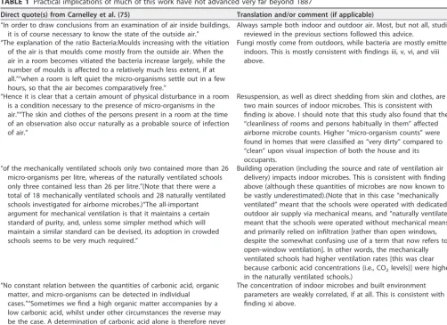
TABLE 1 Practical implications of much of this work have not advanced very far beyond 1887
Direct quote(s) from Carnelley et al. (75) Translation and/or comment (if applicable) “In order to draw conclusions from an examination of air inside buildings,
it is of course necessary to know the state of the outside air.”
Always sample both indoor and outdoor air. Most, but not all, studies reviewed in the previous sections followed this advice.
“The explanation of the ratio Bacteria:Moulds increasing with the vitiation of the air is that moulds come mostly from the outside air. When the air in a room becomes vitiated the bacteria increase largely, while the number of moulds is affected to a relatively much less extent, if at all.”“when a room is left quiet the micro-organisms settle out in a few hours, so that the air becomes comparatively free.”
Fungi mostly come from outdoors, while bacteria are mostly emitted indoors. This is mostly consistent with findings iii, v, vi, and viii above.
“Hence it is clear that a certain amount of physical disturbance in a room is a condition necessary to the presence of micro-organisms in the air.”“The skin and clothes of the persons present in a room at the time of an observation also occur naturally as a probable source of infection of air.”
Resuspension, as well as direct shedding from skin and clothes, are two main sources of indoor microbes. This is consistent with finding ix above. I should note that this study also found that the “cleanliness of rooms and persons habitually in them” affected airborne microbe counts. Higher “micro-organism counts” were found in homes that were classified as “very dirty” compared to “clean” upon visual inspection of both the house and its occupants.
“of the mechanically ventilated schools only two contained more than 26 micro-organisms per litre, whereas of the naturally ventilated schools only three contained less than 26 per litre.”(Note that there were a total of 18 mechanically ventilated schools and 28 naturally ventilated schools investigated for airborne microbes.)“The all-important argument for mechanical ventilation is that it maintains a certain standard of purity, and, unless some simpler method which will maintain a similar standard can be devised, its adoption in crowded schools seems to be very much required.”
Building operation (including the source and rate of ventilation air delivery) impacts indoor microbes. This is consistent with finding x above (although these quantities of microbes are now known to be vastly underestimated).(Note that in this case “mechanically ventilated” meant that the schools were operated with dedicated outdoor air supply via mechanical means, and “naturally ventilated” meant that the schools were operated without mechanical means and primarily relied on infiltration [rather than open windows, despite the somewhat confusing use of a term that now refers to open-window ventilation]. In other words, the mechanically ventilated schools had higher ventilation rates [this was clear because carbonic acid concentrations {i.e., CO2levels}] were higher in the naturally ventilated schools.)
“No constant relation between the quantities of carbonic acid, organic matter, and micro-organisms can be detected in individual cases.”“Sometimes we find a high organic matter accompanies by a low carbonic acid, whilst under other circumstances the reverse may be the case. A determination of carbonic acid alone is therefore never a sufficient indication of the purity or otherwise of a given sample of air.”
The concentration of indoor microbes and built environment parameters are weakly correlated, if at all. This is consistent with finding xi above.
on September 8, 2020 by guest
http://msystems.asm.org/
microbiomes of indoor environments reviewed above. In fact, 7 of these 12 enumerated findings from more-recent investigations using molecular methods (specifically findings iii, v, vi, viii, ix, x, and xi) were identified or suggested in the 1887 paper, albeit in an admittedly broad and somewhat crude manner, and without much complexity. The following ⬎100 years of the use of culture-based methods to sample microbes in buildings continued to build on many of these same themes, but this new body of work in the last 10 to 15 years using modern molecular methods has very rapidly added much needed nuance and complexity to our understanding of indoor microbiomes. However, it remains to be seen how this new knowledge base will change how we design, build, and operate buildings.
SUMMARY
We are just now beginning to understand the complexity with which indoor micro-biomes may affect human health in both positive and negative ways, but much more research is needed to better understand these complicated interactions. Although the use of molecular methods to analyze microbial samples has greatly increased the complexity with which we understand indoor microbes, we still know much more about relatively simple microbial characterizations based on sequence information (e.g., relative abundance of certain taxa and overall measures of diversity and richness) than we do about the function, expression, and viability of the vast numbers of microorganisms present inside buildings. Until molecular methods and statistical tech-niques advance to a state in which more-complex microbial characteristics (e.g., gene expression and function) can easily and cheaply be assessed in environmental samples, the true usefulness of molecular techniques may be best realized when used in conjunction with traditional methods of culturing and viability assessments (15). In fact, looking back at one of the earliest applications of molecular methods to indoor environments, in 2007, Lee et al. stated that “the combination of culture and culture-independent methods provided powerful means for determining both viability and diversity of bacteria in child-care facilities” (10). I would tend to agree.
REFERENCES
1. Kelley ST, Gilbert JA. 2013. Studying the microbiology of the indoor environment. Genome Biol14:202. http://dx.doi.org/10.1186/gb-2013 -14-2-202.
2. Konya T, Scott JA. 2014. Recent advances in the microbiology of the built environment. Curr Sustain/Renew Energy Rep1:35– 42. http:// dx.doi.org/10.1007/s40518-014-0007-4.
3. Adams RI, Bateman AC, Bik HM, Meadow JF. 2015. Microbiota of the indoor environment: a meta-analysis. Microbiome 3:49. http:// dx.doi.org/10.1186/s40168-015-0108-3.
4. Whitfield J. 2005. Is everything everywhere? Science310:960 –961.
http://dx.doi.org/10.1126/science.310.5750.960.
5. Tringe SG, Rubin EM. 2005. Metagenomics: DNA sequencing of envi-ronmental samples. Nat Rev Genet 6:805– 814. http://dx.doi.org/ 10.1038/nrg1709.
6. Wooley JC, Godzik A, Friedberg I. 2010. A primer on metagenomics. P L o S C o m p u t B i o l 6 :e 1 0 0 0 6 6 7 . h t t p : / / d x . d o i . o r g / 1 0 . 1 3 7 1 / journal.pcbi.1000667.
7. Tsai FC, Macher JM. 2005. Concentrations of airborne culturable bac-teria in 100 large US office buildings from the BASE study. Indoor Air 15(Suppl 9):71– 81.http://dx.doi.org/10.1111/j.1600-0668.2005.00346.x. 8. Kloos WE, Musselwhite MS. 1975. Distribution and persistence of
Staphylococcus and Micrococcus species and other aerobic bacteria on human skin. Appl Microbiol30:381–385.
9. Moschandreas DJ, Pagilla KR, Storino LV. 2003. Time and space uniformity of indoor bacteria concentrations in Chicago area residences. Aerosol Sci Technol 37:899 –906. http://dx.doi.org/10.1080/ 02786820300935.
10. Lee L, Tin S, Kelley ST. 2007. Culture-independent analysis of bacterial diversity in a child-care facility. BMC Microbiol7:27.http://dx.doi.org/ 10.1186/1471-2180-7-27.
11. Lax S, Smith DP, Hampton-Marcell J, Owens SM, Handley KM, Scott NM, Gibbons SM, Larsen P, Shogan BD, Weiss S, Metcalf JL, Ursell
LK, Vázquez-Baeza Y, Van Treuren W, Hasan NA, Gibson MK, Colwell R, Dantas G, Knight R, Gilbert JA. 2014. Longitudinal analysis of microbial interaction between humans and the indoor environment. Science345:1048 –1052.http://dx.doi.org/10.1126/science.1254529. 12. Miletto M, Lindow SE. 2015. Relative and contextual contribution of
different sources to the composition and abundance of indoor air bacteria in residences. Microbiome 3:61. http://dx.doi.org/10.1186/ s40168-015-0128-z.
13. Prussin AJ, Marr LC. 2015. Sources of airborne microorganisms in the built environment. Microbiome3:78.http://dx.doi.org/10.1186/s40168 -015-0144-z.
14. Prussin AJ, Garcia EB, Marr LC. 2015. Total concentrations of virus and bacteria in indoor and outdoor air. Environ Sci Technol Lett2:84 – 88. 15. Gibbons SM, Schwartz T, Fouquier J, Mitchell M, Sangwan N, Gilbert
JA, Kelley ST. 2015. Ecological succession and viability of human-associated microbiota on restroom surfaces. Appl Environ Microbiol 81:765–773.http://dx.doi.org/10.1128/AEM.03117-14.
16. Rintala H, Pitkäranta M, Toivola M, Paulin L, Nevalainen A. 2008. Diversity and seasonal dynamics of bacterial community in indoor en-vironment. BMC Microbiol8:56.http://dx.doi.org/10.1186/1471-2180-8 -56.
17. Hewitt KM, Gerba CP, Maxwell SL, Kelley ST. 2012. Office space bacterial abundance and diversity in three metropolitan areas. PLoS One 7:e37849.http://dx.doi.org/10.1371/journal.pone.0037849.
18. Meadow JF, Altrichter AE, Bateman AC, Stenson J, Brown GZ, Green JL, Bohannan BJM. 2015. Humans differ in their personal microbial cloud. PeerJ3:e1258.http://dx.doi.org/10.7717/peerj.1258.
19. Amend AS, Seifert KA, Samson R, Bruns TD. 2010. Indoor fungal composition is geographically patterned and more diverse in temperate zones than in the tropics. Proc Natl Acad Sci U S A107:13748 –13753.
http://dx.doi.org/10.1073/pnas.1000454107.
20. Barberán A, Dunn RR, Reich BJ, Pacifici K, Laber EB, Menninger HL,
on September 8, 2020 by guest
http://msystems.asm.org/
Morton JM, Henley JB, Leff JW, Miller SL, Fierer N. 2015. The ecology of microscopic life in household dust. Proc R Soc B Biol Sci 282: 20151139.http://dx.doi.org/10.1098/rspb.2015.1139.
21. Adams RI, Miletto M, Taylor JW, Bruns TD. 2013. Dispersal in microbes: fungi in indoor air are dominated by outdoor air and show dispersal limitation at short distances. ISME J 7:1262–1273. http:// dx.doi.org/10.1038/ismej.2013.28.
22. Emerson JB, Keady PB, Brewer TE, Clements N, Morgan EE, Awer-buch J, Miller SL, Fierer N. 2015. Impacts of flood damage on airborne bacteria and fungi in homes after the 2013 Colorado front range flood. Environ Sci Technol49:2675–2684.http://dx.doi.org/10.1021/es503845j. 23. Hyvärinen A, Vahteristo M, Meklin T, Jantunen M, Nevalainen A, Moschandreas D. 2001. Temporal and spatial variation of fungal con-centrations in indoor air. Aerosol Sci Technol 35:688 – 695. http:// dx.doi.org/10.1080/02786820117763.
24. He C, Salonen H, Ling X, Crilley L, Jayasundara N, Cheung HC, Hargreaves M, Huygens F, Knibbs LD, Ayoko GA, Morawska L. 2014. The impact of flood and post-flood cleaning on airborne microbiological and particle contamination in residential houses. Environ Int69:9 –17.
http://dx.doi.org/10.1016/j.envint.2014.04.001.
25. Rao CY, Riggs MA, Chew GL, Muilenberg ML, Thorne PS, Van Sickle D, Dunn KH, Brown C. 2007. Characterization of airborne molds, endo-toxins, and glucans in homes in New Orleans after hurricanes Katrina and Rita. Appl Environ Microbiol 73:1630 –1634. http://dx.doi.org/ 10.1128/AEM.01973-06.
26. Chew GL, Wilson J, Rabito FA, Grimsley F, Iqbal S, Reponen T, Muilenberg ML, Thorne PS, Dearborn DG, Morley RL. 2006. Mold and endotoxin levels in the aftermath of hurricane Katrina: a pilot project of homes in New Orleans undergoing renovation. Environ Health Perspect 114:1883–1889.http://dx.doi.org/10.1289/ehp.9258.
27. Tringe SG, Zhang T, Liu X, Yu Y, Lee WH, Yap J, Yao F, Suan ST, Ing SK, Haynes M, Rohwer F, Wei CL, Tan P, Bristow J, Rubin EM, Ruan Y. 2008. The airborne metagenome in an indoor urban environment. PLoS One3:e1862.http://dx.doi.org/10.1371/journal.pone.0001862. 28. Flores GE, Bates ST, Caporaso JG, Lauber CL, Leff JW, Knight R,
Fierer N. 2013. Diversity, distribution and sources of bacteria in residen-tial kitchens. Environ Microbiol15:588 –596.http://dx.doi.org/10.1111/ 1462-2920.12036.
29. Flores GE, Bates ST, Knights D, Lauber CL, Stombaugh J, Knight R, Fierer N. 2011. Microbial biogeography of public restroom surfaces. PLoS One6:e28132.http://dx.doi.org/10.1371/journal.pone.0028132. 30. Dunn RR, Fierer N, Henley JB, Leff JW, Menninger HL. 2013. Home
life: factors structuring the bacterial diversity found within and between h o m e s . P L o S O n e 8 :e 6 4 1 3 3 . h t t p : / / d x . d o i . o r g / 1 0 . 1 3 7 1 / journal.pone.0064133.
31. Hospodsky D, Qian J, Nazaroff WW, Yamamoto N, Bibby K, Rismani-Yazdi H, Peccia J. 2012. Human occupancy as a source of indoor airborne bacteria. PLoS One 7:e34867. http://dx.doi.org/10.1371/ journal.pone.0034867.
32. Meadow JF, Altrichter AE, Kembel SW, Moriyama M, O’Connor TK, Womack AM, Brown GZ, Green JL, Bohannan BJM. 2014. Bacterial communities on classroom surfaces vary with human contact. Micro-biome2:7.http://dx.doi.org/10.1186/2049-2618-2-7.
33. Jeon Y-S, Chun J, Kim B-S. 2013. Identification of household bacterial community and analysis of species shared with human microbiome. Curr Microbiol67:557–563.http://dx.doi.org/10.1007/s00284-013-0401-y. 34. Knights D, Kuczynski J, Charlson ES, Zaneveld J, Mozer MC, Collman
RG, Bushman FD, Knight R, Kelley ST. 2011. Bayesian community-wide culture-independent microbial source tracking. Nat Methods8:761–763.
http://dx.doi.org/10.1038/nmeth.1650.
35. Hewitt KM, Mannino FL, Gonzalez A, Chase JH, Caporaso JG, Knight R, Kelley ST. 2013. Bacterial diversity in two neonatal intensive care units (NICUs). PLoS One 8:e54703. http://dx.doi.org/10.1371/ journal.pone.0054703.
36. Hospodsky D, Yamamoto N, Nazaroff WW, Miller D, Gorthala S, Peccia J. 2015. Characterizing airborne fungal and bacterial concentra-tions and emission rates in six occupied children’s classrooms. Indoor Air 25:641– 652.http://dx.doi.org/10.1111/ina.12172.
37. Qian J, Hospodsky D, Yamamoto N, Nazaroff WW, Peccia J. 2012. Size-resolved emission rates of airborne bacteria and fungi in an occu-pied classroom. Indoor Air22:339 –351.http://dx.doi.org/10.1111/j.1600 -0668.2012.00769.x.
38. Bhangar S, Adams RI, Pasut W, Huffman JA, Arens EA, Taylor JW, Bruns TD, Nazaroff WW. 2016. Chamber bioaerosol study: human
emissions of size-resolved fluorescent biological aerosol particles. Indoor Air26:193–206.http://dx.doi.org/10.1111/ina.12195.
39. Kembel SW, Jones E, Kline J, Northcutt D, Stenson J, Womack AM, Bohannan BJ, Brown GZ, Green JL. 2012. Architectural design influ-ences the diversity and structure of the built environment microbiome. ISME J6:1469 –1479.http://dx.doi.org/10.1038/ismej.2011.211. 40. Kembel SW, Meadow JF, O’Connor TK, Mhuireach G, Northcutt D,
Kline J, Moriyama M, Brown GZ, Bohannan BJM, Green JL. 2014. Architectural design drives the biogeography of indoor bacterial com-m u n i t i e s . P L o S O n e 9 :e 8 7 0 9 3 . h t t p : / / d x . d o i . o r g / 1 0 . 1 3 7 1 / journal.pone.0087093.
41. Meadow JF, Altrichter AE, Kembel SW, Kline J, Mhuireach G, Moriyama M, Northcutt D, O’Connor TK, Womack AM, Brown GZ, Green JL, Bohannan BJ. 2014. Indoor airborne bacterial communities are influenced by ventilation, occupancy, and outdoor air source. Indoor Air24:41– 48.http://dx.doi.org/10.1111/ina.12047.
42. Adams RI, Bhangar S, Pasut W, Arens EA, Taylor JW, Lindow SE, Nazaroff WW, Bruns TD. 2015. Chamber bioaerosol study: outdoor air and human occupants as sources of indoor airborne microbes. PLoS One 10:e0128022.http://dx.doi.org/10.1371/journal.pone.0128022. 43. Dannemiller KC, Gent JF, Leaderer BP, Peccia J. 2016. Influence of
housing characteristics on bacterial and fungal communities in homes of asthmatic children. Indoor Air26:179 –192.http://dx.doi.org/10.1111/ ina.12205.
44. Weikl F, Tischer C, Probst AJ, Heinrich J, Markevych I, Jochner S, Pritsch K. 2016. Fungal and bacterial communities in indoor dust follow different environmental determinants. PLoS One11:e0154131.http:// dx.doi.org/10.1371/journal.pone.0154131.
45. Ramos T, Stephens B. 2014. Tools to improve built environment data collection for indoor microbial ecology investigations. Build Environ 81:243–257.http://dx.doi.org/10.1016/j.buildenv.2014.07.004. 46. Ramos T, Dedesko S, Siegel JA, Gilbert JA, Stephens B. 2015. Spatial
and temporal variations in indoor environmental conditions, human occupancy, and operational characteristics in a new hospital building. P L o S O n e 1 0 :e 0 1 1 8 2 0 7 . h t t p : / / d x . d o i . o r g / 1 0 . 1 3 7 1 / journal.pone.0118207.
47. Dedesko S, Stephens B, Gilbert JA, Siegel JA. 2015. Methods to assess human occupancy and occupant activity in hospital patient rooms. Build Environ90:136 –145.http://dx.doi.org/10.1016/j.buildenv.2015.03.029. 48. Chase J, Fouquier J, Zare M, Sonderegger DL, Knight R, Kelley ST,
Siegel J, Caporaso JG. 2016. Geography and location are the primary drivers of office microbiome composition. mSystems 1:e00022-16.
http://dx.doi.org/10.1128/mSystems.00022-16.
49. Gibbons SM. 2016. The built environment is a microbial wasteland. mSystems1:e00033-16.http://dx.doi.org/10.1128/mSystems.00033-16. 50. Bhangar S, Huffman JA, Nazaroff WW. 2014. Size-resolved fluorescent
biological aerosol particle concentrations and occupant emissions in a university classroom. Indoor Air24:604 – 617.http://dx.doi.org/10.1111/ ina.12111.
51. Bean B, Moore BM, Sterner B, Peterson LR, Gerding DN, Balfour HH. 1982. Survival of influenza viruses on environmental surfaces. J Infect Dis 146:47–51.http://dx.doi.org/10.1093/infdis/146.1.47.
52. Greatorex JS, Digard P, Curran MD, Moynihan R, Wensley H, Wreghitt T, Varsani H, Garcia F, Enstone J, Nguyen-Van-Tam JS. 2011. Survival of influenza A(H1N1) on materials found in households: implications for infection control. PLoS One6:e27932.http://dx.doi.org/ 10.1371/journal.pone.0027932.
53. Coughenour C, Stevens V, Stetzenbach LD. 2011. An evaluation of methicillin-resistantStaphylococcus aureussurvival on five environmen-tal surfaces. Microb Drug Resist17:457– 461.http://dx.doi.org/10.1089/ mdr.2011.0007.
54. Casanova LM, Jeon S, Rutala WA, Weber DJ, Sobsey MD. 2010. Effects of air temperature and relative humidity on coronavirus survival on surfaces. Appl Environ Microbiol 76:2712–2717. http://dx.doi.org/ 10.1128/AEM.02291-09.
55. Tang JW. 2009. The effect of environmental parameters on the survival of airborne infectious agents. J R Soc Interface6(Suppl 6):S737–S746.
http://dx.doi.org/10.1098/rsif.2009.0227.focus.
56. Makison C, Swan J. 2006. The effect of humidity on the survival of MRSA on hard surfaces. Indoor Built Environ15:85–91.http://dx.doi.org/ 10.1177/1420326X06062582.
57. Traoré O, Springthorpe VS, Sattar SA. 2002. A quantitative study of the survival of two species of Candida on porous and non-porous
on September 8, 2020 by guest
http://msystems.asm.org/
environmental surfaces and hands. J Appl Microbiol92:549 –555.http:// dx.doi.org/10.1046/j.1365-2672.2002.01560.x.
58. Jawad A, Heritage J, Snelling AM, Gascoyne-Binzi DM, Hawkey PM. 1996. Influence of relative humidity and suspending menstrua on sur-vival of Acinetobacter spp. on dry surfaces. J Clin Microbiol 34: 2881–2887.
59. Handley BA, Webster AJ. 1993. Some factors affecting airborne survival ofPseudomonas fluorescensindoors. J Appl Bacteriol75:35– 42.http:// dx.doi.org/10.1111/j.1365-2672.1993.tb03404.x.
60. Mbithi JN, Springthorpe VS, Sattar SA. 1991. Effect of relative humid-ity and air temperature on survival of hepatitis A virus on environmental surfaces. Appl Environ Microbiol57:1394 –1399.
61. McEldowney S, Fletcher M. 1988. The effect of temperature and rela-tive humidity on the survival of bacteria attached to dry solid surfaces. Lett Appl Microbiol 7:83– 86. http://dx.doi.org/10.1111/j.1472 -765X.1988.tb01258.x.
62. Karim YG, Ijaz MK, Sattar SA, Johnson-Lussenburg CM. 1985. Effect of relative humidity on the airborne survival of rhinovirus-14. Can J Micro-biol31:1058 –1061.http://dx.doi.org/10.1139/m85-199.
63. Sattar SA, Ijaz MK, Johnson-Lussenburg CM, Springthorpe VS. 1984. Effect of relative humidity on the airborne survival of rotavirus SA11. Appl Environ Microbiol47:879 – 881.
64. Dick EC, Jennings LC, Mink KA, Wartgow CD, Inhorn SL. 1987. Aerosol transmission of rhinovirus colds. J Infect Dis 156:442– 448. http:// dx.doi.org/10.1093/infdis/156.3.442.
65. Wong BCK, Lee N, Li Y, Chan PKS, Qiu H, Luo Z, Lai RWM, Ngai KLK, Hui DSC, Choi KW, Yu ITS. 2010. Possible role of aerosol transmission in a hospital outbreak of influenza. Clin Infect Dis51:1176 –1183.http:// dx.doi.org/10.1086/656743.
66. Li Y, Leung GM, Tang JW, Yang X, Chao CYH, Lin JZ, Lu JW, Nielsen PV, Niu J, Qian H, Sleigh AC, Su H-JJ, Sundell J, Wong TW, Yuen PL. 2007. Role of ventilation in airborne transmission of infectious agents in the built environment—a multidisciplinary systematic review. Indoor Air 17:2–18.http://dx.doi.org/10.1111/j.1600-0668.2006.00445.x.
67. Beggs CB, Shepherd SJ, Kerr KG. 2010. Potential for airborne trans-mission of infection in the waiting areas of healthcare premises: sto-chastic analysis using a Monte Carlo model. BMC Infect Dis10:247.
http://dx.doi.org/10.1186/1471-2334-10-247.
68. Eggleston PA, Rosenstreich D, Lynn H, Gergen P, Baker D, Kattan M,
Mortimer KM, Mitchell H, Ownby D, Slavin R, Malveaux F. 1998. Relationship of indoor allergen exposure to skin test sensitivity in inner-city children with asthma. J Allergy Clin Immunol102:563–570.http:// dx.doi.org/10.1016/S0091-6749(98)70272-6.
69. Quansah R, Jaakkola MS, Hugg TT, Heikkinen SA, Jaakkola JJ. 2012. Residential dampness and molds and the risk of developing asthma: a systematic review and meta-analysis. PLoS One 7:e47526. http:// dx.doi.org/10.1371/journal.pone.0047526.
70. Dick S, Friend A, Dynes K, AlKandari F, Doust E, Cowie H, Ayres JG, Turner SW. 2014. A systematic review of associations between environ-mental exposures and development of asthma in children aged up to 9 years. BMJ Open 4:e006554. http://dx.doi.org/10.1136/bmjopen-2014 -006554.
71. Fujimura KE, Demoor T, Rauch M, Faruqi AA, Jang S, Johnson CC, Boushey HA, Zoratti E, Ownby D, Lukacs NW, Lynch SV. 2014. House dust exposure mediates gut microbiome Lactobacillus enrichment and airway immune defense against allergens and virus infection. Proc Natl A c a d S c i U S A 1 1 1 :8 0 5 – 8 1 0 . h t t p : / / d x . d o i . o r g / 1 0 . 1 0 7 3 / pnas.1310750111.
72. Dannemiller KC, Mendell MJ, Macher JM, Kumagai K, Bradman A, Holland N, Harley K, Eskenazi B, Peccia J. 2014. Next-generation DNA sequencing reveals that low fungal diversity in house dust is associated with childhood asthma development. Indoor Air 24:236 –247.http:// dx.doi.org/10.1111/ina.12072.
73. Lynch SV, Wood RA, Boushey H, Bacharier LB, Bloomberg GR, Kat-tan M, O’Connor GT, Sandel MT, Calatroni A, Matsui E, Johnson CC, Lynn H, Visness CM, Jaffee KF, Gergen PJ, Gold DR, Wright RJ, Fujimura K, Rauch M, Busse WW, Gern JE. 2014. Effects of early-life exposure to allergens and bacteria on recurrent wheeze and atopy in urban children. J Allergy Clin Immunol 134:593– 601.e12. http:// dx.doi.org/10.1016/j.jaci.2014.04.018.
74. Dannemiller KC, Gent JF, Leaderer BP, Peccia J. 2016. Indoor micro-bial communities: influence on asthma severity in atopic and nonatopic children. J Allergy Clin Immunol 138:76 – 83.e1. http://dx.doi.org/ 10.1016/j.jaci.2015.11.027.
75. Carnelley T, Haldane JS, Anderson AM. 1887. The carbonic acid, organic matter, and micro-organisms in air, more especially of dwellings and schools. Philos Trans R Soc B Biol Sci178:61–111.http://dx.doi.org/ 10.1098/rstb.1887.0004.