Anticancer and Antioxidant Properties of Flavored Green Tea Extracts
Bindhu Alappat*
Jaclyn A. Sarna
Chau Truong
Department of Chemistry
Saint Xavier University
Chicago, IL 60655
Abstract
Brewed green tea has been associated with just about everything healthy -immunity boosting to prevention of chronic diseases. Speculations about the benefits of green tea range back to ancient times, but their bio activities are yet to be established. The antioxidants in green tea (catechins) are shown to slow the growth of cancer cells, reduce the size of tumors, and soften the sharp side effects of chemotherapy. In Asian countries green tea is consumed as plain brewed tea, but in western countries the popularity of tea is for the flavored green tea available in the market. All existing research work documents health benefits of plain green tea, but studies on flavored teas are not as widespread. In this paper, various flavored green teas were analyzed and compared with plain green tea for anticancer, and antioxidant capabilities. Jasmine and blueberry showed the strongest anticancer properties whereas the most antioxidant was Jasmine.
Keywords:
Catechin, High Performance Liquid Chromatography, 2,2-diphenyl-1-picrylhydrazyl (DPPH), UV-Vis Spectrophotometer, radical scavenging effect, IC50, breast cancer cell, fluorescence1. Introduction
Tea is one of the most popular beverages in the world and is consumed by over two-thirds of the world’s population. Tea has been extensively studied for its wide range of health benefits, including anti-diabetic [Park, 2014], anti-oxidant [kumaran, 2009], anti-cancer [Lecumberri, 2013], and anti-microbial capabilities [Bancirova, 2010]. Other documented benefits vary from enhanced metabolism leading to weight loss; promoting digestion and ingestion of fatty food; reduction of total cholesterol but raise High Density Lipid (HDL) and prevention of plaque formation and enhanced oral health [Yamamoto, 1997; Yukihiko, 2001]. The leaves of the plant, Camellia Sinensis, are processed in different ways to produce four different types of tea: white, green, oolong, and black [Dattner, 2003]. These specific treatments produce teal eaves with varying chemical compositions and potential health benefits. Green tea has a high concentration of polyphenols. Specifically, it contains a large amount of catechins- compounds believed to be responsible for tea’s effect in our biological activities [Cho, 2007]. Although medicinal and pharmacological effects of plain green tea are well studied, the chemistry and health benefits of newly marketed flavored green teas are not yet documented. A systematic investigation of the chemical components of a variety of commercial flavored teas, such as mango, mint, pomegranate, blueberry, lemon, jasmine and peach are presented in this paper. The purpose of this project is to determine whether any flavored green teas showed significantly higher antioxidant, and anticancer abilities compared to plain green tea.
Green tea was chosen as the sole focus of the experiments due to both its catechin content and prevalence. Green tea research generally focuses on specific catechins. These compounds are proanthocyanidin derivatives, or more broadly, flavanols [Yamamoto, 1997]. The most abundant catechin is epigallocatechingallate, or EGCG [Cho, 2007], but green tea contains as many as twelve catechins [Babich, 2007]. Eight prominent catechins, with abbreviations, are Epicatechin (EC),(+)-catechin (C), Epigallocatechin (EGC), Gallocatechin (GC), (-)-Epicatechingallate(ECG),(-)-Catechingallate(CG),(-)-Epigallocatechingallate(EGCG),and(-)-Gallocatechingallate (GCG). These polyphenols can have varying effects, depending on the compounds they react with. When they react with free radicals, such as 2,2-diphenyl-1-picrylhydrazyl(DPPH), they are oxidized while the DPPH is reduced [Scherer, 2009; Huang, 2005]. However, in the case of EGCG reacting with cancer cells, the catechin oxidizes itself and attacks cysteine residues of proteins in cancerous cells [Yang, 2007]. When EGCG binds to proteins in cancerous cells, it can inhibit their growth, induce apoptosis (programmed cell death), or inhibit angiogenesis, which is the cancer cells causing blood vessels to form and connect to it [Ishii, 2008]. It is not known, however, how the catechin differentiates between normal and cancerous cells and causes apoptosis only in the latter.
The antioxidant capabilities of flavored teas’ are well investigated. A variety of radicals are commercially available for antioxidant research. 2,2-diphenyl-1-picrylhydrazyl (DPPH) is an organic nitrogen free radical used to test antioxidant capabilities [Henning, 2003 ]. DPPH represents any kind of free radical in the body, whether it is nitrogen based or oxygen based. A compound is a good antioxidant if it can easily donate an electron to the electron deficient radical, stopping the chain of molecules stealing electrons from each other. Free radicals do occur in the body, but available antioxidants can prevent them from doing too much damage. When free radicals are released, they are able to destroy the cell membrane. Once this amphipathic envelope that encloses the cell is destroyed, it will release cell organelles, cell composition, and most importantly the nucleus. Free radicals can continue to disrupt the nuclear envelope which causes the release of free DNA, the genetic blueprint, to the outer space there by completely destroying a healthy cell [Huang, 2005; Henning, 2003]. Therefore, it is important to scavenge these free radicals in body. Chemical composition of various flavored green tea and their anticancer and antioxidant properties are discussed in this paper. Identification and quantification of various catechins in flavored teas were performed using High Performance Liquid Chromatography (HPLC). DPPH method was used to study the antioxidant properties of flavored tea extracts. This was followed by an investigation of their anticancer properties and is presented in relation to their catechin content.
2. Materials and Methods
2.1. Tea Samples
A variety of flavored green tea-Mango, Jasmine, Peach, Mint, Pomegranate, Lemon, and Blueberry-were used for the study. Each flavored type had a distinctive scent, taste, and ingredient which made them unique. All green tea samples were purchased locally but produced by same company R. C. Bigelow Inc, Fairfield, CT.
2.2. Catechin Standards
Catechin, Epicatechin, Epicatechingallate, Epigallocatechin, Gallocatechingallate, Epigallocatechingallate were all purchased from Nacalai Tesque, China. DPPH (CAS Number1898-66-4) was purchased from Sigma-Aldrich.
2.3. Instrument Details
HPLC: Waters 600E with UV/Vis detector, column heater and auto sampler. Colum- Waters Symmetry, 4.6mm X 250 mm, 5µ; Guard column-Symmetry.
UV-Vis Spectrometer: Agilent 8453. Breast cancer cells (MDA-MB-231) were donated by University of Nebraska, Medical School.
2.4. Other Reagents
All reagents and solvents were purchased from commercial sources and used without further purification. Phosphoric acid: Burdick &Jackson75-05-8; Acetonitrile: Mallinckrodt Chemicals 7664-38-2
2.5. HPLC Method
Preparation of standards: Catechin standards for HPLC were prepared by accurately weighing 2.5mg of standard sample into an 8mL amber vial. Mix with 5.0mL 0.1% H3PO4 and sonicated for10 minutes and allowed to cool.
2.6. Preparation of Extracts
Tea extracts were prepared by accurately weighing 100 mg of sample into a 100mL volumetric flask mixing it with 75mL of 0.1% H3PO4. Samples were sonicated for1 hour and allowed to cool before making up the volume.
All samples were filtered through 0.45µ PTFE filter for HPLC use. Chromatograms were run at 280nm with a column temperature of 220C using gradient method. The injection volume was maintained at 10µL and the mobile phases used were 0.1% H3PO4 (A); acetonitrile (B); miliQ water (C) and methanol (D).
2.7. Antioxidant Study
Eight different teas were compared in this experiment: blueberry, jasmine, lemon, mango, mint, peach, pomegranate, and plain green tea. Each tea sample was prepared the same way by steeping 1.000g of tea in 50 mL hot water (80C) for three minutes. The filtered solution was then dried under nitrogen gas on an 80°C hotplate and reconstituted in 50 mL of methanol. Each tea sample was prepared in triplicate. In addition, tea extracts with varying amount of tea -0.25g, 0.50 g, 1.00g, 2.00g, and 3.00g-were also prepared and used for IC50 calculations. The DPPH solution was prepared by dissolving 0.0025g of DPPH in 100mL methanol. For antioxidant study, samples were prepared by mixing 0.1 mL aliquots of methanolic tea extracts with 3.9 ml of DPPH solution. The blank was 100% DPPH concentration. The mixtures of tea with DPPH were incubated for 60 minutes at 37ºC. Sample preparations and antioxidant studies were all performed in a dark room since DPPH was sensitive to light. After an hour of incubation, the UV-Vis spectra were recorded at 515nm for each tea extracts, blank DPPH solution and tea-DPPH solutions. Percent inhibition or the radical scavenging effect [Sanchez-Moreno, 1999; Scherer, 2009] was calculated using the formula I%=100*(Absi-Absf)/Absi, where Absi is the absorbance of the
DPPH stock solution and Absf is the absorbance of the tea and DPPH solution after 60 minutes of incubation. The
higher the percent inhibition, the more antioxidant the particular flavored tea was. Further, IC50 calculations -efficiency concentration at 50% inhibition-were done graphically using a calibration curve in the linear range by plotting the extract concentration vs. the corresponding scavenging effect [Sanchez-Moreno, 1999].
2.8. Anticancer study
Six tea solutions were compared for their anti-cancer activity in this experiment: blueberry, jasmine, mango, mint, pomegranate, and plain green tea. 1.000g of tea was steeped in water at 80C for three minutes. The filtered solution was then evaporated and labeled. The flavored tea extracts were dissolved in ethanol to make 2µg/mL solutions. 20µL of this solution was added to 180µL of media, making a 200ng/mL concentration of the flavored tea. A drug plate was generated using10µL of the above solution transferred to the drug plate in triplicate and then half dilutions were made across the plate (the concentrations are 200ng/mL to 0.4ng/mL). Breast cancer cells (MDA-MB-231) were plated and allowed to attach overnight. The tea extracts from the drug plate above was transferred to the assay plate and incubated for 72 hours. At the end of the incubation period 10µL of presto blue viability reagent was added and the plate was incubated again for an additional 15 minutes. Fluorescence was measured on spectramax M5 plate reader ex=560nm and em=590nm [Pesseto, 2012].
3. Results and Discussion
3.1. HPLC Analysis
3.2. Antioxidant Analysis
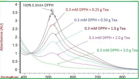
Antioxidant capacity of seven flavored teas was studied in terms of percent absorbance. For each trial, jasmine tea gave the highest percent inhibition average about 85%. The second most antioxidant teas were blueberry and pomegranate while the least antioxidant tea was mango (27% inhibition) followed by Mint. Figure 3 belongs to Jasmine tea of different concentrations mixed with 0.3mM DPPH. The top black colored line indicates absorbance of 100% DPPH with no tea; therefore, it has the highest value of 3.4328 at 517 nm. The noise at the highest absorbance may be due to undissolved particles which scatter the UV-Vis light. As DPPH reacted with 0.25 g, the absorbance decreased due to the tea scavenging and reduced the radical to 2.7565. When DPPH reduced, the color turned to yellow/colorless, hence the absorbance decreased. Various colored lines in the figure indicate various amount of tea reacted with DPPH. It is clear that as the tea weight increased to 3.0g, the absorbance nearly reduced to zero. An absorption measurement of tea alone did not show any peak at 517nm, confirming no interference from tea at that wavelength for any flavor.
Figure 4 contains the absorbance data of various concentrations of Jasmine flavored tea at 517nm. The tea concentrations were calculated and plotted against I%. Based on the graph, at the 50% inhibition point, it can be extrapolated to calculate the exact tea concentration which inhibits 50% of DPPH activity. For brevity only Jasmine tea which has an IC50 of 24.97 mg/mL is shown in figure 4.
Figure 5 represents the scavenging effect of all flavored tea measured against all DPPH concentrations studied. The first row of 0.3258 mM DPPH and 0.25 g of various flavored teas, the jasmine tea show to have high scavenging effects [I%] toward free radicals, in second is the blueberry, and third is the pomegranate. As for the mango, mint, peach, and lemon, they show to have smaller scavenging effects than the first three flavored green tea. The higher the scavenging effect, the more active that flavored tea can reduce the radicals. The lower the I% value, the less it can reduce the DPPH. In the second row of data with 0.2000mM DPPH and tea, the same trend appears. However, as the DPPH concentrations decreased to 0.1242mM and 0.0800mM, the data changed. The blueberry, pomegranate, and jasmine scavenging effect are comparable. They are relatively the same and the one with highest scavenging effect is jasmine, pomegranate, and blueberry respectively.
During the preparation of blueberry and pomegranate green tea, we noticed deep vibrant dark blue and purple color for the extracts. The colors are from anthocyanin, a chemical component which is known to have antioxidant property. This may explain why the blueberry and pomegranate has such a high antioxidant property. The catechins in tea and anthocyanin may work together increasing their overall antioxidant ability and the scavenging effect. However, the jasmine flavored green tea is colorless, yet it shown to have high I% value. This might be due to the same synergistic effects of chemical components in jasmine with the catechins in green tea; there by increasing the scavenging effect. The lower I% of mango, mint, peach, and lemon teas might be due to fact that the chemical components in those flavors do not have any antioxidant activity; therefore, the scavenging effect is only due to the green tea catechins. A further investigation is needed in the future, to completely understand the mechanism.
3.3. Anticancer Study
By binding to certain proteins, EGCG can alter their conformation and impede their function. The researchers examined glyceraldehyde-3-phosphate dehydrogenase, which catalyzes the sixth step of glycolysis. By forming covalent bonds with this enzyme, EGCG can prevent the breakdown of glucose into pyruvate and cause the cell to commit programmed cell death due to nutrient deprivation [Carvalho, 2010]. It is not known, however, how the catechin differentiates between normal and cancerous cells and causes apoptosis only in the latter. Furthermore, EGCG also inhibits the activation of protein kinases, blocks the activation of transcription factors, inhibits cell proliferation, modulates cell cycle regulation, interferes with receptor binding, and suppresses invasiveness [Fujiki, 2012]. This catechin and others can cause cancer cell death through one or multiple of these pathways while leaving healthy cells alone.
4. Conclusion
Based on these antioxidant and anticancer studies, we can conclude that extract from the jasmine tea exhibited the highest activity on both studies. Blueberry and pomegranate green tea extracts, had deep vibrant color (dark blue and purple) in solution due to the presence of anthocyanin. This may explain why the blueberry and pomegranate has such high antioxidant property. However, the jasmine flavored green tea, which was colorless, had the highest I% value. This might be due to the chemical components within jasmine that could have synergistic effects with the catechins in green tea; there by increasing the scavenging effect. Similarly, the reason for lower I% of mango, mint, peach, and lemon could be due to antagonistic effect of chemical components in them with the tea catechins. The jasmine plant has a history of being used medicinally and it is a good candidate for future research. It has been used for psychiatric disorders and other illnesses [Ferreres, 2014]. Research in the future will focus on determining the major compounds in jasmine and to see which compounds have synergistic antioxidant and anticancer effects with the catechins in green tea.
5. Acknowledgements
6. References
Babich, H., Zuckerbraun, H.L., & Weinerman, S. M. (2007). In vitro cytotoxicity of (-) catechingallate, a minor polyphenol in green tea. Toxicology Letters, 171, 171-180
Bancirova, M. (2010).Comparison of the antioxidant capacity and the antimicrobial activity of black and green tea. Food Research International, 43, 1379-1382.
Bonoli, M., Pelillo, M., Toschi, T.G.& Lercker,G.(2003). Analysis of green tea catechins: comparative study between HPLC and HPCE. Food Chemistry, 81, 631-638.
Carvalho, M., Jerónimo,C.,Valentão,P., Andrade,P.B., & Silva,B.M. (2010). Green tea: A promising anticancer agent for renal cell carcinoma. Food Chemistry, 122, 49-54.
Chen,Q., Bryant,V.C., Lopez, H., Kelly, D.L., Luo,X. & Natarajan,A. (2011).2,3-Substituted quinoxalin-6-amine analogs as antiproliferatives: A structure activity relationship study. Bioorg Med Chem Lett, 19, 1929-1932.
Cho,K.,Sukhthankar,M., Lee,S.,Yoon,J., Baek,S.J.(2007). Green tea Catechin(-)-epicatechingallate induces tumor suppressor protein ATF3 via EGR-1activation. European Journal of Cancer, 43, 2402-2412.
Dattner, C., & Boussahba, S.(2003).The Book Of Green Tea. New York. Universe Print.
Ferreres, F., Grosso, C., Gil-Izquierdo, A., Valentao, P., & Andrade, P. B. (2014). Assessing Jasminum grandiflorum L. authenticity by HPLC-DAD-ESI/MSn and effects on physiological enzymes and oxidative species. Journal of Pharmaceutical and Biomedical Analysis, 88, 157-161.
Fujiki, H., & Suganuma, M. (2012). Green tea: An effective synergist with anticancer drug for tertiary cancer prevention. Cancer letters, 324, 119-125.
Furukawa, A., Oikawa, S., Murata, M., Hiraku, Y., & Kawanishi, S. (2003). (−) Epigallocatechingallate causes oxidative damage to isolated and cellular DNA. Biochemical Pharmacology, 66, 1769-1778.
Henning, S.M., Fajardo-Lira, C., Lee, H.W., Youssefian, A. A.,Go V.L.W. & Hebber, D.(2003). Catechin content of 18 teas and a green tea extract supplement correlates with the antioxidant capacity. Nutrition and cancer, 45, 226-235.
Huang, D., Ou, B. & Prior,R. L. (2005). The chemistry behind antioxidant capacity assays. J. Agric. Food Chem., 53, 1841–1856.
Ishii, T., Mori, T., Tanaka,T., Mizuno,D., Yamaji, R., kumazawa, S., Nakayama,T., & Akagawa, W. (2008). Covalent modification of proteins by green tea polyphenol (-)-epigallo-catechin-3-gallate through autoxidation. Free Radical Biology and Medicine, 45, 1384-1394.
Kumaran, V. S., Arulmathi, K., & Kalaiselvi,P. (2009).Senescence mediated redox imbalance in cardiac tissue: Antioxidant rejuvenating potential of green tea extract. Nutrition, 25, 847-854.
Kuzuhara, T., Suganuma, M., &Fujiki, H. (2008).Green tea catechin as a chemical chaperone in cancer prevention. Cancer Letters, 261, 12-20.
Lecumberri, E., Dupertuis, Y. M., Miralbell, R.,& Pichard,C. (2013).Green tea polyphenol epigallocatechin-3-gallate (EGCG) as adjuvant in cancer therapy. Clinical Nutrition, 32, 894-903.
Park,J., Bae, J., Im,S.,& Song, D. (2014).Green tea and type2 diabetes. Integrative Medicine Research, 3, 4-10. Pessetto, Z.Y., Yan, Y., Bessho, T., & Natarajan, A. (2012). Inhibition of BRCT (BRCA1)-phosphoprotein
interaction enhances the cytotoxic effect of olaparib in breast cancer cells: a proof of concept study for synthetic lethal therapeutic option. Breast Cancer Research & Treatment, 134,511-517.
Sanchez-Moreno., C., Larrauri, J. A. & Saura-Calixto, F.(1999). Free radical scavenging capacity and inhibition of lipid oxidation of wines, grape juices and related polyphenolic constituents. Food Res. Int., 32, 407-412.
Scherer, R.& Godoy, H.T.(2009). Antioxidant activity index (AAI) by the 2, 2-diphenyl-1- picrylhydrazyl method. Food Chemistry, 112, 654-658.
Yamamoto, T., Juneja, L. R., Chu, D. & Kim, M. (1997). Chemistry and Applications of Green Tea, CRC Press: New York.
Yang, C.S. (2007). Tea and cancer prevention: molecular mechanisms and human relevance. Toxicol. Appl. Pharmacol., 224, 265-273.
Figure 2: Calibration Curve of Catechin using various Concentrations. Integrated Intensity under the Curve is plotted against the Concentration
Figure 4: UV-Vis Absorbance of DPPH with different Jasmine Concentrations and IC 50
Figure 5: Scavenging Effect [I%] of 0.25 g of Various Flavored Tea
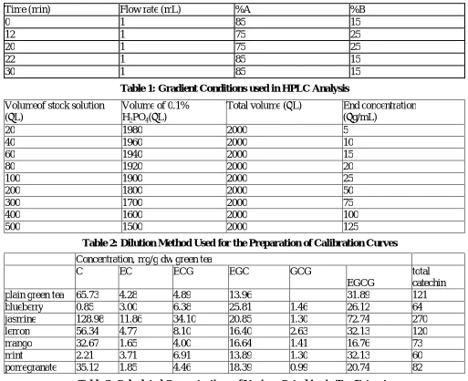
Time (min) Flow rate (mL) %A %B
0 1 85 15
12 1 75 25
20 1 75 25
22 1 85 15
30 1 85 15
Table 1: Gradient Conditions used in HPLC Analysis
Volumeof stock solution (µL)
Volume of 0.1% H3PO4(µL)
Total volume (µL) End concentration (µg/mL)
20 1980 2000 5
40 1960 2000 10
60 1940 2000 15
80 1920 2000 20
100 1900 2000 25
200 1800 2000 50
300 1700 2000 75
400 1600 2000 100
500 1500 2000 125
Table 2: Dilution Method Used for the Preparation of Calibration Curves
Concentration, mg/g dw green tea
C EC ECG EGC GCG
EGCG
total catechin
plain green tea 65.73 4.28 4.89 13.96 31.89 121
blueberry 0.85 3.00 6.38 25.81 1.46 26.12 64
jasmine 128.98 11.86 34.10 20.85 1.30 72.74 270
lemon 56.34 4.77 8.10 16.40 2.63 32.13 120
mango 32.67 1.65 4.00 16.64 1.41 16.76 73
mint 2.21 3.71 6.91 13.89 1.30 32.13 60
pomegranate 35.12 1.85 4.46 18.39 0.99 20.74 82
![Figure 5: Scavenging Effect [I%] of 0.25 g of Various Flavored Tea](https://thumb-us.123doks.com/thumbv2/123dok_us/8361950.1671709/9.612.102.517.155.501/figure-scavenging-effect-i-g-various-flavored-tea.webp)