How to cite this paper: Taguchi, Y., Tanabe, H., Fuchigami, K. and Tanaka, M. (2015) Microencapsulation of Ascorbic Acid
Tripalmitin by Using Dry Coating Method
Yoshinari Taguchi1, Hidetoshi Tanabe1, Kiyomi Fuchigami2, Masato Tanaka1* 1Graduate School of Science and Technology, Niigata University, Niigata, Japan
2Department of Research & Development, Shofu Inc., Kyoto, Japan
Email: *[email protected]
Received 7 January 2015; accepted 26 January 2015; published 30 January 2015
Copyright © 2015 by authors and Scientific Research Publishing Inc.
This work is licensed under the Creative Commons Attribution International License (CC BY). http://creativecommons.org/licenses/by/4.0/
Abstract
It was tried to microencapsulate the ascorbic acid powder as a redox initiator with tripalmitin by use of the dry coating method. In the experiment, the feed ratio of the amount of tripalmitin to that of ascorbic acid, the coating time and the concentration of ethyl alcohol of pulverizing solvent were changed stepwise. The characteristics of microcapsules such as the content of core material, the yield, the water proof degree, the microencapsulation efficiency and the mean diameter were estimated. The yield, the microencapsulation efficiency and the water proof degree gradually in-creased with the feed ratio, the coating time and the concentration of pulverizing solvent. The mi-crocapsules showed the thermal responsibility and induced polymerization of methyl methacry-late monomer.
Keywords
Ascorbic Acid, Tripalmitin Microcapsules, Dry Coating, Redox Initiator, Thermal Responsibility
1. Introduction
Microcapsules with the many functions have been prepared and applied to the various fields such as drugs, tex-tiles, information recording materials, paintings, cosmetics, food materials, adhesives, medicine, agriculture and so on [1] [2]. The purposes of microencapsulation are to protect the core material from environment, to release the core material according to occasion demands, to handle the gaseous and the liquid core materials as the solid particles and to modify the surface of core material [2]. Moreover, we can give the microcapsules the various functions by how to select the physicochemical properties of the core and the shell materials and how to com-bine these materials. For examples, there are the combinations of the hydrophilic core materials with the
phobic and/or the hydrophilic shell materials and the combinations of the inorganic core materials with the or-ganic and/or the inoror-ganic shell materials. Also, we can select the shell materials with the various physicochem-ical properties such as magnetism, electric conductivity, thermal conductivity, chemphysicochem-ical resistance, biodegrada-bility and so on [3]-[10].
Ascorbic acid is well known as a redox initiator for polymerization of styrene and methyl methacrylate. If the redox initiator could be microencapsulated and added into these monomers beforehand, polymerization could be induced by breaking the microcapsules due to appropriate stimuli. In dental treatment using polymerization stated above, it has been strongly desired to microencapsulate powdery ascorbic acid with the hydrophobic ma-terial. Namely, if ascorbic acid as a redox initiator for polymerization could be microencapsulated with the hy-drophobic and chemical resistance materials, the microcapsules could be added into monomer such as styrene and methyl methacrylate beforehand and polymerization should be induced by breaking microcapsules due to heating. As a result, it may be expected that dental treatment has to be extremely improved.
In general, it is well known that the preparation method by using the water phase is not suitable to microen-capsulation of water soluble core material [11]-[15]. For example, as the water soluble core materials dissolve in the water phase during the microencapsulation process, the content of core materials may be decreased. Accor-dingly, it is desirable that the preparation method without the water phase can be applied for microencapsulation of the water soluble materials and the hydrophilic materials. So, it is necessary to develop the method without water for preparing the microcapsules containing the water soluble materials or the hydrophilic materials.
In this study, it is tried to microencapsulate powdery ascorbic acid with tripalmitin as the hydrophobic thermal responsible shell material by using the dry coating method, in which the core and the shell materials are mixed in the mill pot together with pulverizing solvent and the surface of core material is coated with the shell material.
The purposes of this study are to establish the preparation method of microcapsules containing the water so-luble ascorbic acid, to investigate how the operational conditions such as the added amount of shell material, the concentration of ethyl alcohol of pulverizing solvent and the coating time affect the characteristics of microcap-sules and to apply the microcapmicrocap-sules to polymerization of methyl methacrylate.
2. Experimental
2.1. MaterialsWater soluble core material was L-(+)-ascorbic acid (LAA) with the mean diameter of 20 μm (Wako Pure Chemical Industries, Ltd.). Tripalmitin (TP) with the melting point of 80˚C was used as the hydrophobic shell material, which was purchased from Kanto Chemical Co., Inc. Ethyl alcohol (EtOH) (Kanto Chemical Co., Inc.) was used as the pulverizing solvent for the shell material. Methyl methacrylate monomer (Kanto Chemical Co., Inc.) was used to investigate whether the microcapsules could induce polymerization or not.
2.2. Preparation of Microcapsules
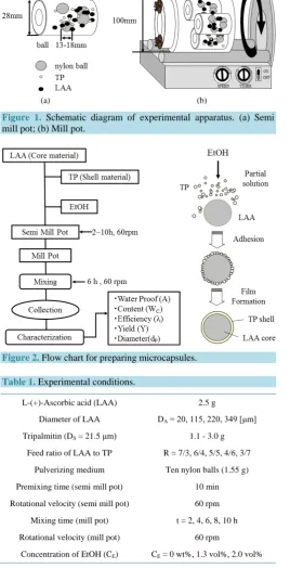
Figure 1 and Figure 2show the schematic diagram of experimental apparatus and the flow chart for preparing
the microcapsules, respectively. The unmicroencapsulated active materials are easily deactivated by oxygen or humidity in environment. If the active materials could be microencapsulated by the materials with the barrier ability to oxygen or humidity, the activity of microencapsulated materials should be kept for long time. In mi-croencapsulation of the core materials deactivated easily by oxygen or humidity, it is necessary to develop the microencapsulation method without air and humidity. Taking these things into consideration, we have adopted the dry coating method which is performed under nitrogen atmosphere using only the pulverizing solvent.
First, in order to make sure of the shell formation, LAA and TP were premixed for ten min at the revolution speed of 60 rpm in the semi mill pot as shown inFigure 1(a) by adding the given volume of EtOH. In the semi mill pot, ten nylon balls with the diameter of 7 mm were added. Then, the three semi mill pots containing the mixture of LAA and TP were set in the mill pot to increase the production amount of microcapsules as shown in
Figure 1(b)and mixed for the given time at the revolution speed of 60 rpm. Microencapsulation may progress
in turn partial solution of TP with EtOH, adhesion of TP and film formation on the surface of LAA particles. In the preparation method of microcapsules presented here, the concentration of EtOH (CE: wt% to TP), the added
(a) (b)
[image:3.595.185.454.106.631.2]Figure 1. Schematic diagram of experimental apparatus. (a) Semi mill pot; (b) Mill pot.
Figure 2. Flow chart for preparing microcapsules.
Table 1. Experimental conditions.
L-(+)-Ascorbic acid (LAA) 2.5 g
Diameter of LAA DA = 20, 115, 220, 349 [μm]
Tripalmitin (DS = 21.5 μm) 1.1 - 3.0 g
Feed ratio of LAA to TP R = 7/3, 6/4, 5/5, 4/6, 3/7
Pulverizing medium Ten nylon balls (1.55 g)
Premixing time (semi mill pot) 10 min
Rotational velocity (semi mill pot) 60 rpm
Mixing time (mill pot) t = 2, 4, 6, 8, 10 h
Rotational velocity (mill pot) 60 rpm
Concentration of EtOH (CE) CE = 0 wt%, 1.3 vol%, 2.0 vol%
2.3. Characterization of Microcapsules
Microcapsules prepared as stated above were characterized about the following things:
2.3.1. Observation of Microcapsules and LAA
[image:3.595.187.440.241.515.2]2.3.2. Mean Diameters of Microcapsules and LAA
The diameters of microcapsules (dp) and LAA particles (DA) were measured by the sieve classification method.
2.3.3. Water Proof Degree
First, LAA of a given weight was dissolved into distilled water of 40 cm3 and then, the concentration of NaOH to neutralize the LAA aqueous solution was measured by titrating with 0.02 mol/L NaOH aqueous solution. From these results, the correlating curve between the concentration of LAA and that of NaOH was obtained be-forehand. Then, the microcapsules of 0.5 g were added into distilled water of 40 cm3 to be dispersed for 30min by mixing gently with the magnetic stirrer, under which conditions any microcapsules were not broken mechan-ically. After this operation, the given volume (10 cm3) of aqueous solution, in which the microcapsules were dispersed, was sampled out and then, the concentration of NaOH to neutralize this solution was measured. By comparing the measured concentration of NaOH with the correlating curve, the concentration of LAA leaking from the microcapsules was estimated. Moreover, the remaining microcapsules were redispersed in distilled water of 40 cm3 and broken by heating up to 80˚C to completely dissolve LAA microencapsulated. Thus, the weight of LAA not leaking from the microcapsules was estimated in the same manner as stated above. From these values, the water proof degree (A) was calculated by using the following Equation (1).
weight of LAA remaining in microcapsules A
initial weight of LAA microencapsulated
= (1)
2.3.4. Content, Microencapsulation Efficiency, Yield
The content (WC) of core material, the microencapsulation efficiency (λ), the yield (Y) were calculated by using
the following equations from the measured values.
C
total weight of LAA microencapsulated W
weight of microcapsules
= (2)
total weight of LAA microencapsulated weight of LAA in feed
λ = (3)
weight of microcapsules prepared Y
weight of TP and LAA in feed
= (4)
2.3.5. Application of Microcapsules to Polymerization of MMA
The microcapsules (0.5 g) were added into the test tube, in which 10 cm3 of MMA monomer dissolving potas-sium persulfate (KPS) was poured beforehand. It was observed whether polymerization could be induced by heating the test tube at 130˚C and breaking the microcapsules or not. For comparison, the same experiments as stated just above were conducted by adding only LAA with heating at 50˚C and by adding the microcapsules with heating at 50˚C, respectively. The microcapsules were not broken at 50˚C.
3. Results and Discussion
3.1. Effect of Diameter Ratio of LAA to TP
Figure 3shows the SEM photographs of LAA particles with the different diameters. The mean diameters (DA)
of LAA particles are 250 µm (a), 200 µm (b), 150 µm (c), 21.5 µm (d), respectively. While, the mean diameter (DS) of TP particles was 20 µm. The LAA particles are irregular and wide distribution.
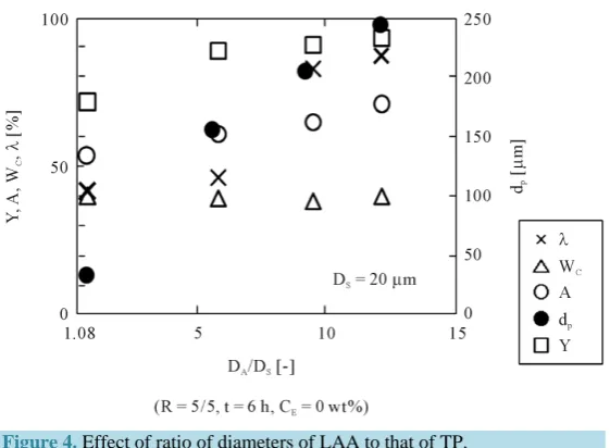
Figure 4shows the effect of ratio of mean diameters (DA) of LAA particles to those (DS) of TP particles on
(a) (b)
[image:5.595.153.501.73.564.2](c) (d)
[image:5.595.171.455.80.329.2]Figure 3. SEM photographs of LAA particles. (a) DA = 250 [μm]; (b) DA = 200 [μm]; (c) DA = 150 [μm]; (d) DA = 21.5 [μm].
Figure 4. Effect of ratio of diameters of LAA to that of TP.
On the other hand, the content (WC) was kept almost constant (WC = 0.4) and close to the calculated content
(WC = 0.42) based on the feed ratio of LAA and TP. The mean diameters of microcapsules increased according
to the diameters of LAA particles.
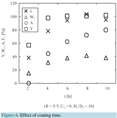
3.2. Effect of Coating Time
Figure 5 shows the SEM photographs of microcapsules taken each at elapsing time where the diameter ratio
(DA/DS) of LAA particles to TP particles was 10.0 and the feed ratio (R) of LAA to TP was 5/5. It was found
that the adhesion amount of TA on the LAA particles increased with the coating time and the denser shell was formed.
Figure 6shows the dependences of the yield (Y), the water proof degree (A), the content (WC) and the
[image:5.595.174.455.360.566.2]2 h 4 h 6 h 8 h 10 h
(a)
(b)
[image:6.595.78.542.78.271.2](DA/DS = 10, CE = 0, R = 5/5)
[image:6.595.199.428.314.544.2]Figure 5.SEM photographs of microcapsules (effect of coating time).
Figure 6.Effect of coating time.
from 40% to ca. 100% after 6 h. The water proof degree (A) increased from 0 at t = 2 h to 80% at t = 10 h. The content (WC) increased from 18 at t = 2 h to ca. 40%. The yield (Y) increased from 59% at t = 2 h to 100% at t =
4 h.
3.3. Effect of Concentration of Pulverizing Solvent
Figure 7shows the SEM photographs of microcapsules prepared by changing the concentration of pulverizing
solvent at t = 10 h where the diameter ratio of LAA particles to TP particles was 10.0 and the feed ratio of LAA to TP was 5/5. With increasing the concentration of pulverizing solvent, the adhesion amount of TA increased and the denser shell was formed.
Figure 8shows the dependence of the yield (Y), water proof degree (A), the content (WC) and the
microen-capsulation efficiency (λ) on the concentration of pulverizing solvent. The yield (Y) reached 100% at CE = 1.3
vol%. The content (WC) gradually increased and reached 40% at CE = 1.3 vol%. The water proof degree (A) and
(R = 5/5, t = 10 h, DA/DS = 10.0)
[image:7.595.202.428.81.346.2]Figure 7. SEM photographs of microcapsules (effect of con-centration of EtOH).
Figure 8. Effect of concentration of EtOH.
from 61% at CE = 0 wt% to 82% at CE = 2.0 wt%, respectively.
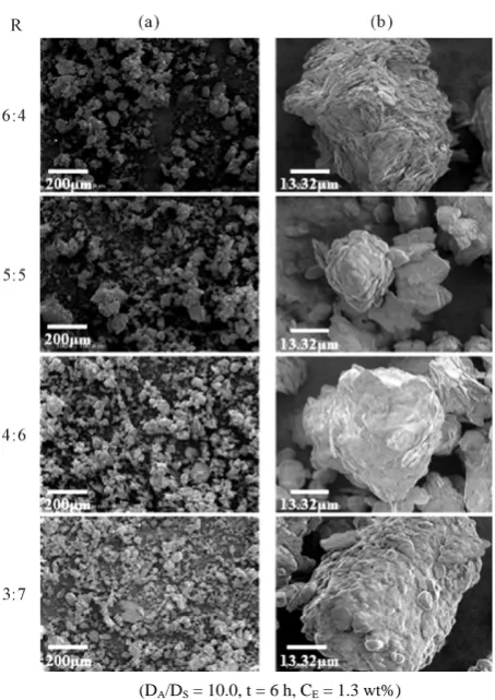
3.4. Effect of Feed Ratio
Figure 9shows the SEM photographs of microcapsules prepared by changing the feed ratio (R) of TP to LAA at
t = 6 h, DA/DS = 10 and CE = 1.3 wt%. It was found that the larger the ratio, the denser the shell become.
Figure 10shows the dependences of the yield (Y), the water proof degree (A), the content (WC), the
micro-encapsulation efficiency (λ) on the feed ratio (R). The yield (Y) become 100% at each feed ratio. The microen-capsulation efficiency (λ) over 90% could be obtained at each feed ratio. The content (WC) increased with
[image:7.595.201.429.381.579.2](DA/DS = 10.0, t = 6 h, CE = 1.3 wt%)
Figure 9. SEM photographs of microcapsules (effect of feed ratio).
Figure 10. Effect of feed ratio.
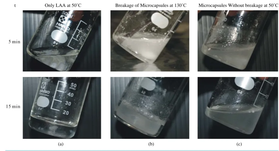
[image:8.595.156.443.83.611.2]3.5. Application of Microcapsules to Polymerization of Methyl Methacrylate Monomer
Figure 11shows the photographs of the polymerization system of MMA monomer, where the microcapsules
prepared at DA/DS = 10, CE = 1.3 wt%, R = 5/5, t = 10 h were used. The monomer phase was solidified by
[image:8.595.201.431.433.628.2](a) (b) (c)
Figure 11. Application of microcapsules to polymerization.
TP. From this result, it was confirmed that LAA was microencapsulated well and could induce polymerization of MMA monomer.
4. Conclusions
It was tried to microencapsulate LAA with TP by using the dry coating method. The fundamental results were obtained as follows.
1) LAA was microencapsulated well with TP.
2) The yield, the content, the water proof degree and the microencapsulation efficiency increased with the coating time.
3) The yield, the microencapsulation efficiency, the content and the water proof degree increased with the mean diameter ratio of LAA particles to TP particles, the feed ratio of LAA to TP and the concentration of pul-verizing solvent.
4) It was confirmed that the microcapsules were broken due to heating at T = 130˚C and could induce poly-merization of methyl methacrylate monomer.
References
[1] Kondo, T. (1967) Saishin Maikurokapseruka Gijutsu (Microencapsulation Technique). TES, Tokyo.
[2] Tanaka, M. (2008) Key Point of Preparation of Nano/Microcapsules. Techno System Publishing Co. Ltd., Tokyo.
[3] Tanaka, M. and Hayashi, K. (1989) Preparation of Polystyrene Particles Coated with Ferrite Powder by Suspension Polymerization. Kagaku Kogaku Ronbunshu, 15, 1144-1152. http://dx.doi.org/10.1252/kakoronbunshu.15.1144 [4] Tanaka, M. and Hosogai, K. (1990) Suspension Polymerization of Styrene with Circular Loop Reactor. Journal of
Ap-plied Polymer Science, 39, 955-966. http://dx.doi.org/10.1002/app.1990.070390414
[5] Tanaka, M., Saito, A., Hosogai, K. and Kimura, I. (1992) Preparation of Fine Polymer Particles Coated Uniformly with Magnetite Powder by Suspension Polymerization. Kagaku Kogaku Ronbunshu, 18, 330-337.
http://dx.doi.org/10.1252/kakoronbunshu.18.330
[6] Tanaka, M., Hosogai, K., Yuda, T., Kimura, I. and Saito, N. (1992) Preparation of Composite Particles Composed of Polystyrene and Carbon Silicide by Suspension Polymerization. Journal of the Japan Society of Colour Material, 65, 484-491. http://dx.doi.org/10.4011/shikizai1937.65.484
Japan, 32, 229-236. http://dx.doi.org/10.4164/sptj.32.229
[8] Taguchi, Y., Saito, N., Kimura, I. and Tanaka, M. (1999) Preparation of Fine Composite Particles Composed of Inor-ganic Solid Powders and OrInor-ganic Polymers by Utilizing Liquid-Liquid Dispersion. Colloids and Surfaces A: Physio-chemical and Engineering Aspects, 153, 401-404. http://dx.doi.org/10.1016/S0927-7757(98)00462-2
[9] Taguchi, Y. and Tanaka, M. (2001) Preparation of Composite Particles Composed of Two Kinds of Solid Powders and Waste Polymer by Semi-Chemical Recycle Method. Journal of Chemical Engineering of Japan, 34, 1177-1181. http://dx.doi.org/10.1252/jcej.34.1177
[10] Taguchi, Y. and Tanaka, M. (2003) Preparation of Composite Particles Made from Solid Powders and Wasted Plastics by Semi Chemical Recycle Method. Journal of Applied Polymer Science, 88, 483-488.
http://dx.doi.org/10.1002/app.11920
[11] Kofuji, K., Qian, C.J., Murata, Y. and Kawashima, S. (2005) Preparation of Chitosan Microparticles by Water-in- Vegetable Oil Emulsion Coalescence Technique. Reactive & Functional Polymers, 62, 77-83.
http://dx.doi.org/10.1016/j.reactfunctpolym.2004.09.002
[12] Chen, M.C., Mi, F.L., Liao, Z.X., Sonaje, K., Chung, M.F., et al. (2013) Recent Advances in Chitosan-Based Nanopar-ticles for Oral Delivery of Macromolecules. Advanced Drug Delivery Reviews, 65, 865-879.
http://dx.doi.org/10.1016/j.addr.2012.10.010
[13] Filho, L.P., Kokol, V. and Voncica, B. (2013) Synthesis and Characterization of Ethyl Cellulose Microcapsules Con-taining Model Active Ingredients. Macromolecular Symposia, 328, 45-55. http://dx.doi.org/10.1002/masy.201350605 [14] Betz, M. and Kulozik, U. (2011) Microencapsulation of Bioactive Bilberry Anthocyanins by Means of Whey Protein
Gels. Procedia Food Science, 1, 2047-2056. http://dx.doi.org/10.1016/j.profoo.2011.10.006
[15] Betz, M., Steiner, B., Schantz, M., Oidtmann, J., Mader, K., Richling, E. and Kulozik, U. (2012) Antioxidant Capacity of Bilberry Extract Microencapsulated in Whey Protein Hydrogels. Food Research International, 47, 51-57.