Development 139, 3021-3031 (2012) doi:10.1242/dev.080127 © 2012. Published by The Company of Biologists Ltd
INTRODUCTION
Genome-encoded miRNAs bind to specific sites on the 3⬘ untranslated region (3⬘UTR) of their target mRNAs, to impart post-transcriptional silencing (Bartel, 2004). This regulatory layer acts in concert with transcription factors to refine gene expression and confer robustness to developmental transitions (Stark et al., 2005; Hornstein and Shomron, 2006; Li et al., 2009). Total inactivation of miRNA maturation causes pancreas agenesis (Lynn et al., 2007), indicating that miRNAs are essential for early pancreas development. Specific miRNAs were shown to act in endocrine tissues; for example, miR-375 is highly expressed in the endocrine pancreas (Poy et al., 2004; Avnit-Sagi et al., 2009), and loss of its function disrupts islet morphogenesis and endocrine cell differentiation (Kloosterman et al., 2007; Poy et al., 2009).
miR-7 is another miRNA that is highly and specifically expressed in the endocrine pancreas of zebrafish, mouse and human (Wienholds et al., 2005; Landgraf et al., 2007; Bravo-Egana et al., 2008; Correa-Medina et al., 2009). miR-7 is an evolutionarily conserved miRNA, encoded by a single gene in flies and by three different genomic loci in humans and mice (mouse: mmu-mir-7a-1 at Chrmmu-mir-7a-13, mmu-mir-7a-2 at Chr7 and mmu-mir-7b at Chrmmu-mir-7a-17). The duplication of the miR-7 gene in vertebrates hampers genetic loss-of-function analysis. As a consequence, the role of miR-7 in endocrine pancreas development is still unclear.
The endocrine differentiation program is initiated by neurogenin 3 (Ngn3), which induces a complex network of transcription factors, to specify the different endocrine lineages (Gradwohl et al., 2000; Gu et al., 2002; Martin et al., 2007; Bonal and Herrera, 2008; Lyttle et al., 2008). This process results in the differentiation of mature hormone-producing cells (Murtaugh and Melton, 2003).
Pax6, a paired-domain transcription factor acting downstream of Ngn3, is pivotal in differentiation of hormone-producing cell types: -, -, - and PP cells. However, Pax6 negatively regulates the formation of ghrelin-expressing -cells (Sander et al., 1997; St-Onge et al., 1997; Ashery-Padan et al., 2004; Heller et al., 2005; Dames et al., 2010). In both humans and mice, two Pax6 alleles are required in order to maintain proper glucose homeostasis, i.e. loss of only one allele results in glucose intolerance (Yasuda et al., 2002; Ding et al., 2009). Similarly, the development of other organs is sensitive to Pax6 haplo-insufficiency, including the iris and the lens (van Raamsdonk and Tilghman, 2000; Collinson et al., 2001; Baulmann et al., 2002; Kroeber et al., 2010). However, high levels of Pax6 are also not tolerated. Thus, Pax6 overexpression in mice causes eye abnormalities (Schedl et al., 1996) and induces apoptosis in the brain and in the endocrine pancreas (Yamaoka et al., 2000; Berger et al., 2007). Taken together, it appears that Pax6 expression is tightly controlled to ensure appropriate levels of expression.
In this study, we characterized miR-7 expression and identified its functions in endocrine cell differentiation. We show that miR-7 directly represses Pax6 mRNA expression and fine-tunes its levels. Together, miR-7 and Pax6 are wired into a network that regulates endocrine cell differentiation. Our results demonstrate the importance of miRNAs in refining the development of hormone-producing cells.
MATERIALS AND METHODS Animals
Mice were housed and handled in accordance with protocols approved by the Institutional Animal Care and Use Committee of Weizmann Institute of Science. Conditional miR-7 transgenic mice were generated as previously described (Srinivas et al., 2001). Briefly, a 500 bp fragment flanking the miR-7a-1 gene was cloned into the Rosa26 locus downstream of the PGK promoter and a transcriptional STOP cassette and upstream of IRES-EGFP-polyadenylation signal. Correct homologous recombination into the ROSA26 locus was identified by Southern blot analysis to embryonic stem cell colonies (129/SvEv). Scanning 209 colonies identified 29 positive colonies and a mouse line was derived through blastocyst injection (C57BL6/J background). Rosa-miR-7 mice were crossed to a 1Department of Molecular Genetics, Weizmann Institute of Science, Rehovot 76100,
Israel. 2Integrated DNA Technologies, 1710 Commercial Park Coralville, IA 52241, USA.
*Author for correspondence ([email protected])
Accepted 1 June 2012 SUMMARY
Genome-encoded microRNAs (miRNAs) provide a post-transcriptional regulatory layer that is important for pancreas development. However, how specific miRNAs are intertwined into the transcriptional network, which controls endocrine differentiation, is not well understood. Here, we show that microRNA-7 (miR-7) is specifically expressed in endocrine precursors and in mature endocrine cells. We further demonstrate that Pax6 is an important target of miR-7. miR-7 overexpression in developing pancreas explants or
in transgenic mice led to Pax6 downregulation and inhibition of - and -cell differentiation, resembling the molecular changes
caused by haploinsufficient expression of Pax6. Accordingly, miR-7 knockdown resulted in Pax6 upregulation and promoted - and
-cell differentiation. Furthermore, Pax6 downregulation reversed the effect of miR-7 knockdown on insulin promoter activity. These
data suggest a novel miR-7-based circuit that ensures precise control of endocrine cell differentiation.
KEY WORDS: -Cell, microRNA, miR-7, Pax6, Pancreas development, Mouse
Pancreas-enriched miRNA refines endocrine cell
differentiation
Sharon Kredo-Russo1, Amitai D. Mandelbaum1, Avital Ness1, Ilana Alon1, Kim A. Lennox2, Mark A. Behlke2 and Eran Hornstein1,*
D
E
V
E
LO
P
M
E
N
Pdx1-Cre transgene (Gu et al., 2002) and mated to homozygosity. Other mouse strains used in this study were Ngn3-CreER, serving as Ngn3 nulls (Wang et al., 2010), and a Pax6 null allele (St-Onge et al., 1997).
Organ culture
Dorsal pancreatic rudiments of E12.5 ICR mouse embryos were dissected from the adjacent mesenchyme, using a tungsten needle. The explants were cultured in M199 medium supplemented with 10% fetal bovine serum (Gibco), 2 mM L-glutamine and 100 U/ml penicillin/streptomycin. Individual explants were plated in 30 l inverted ‘hanging drops’ on a 35-mm Petri dish cover (NUNC), with medium containing either anti-microRNA antagomirs (Dharmacon) or cholesterol-conjugated miRNA mimics (IDT) at 1 M concentration. The exact oligo sequences are in supplementary material Table S2. Explants were further grown for up to 48 hours at 37°C with a 5% CO2 in a humidified incubator as previously described (Kredo-Russo and Hornstein, 2011). BrdU (3 g/ml) was added to the medium 1 hour before harvest for analysis of proliferation.
Pancreas histology and quantification analysis
Immunofluorescence protocols for paraffin sections have been described previously (Melkman-Zehavi et al., 2011) and whole-explant staining has been described previously (Kredo-Russo and Hornstein, 2011). The primary antibodies used were rabbit anti-Pax6 (1:300, Covance), guinea pig anti-insulin (1:200, Dako), rabbit anti-glucagon (1:200, Dako), rabbit anti-Cpa1 (1:100, Sigma), mouse anti-Ngn3 [1:500, Developmental Studies Hybridoma Bank (DSHB)] goat anti-ghrelin (1:100, Santa Cruz) and goat anti-Hnf1b (1:200, Santa-Cruz). Secondary antibodies were Cy2-, Cy3- or Cy5-conjugated donkey anti-guinea pig, anti-mouse and anti-rabbit IgG (1:200, Jackson ImmunoResearch). Nuclei were stained with DAPI (1:10,000, Molecular Probes). Whole-mount BrdU analysis that includes a 2 hour DNase I treatment was carried out as previously described (Tkatchenko, 2006).
Fluorescent confocal images were captured with a Zeiss LSM 510 microscope, using an optical depth of 1 m, with at least six to eight optical sections at 5 m intervals throughout the whole organ.
Morphometry of the explants was performed by quantification of the immunostained area from the entire explant sections from a minimum of three mutants and three wild-type matched littermates,. Total tissue area and total hormone-positive area, were calculated using ‘Niss-elements’ software (Nikon).
For cell number quantification at E15.5, hormone-positive cells were manually counted every fifth section throughout the whole pancreas anlagen. Data are the average number of cells/section in multiple sections and were analyzed for four or more individual animals per genotype. Cell number analysis of total hormone-positive cells in whole E12.5 explants was performed manually, by counting cells in six stacked z-section confocal images, spanning the whole explants.
Quantitative PCR for miRNA and mRNA
Extraction of total RNA was carried out using the miRNeasy Mini Kit (QIAGEN). Synthesis of cDNA obtained by using an oligo d(T) primer (Promega) and SuperScript II reverse transcriptase (Invitrogen). Synthesis of cDNA of miRNA obtained by using Taqman MicroRNA qPCR Assays (Applied Biosystems). qPCR analysis of mRNA was performed on LightCycler 480 System (Roche) using Kapa SYBR Green qPCR kit (Finnzymes). miRNA and mRNA levels were normalized to the expression of small RNAs (sno234 and U6) or mRNA (Gapdh and Hprt), respectively. Primer sequences are described in supplementary material Table S2.
miRNA in situ hybridization
Paraffin sections of E12.5-E15.5 pancreata were hybridized with DIG-labeled LNA probes (Exiqon) overnight at 48°C (miR-7), 54°C (U6, control) or 60°C (miR-375) as previously described (Pena et al., 2009) and developed with TSA kit (PerkinElmer) as previously described (Silahtaroglu et al., 2007). When in situ hybridization was combined with immunofluorescence, primary antibodies were added to the anti-Dig-POD incubation (1:500, Roche).
Cell culture, luciferase reporter assay and western blotting HEK-293T cells (American Type Culture Collection) and MIN6 cells (a gift from Jun-ichi Miyazaki, Osaka University, Japan) were grown in Dulbecco’s modified Eagle medium (DMEM) with 10% FBS, 2 mM L
-glutamine, 100 U/ml penicillin/streptomycin at 37°C; 5% CO2 in a humidified incubator. Experiments on MIN6 cells were performed between passages 18 to 28.
A 742 bp fragment of the mouse Pax6 3⬘UTR sequence (chr2 105536551-105537201) was subcloned into psiCHECK-2 Vector (Promega) and transfected into HEK-293T cells using jetPEI (Polyplus Transfection, Illkirch, France), following manufacturers’ instructions. Dual-Reporter luciferase assay was performed 48 hours later, according to the manufacturer’s instructions (Promega). miR-7 overexpression was achieved using expression vectors miRVec-miR-7 or miRVec control (a kind gift from Reuben Agami, The Netherlands Cancer Institute, Amsterdam). 7 knockdown was carried out using oligos against miR-7 or against scrambled sequence, as negative control oligos (50 nM, Ambion), using Lipofectamine 2000 Reagent (Invitrogen). For western blots, cellular lysate was subject to 10% SDS-PAGE and immunoblotted with rabbit anti-Pax6 (1:5000, Chemicon), mouse anti-GAPDH (1:10,000, Ambion) and quantified with ImageJ software.
For analysis of insulin transcription, fireflyluciferase reporter driven by the rat insulin promoter and an A20-Renillaluciferase construct (gift of Michael Walker), were transfected using Lipofectamine 2000 Reagent (Invitrogen) to MIN6 cells. Anti miR-7 oligo (100 nM) and Pax6 siRNA (10 nM) are from IDT; overexpression of miR-7 was achieved using a miRVec vector.
Statistical analysis
Analysis was performed using either Student’s t-test or two-way ANOVA by the JMP software. Results are given as mean±s.e.m. The null hypothesis was rejected at the 0.05 level (**) or 0.01 (*). Gene Ontology analysis was performed using DAVID (Dennis et al., 2003).
RESULTS
miR-7 is expressed in endocrine cells of the pancreas
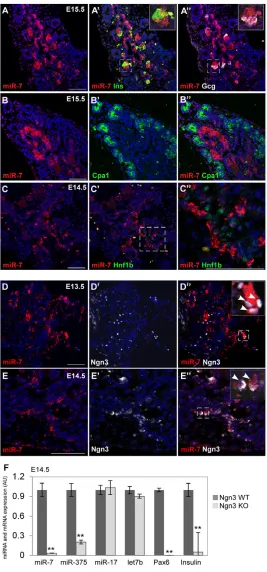
To identify the spatial expression pattern of miR-7, we established a method for fluorescent miRNA in situ hybridization combined with immunofluorescent protein detection in mammalian pancreata. In situ hybridization on E12.5-E15.5 pancreatic sections was carried out using a digoxigenin (DIG)-labeled LNA probe. As positive and negative controls we used ubiquitously expressed U6 and scrambled oligos, respectively (supplementary material Fig. S1A-D). Our analysis uncovered miR-7 expression in a subset of clustered epithelial cells, within the ‘trunk’ compartment (Zhou et al., 2007) of the developing pancreas (Fig. 1A-E; supplementary material Fig. S1A,B). Furthermore, miR-7 was colocalized with insulin and glucagon in differentiating - and -cells, respectively (E13.5-E15.5; Fig. 1A-A⬙; supplementary material Fig. S1E,F). miR-7 and Cpa1 expression domains were mutually exclusive at E15.5 (Fig. 1B-B⬙), as were miR-7 and Hnf1at E14.5 (Fig. 1C-C⬙). These data indicate that miR-7 is not expressed in differentiated acinar or duct cells. To examine miR-7 expression in endocrine precursor cells, we performed immunostaining of Ngn3. At E12.5, E13.5 and E14.5, miR-7 was colocalized with many Ngn3-positive cells (Fig. 1D-E⬙; supplementary material Fig. S1G), suggesting that miR-7 is induced in newly born endocrine cells. Independent genetic support to this study came from the analysis of null pancreata. It was previously shown that Ngn3-deficient embryos completely lack endocrine hormone-producing cells (Gradwohl et al., 2000). Consistent with this, the expression of endocrine markers, such as Pax6 and insulin, was downregulated in Ngn3-null pancreata (Fig. 1F). As miR-7 expression was also abrogated in E14.5 Ngn3-null pancreas, we conclude that this
D
E
V
E
LO
P
M
E
N
miRNA is specifically expressed within the endocrine lineage. Furthermore, this regulation was specific to miR-7, as the expression of 17 and Let-7b was not changed. Notably, miR-375, another pancreatic miRNA (Poy et al., 2004; Avnit-Sagi et al., 2009; Poy et al., 2009), was also downregulated in Ngn3-null pancreata, yet some residual expression was maintained, unlike miR-7. Altogether, this analysis reveals the endocrine-specific expression pattern of miR-7, wherein miR-7 is induced in Ngn3+
precursors and is maintained in the differentiated endocrine cells.
Pax6 is a miR-7 target
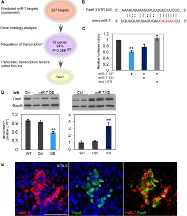
To identify potential miR-7 targets that play a role in pancreas development, we employed two unbiased bioinformatic approaches. First, we analyzed ‘gene ontology’ (GO) terms related to miR-7 targets [DAVID (Dennis et al., 2003)]. Among the 237 predicted miR-7 targets [TargetScan (Lewis et al., 2005)], we found that GO term ‘Regulation of transcription’ is the most significantly enriched (52 genes, P<2.35E–7). Intriguingly, within this list, Pax6
[image:3.612.52.319.58.630.2]was the only established pancreatic transcription factor
Fig. 1. miR-7 is expressed in the endocrine cells of the pancreas.(A-C⬙) miR-7 fluorescent in situ hybridization combined with protein immunofluorescence analysis of E13.5-E15.5 pancreas sections. (A-A⬙) miR-7 (red) colocalization with insulin (Ins; green, A⬘) or glucagon (Gcg; white, A⬙). Blue, nuclei. Insets are higher magnifications of the field marked by the dashed square. (B-B⬙) miR-7 (red) is not expressed in acinar cells marked by Cpa1 at E15.5 (green, B⬘and B⬙) or in duct cells marked by HNF1b at E14.5 (green, C⬘). C⬙is a higher magnification of the field marked by the dashed square in C’. (D-E⬙) miR-7 is expressed in many Ngn3-positive cells at E13.5 (white, D⬘,D⬙) as well as at E14.5 (E⬘,E⬙). Higher magnification insets with arrowheads indicating representative cells that co-express miR-7 and Ngn3. Scale bars: 50m. (F)miR-7 expression is dependent on Ngn3. qPCR analysis of miR-7, miR-375, miR-17, let-7b in E14.5 Ngn3 knockout (KO) pancreatic buds, relative to Ngn3 heterozygous controls (WT). Data are normalized to sno234. qPCR data of Pax6 and insulin expression in the same samples, normalized to Hprt and Gapdh, and then presented relative to control. Error bars represent ±s.e.m. **P<0.05.
D
E
V
E
LO
P
M
E
N
(supplementary material Table S1 and Fig. 2A). Furthermore, the binding site for miR-7 at the Pax6 mRNA 3⬘untranslated region (3⬘UTR) is predicted to be strong and conserved (Fig. 2B). Independently, we built an interaction map of miRNAs with the 3⬘UTRs of transcription factors that are known to control pancreas development, including Pdx1, Ngn3, Nkx2.2, Nkx6.1, MafB, Pax4, Pax6, Arx, Hnf1b and Hnf6 (for a comprehensive list see supplementary material Table S1). This approach provided a wealth of potential interactions; however, Pax6 was the only miR-7 predicted target. As Pax6 expression is known to be tightly regulated in many organs (van Raamsdonk and Tilghman, 2000; Baulmann et al., 2002; Yasuda et al., 2002; Ding et al., 2009), we hypothesized that miR-7 may be a new endocrine regulatory gene upstream of Pax6.
To determine whether miR-7 directly targets Pax6 3⬘UTR, we performed a heterologous reporter assay. The whole 3⬘UTR of Pax6 (742 bp) was cloned into a dual-luciferase reporter vector and introduced into HEK-293T cells along with expression vector for miR-7 (miRvec-7), or a miRNA control vector (harboring a random and non-targeting miRNA-like sequence, ‘Ctrl’). Overexpression of miR-7 (‘miR-7 OE’) significantly decreased
[image:4.612.51.425.308.739.2]luciferase activity, relative to the negative control (Fig. 2C; to 62%). The addition of an anti-miRNA oligo partially blocked this repression, supporting the functionality of the predicted miR-7 binding site. Moreover, introduction of a mutation into the 3⬘UTR sequence, in which six nucleotides of the potential ‘seed’-binding site were deleted (marked in red, Fig. 2B), completely abolished 7-dependent repression (Fig. 2C). To determine whether miR-7 represses the expression of endogenous Pax6, we transfected a -cell line (MIN6) with miRvec-7. Overexpression of miR-7 resulted in a decrease in PAX6 protein level to 60% of its level in untreated wild-type cells (‘WT’) or control miRvec-treated cells (‘Ctrl’), as measured by western blots. Conversely, inhibition of miR-7 by anti-miR-7 oligos (termed ‘miR-7-KD’) significantly upregulated PAX6 protein levels, relative to untreated cells (‘WT’), or to negative control of scrambled oligos (‘Ctrl’, Fig. 2D). Taken together, these results indicate that Pax6 is a bona fide target of miR-7 in -cells. To test the possible existence of a reciprocal feedback loop, in which Pax6 regulates miR-7 expression, we quantified miR-7 levels in E14.5 Pax6-null embryos (‘Pax6 KO’). However, qPCR analysis revealed comparable miR-7 levels between Pax6 KO and littermate controls (supplementary material
Fig. 2. Pax6 is a miR-7 target. (A)Gene ontology analysis of miR-7 predicted targets depicts the term ‘Regulation of transcription’ as significantly enriched (P<2.35E–7). Within this list, Pax6 is the only characterized factor known to control pancreas development (see also supplementary material Table S1). (B)Predicted base pairing of mature miR-7 sequence and Pax6 3⬘UTR. Seed-sequence is coded in red. (C)The relative luciferase activity of a reporter that harbors the Pax6 3⬘UTR (742 bp). The luciferase reporter expression is repressed by miR-7 overexpression (miR-7 OE), whereas introduction of ‘anti-miR’ oligos (miR-7 KD) partially abrogates this repression. A reporter that harbors Pax6 3⬘UTR with deleted miR-7 seed sequence is completely insensitive to miR-7OE (‘mut UTR’). Data are normalized to the activity of firefly luciferase co-expressed from the dual reporter and to a negative control miRNA vector (‘Ctrl’). n3 independent experiments, in triplicates each. **P<0.05. (D)Representative western blots of Pax6 detection in MIN6 cells treated with miR-7 KD oligos or miR-7 OE plasmid, relative to untreated cells or negative controls. Quantification of band densitometry of four independent experiments in duplicates (ANOVA test, **P<0.05). (E)Colocalization of miR-7 in situ hybridization (red) and Pax6 immunofluorescence (green) in E15.5 pancreas sections. Blue, nuclei. Scale bar: 50m.
D
E
V
E
LO
P
M
E
N
Fig. S1I), suggesting that Pax6 does not control miR-7 expression. Next, co-expression of miR-7 and Pax6 in E15.5 endocrine cells, was revealed by combining in situ hybridization with immunofluorescence (Fig. 2E). This co-expression conceivably allows for miRNA:target interactions in the context of endocrine cell differentiation. Therefore, we conclude that miR-7 acts upstream of Pax6 and thus may function in the development of the endocrine pancreas.
miR-7 controls endocrine differentiation in cultured explants
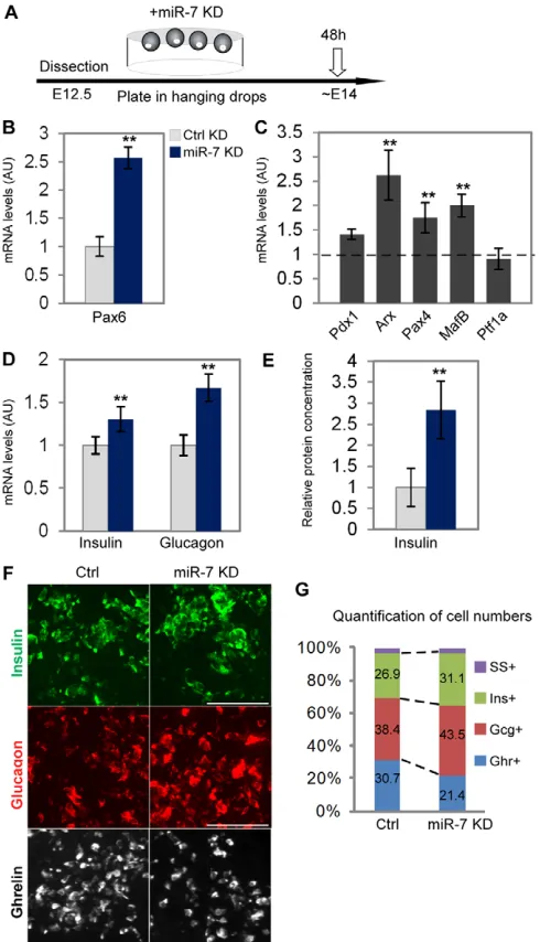
To determine the functional role of miR-7 in the endocrine lineage, we carried out loss-of-function experiments in a primary pancreatic explant system (diagram in Fig. 3A). E12.5 pancreatic buds were cultured for 48 hours under defined conditions, providing an ex vivo model for development (Gunawardana et al., 2005; Kredo-Russo and Hornstein, 2011). We detected typical Ngn3, insulin and miR-7 expression that recapitulated in vivo differentiation, including the expected differentiation of - and -cells (supplementary material Fig. S2A,B). To manipulate miR-7 expression in the explant culture, we used cholesterol-conjugated 2⬘-O-methyl (2⬘OMe) ‘antagomirs’ against miR-7 (‘miR-7 KD’). As ‘non-targeting’ negative controls, we used antagomirs against the liver-specific miR-122, which is not expressed in the pancreas (‘Ctrl-KD’). First, we verified that a Cy-bound oligo is efficiently taken up by the explants (supplementary material Fig. S2C). Next, the functionality of miR-7 KD oligos was confirmed by co-transfecting them into HEK-293T with a miR-7 luciferase reporter that harbors multiple miR-7 binding sites on its 3⬘UTR (Kefas et al., 2008). Reporter luciferase activity was strongly suppressed by miR-7 overexpression, and this was reversed by co-transfecting miR-7 KD together with miR-7 OE oligos (supplementary material Fig. S3), verifying the specificity of the miR-7 KD system. We then used this system to study miR-7-Pax6 interactions in pancreatic explants.
In explants treated with miR-7 KD, Pax6 mRNA levels were upregulated by 2.5-fold, relative to control (Fig. 3B). This result suggests functional regulation of Pax6 levels by miR-7, during pancreas development. To explore the downstream implications of this regulation, we quantified the expression levels of a set of transcription factors that are essential for - and -cell differentiation. This analysis revealed upregulation of Arx, which is specifically expressed in -cells (Collombat et al., 2003), and of -cell factors, Pax4 and MafB. However, the levels of the exocrine transcription factor Ptf1a were unchanged (Fig. 3C). Next, we tested insulin and glucagon expression. Intriguingly, upon miR-7 KD, insulin and glucagon levels increased both in the RNA and protein levels. qPCR analysis revealed upregulation of insulin mRNA by 22% and glucagon mRNA by 61% (Fig. 3D). Accordingly, an increase in insulin protein content was demonstrated by ELISA measurement in miR-7 KD explants, relative to control (Fig. 3E). Furthermore, comprehensive morphometric analysis revealed a 20% increase in the insulin-positive area and in the number of insulin-insulin-positive cells. Similarly, 40% increase in the glucagon-positive area and in the number of glucagon-positive cells were found in miR-7 KD pancreata, relative to controls (Fig. 3F,G; supplementary material Fig. S3). However, Somatostatin expression in miR-7 KD was comparable with control (supplementary material Fig. S3).
To understand the potential causes for the increase in insulin-and glucagon-positive cells, we studied the proliferation capacity of endocrine cells. The total numbers of BrdU-positive nuclei and the percentage of proliferating insulin-positive and
glucagon-positive cells, measured by either Ki67 or BrdU, was comparable in miR-7 KD and control explants (supplementary material Fig. S4). In addition, the numbers of Ngn3-positive progenitors, was not
Fig. 3. miR-7 knockdown controls endocrine differentiation in explant culture. (A)A scheme describing the experimental set up, whereby cholesterol-conjugated oligos are introduced into the medium of E12.5 dorsal pancreatic buds that were grown in hanging drops for 48 hours. (B)Pax6 mRNA levels are upregulated upon miR-7
knockdown (7 KD), relative to knockdown of control miRNA, miR-122 (Ctrl KD). (C)qPCR analysis reveals an increase in Arx, Pax4 and MafB levels, relative to Ctrl KD. (D)Insulin and glucagon mRNA expression increases upon miR-7 KD. (E)Insulin protein levels are upregulated upon miR-7 KD, as measured by ELISA assay. Error bars represent ±s.e.m. **P<0.05. (F)Representative immunostaining of insulin (green), glucagon (red) and ghrelin (white), taken for quantification. Scale bars: 50m. (G)Quantification of endocrine cell types (SS, somatostatin; Ghr, ghrelin; Gcg, glucagon; Ins, insulin). The percentage of cells in each individual population was calculated from the total number of counted cells in serial sections of the whole pancreas anlagen. At least three explants per treatment, average of three to eight independent experimental repeats. qPCR data are normalized to Hprt and Gapdh mRNA, and then to Ctrl KD treatment.
D
E
V
E
LO
P
M
E
N
[image:5.612.316.561.61.488.2]affected by miR-7 KD (supplementary material Fig. S3), suggesting that the increase in insulin- and glucagon-positive cells emerges neither from enhanced proliferation, nor from changes in the size of the Ngn3 precursor pool.
Although Pax6 positively regulates insulin and glucagon expression, it negatively regulates ghrelin expression and differentiation of -cells (Heller et al., 2005; Dames et al., 2010). Accordingly, miR-7 KD resulted in reduced numbers of ghrelin-positive cells (Fig. 3F,G; supplementary material Fig. S3),
supporting the view that miR-7 knockdown acts upstream of Pax6 to promote differentiation into insulin- and glucagon-positive cells at the expense of ghrelin-positive cells.
miR-7 overexpression ex vivo resembles Pax6 haploinsufficiency
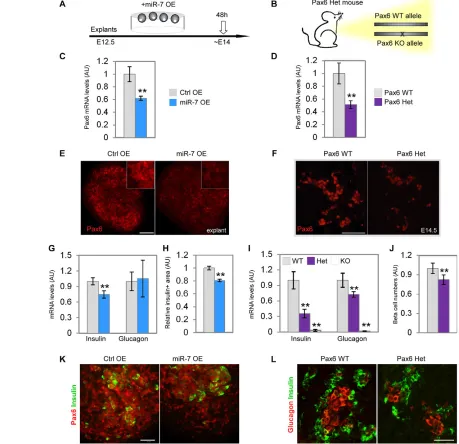
[image:6.612.54.513.169.613.2]To determine the effect of miR-7 overexpression in the explant system, synthetic ‘mimic’ oligos of miR-7 were used (Fig. 4A, ‘miR-7 OE’). As a control, we used a C. elegans miRNA, which is not
Fig. 4. miR-7 overexpression in pancreatic explants phenocopies Pax6 heterozygosity.(A,B)Overexpression of miR-7 in embryonic cultured explants and heterozygous (mono-allelic) expression of Pax6 in vivo. (C-F)miR-7 overexpression downregulates Pax6 in explants, relative to control miR-67 mimic oligos (C, ‘miR-7 OE’, ‘Ctrl OE’, respectively). Similarly, Pax6 expression is reduced in E15.5 heterozygous pancreas (‘Het’), relative to wild-type littermate pancreas (D, ‘WT’, two functional alleles). Pax6 mRNA qPCR analysis (C,D) and Pax6 immunostaining (E,F). (G,I)qPCR study of insulin and glucagon mRNA reveals higher sensitivity of insulin expression to miR-7 OE (G) or Pax6 heterozygosity (I). Note that complete knockout of Pax6 (Pax6 KO), results in abolishment of insulin and glucagon expression. (H)miR-7 OE in explants results in reduction of insulin-positive area normalized to the total explant area. Quantification is performed on serial stained sections of the whole pancreas anlagen and quantified for at least three explants per treatment. (K)Representative immunostaining of insulin (green) and Pax6 (red) taken for quantification. (J,L)Accordingly, -cell numbers are reduced in E15.5 Pax6 ‘Het’. Representative immunostaining of insulin (L, green) taken for analysis. Hormone-positive cells are counted every fifth section throughout the pancreas and the average number of positive cells per organ, from several animals, is normalized to wild-type controls. (WT, n1912 cells; heterozygous, n1751 cells counted from 16 sections each). qPCR data were normalized to Hprt and Gapdh, and then
presented relative to control. n≥5 embryos per genotype, three independent litters. Error bars represent ±s.e.m. **P<0.05.
D
E
V
E
LO
P
M
E
N
expressed in mammals (miR-67, ‘Ctrl OE’). In explants treated with miR-7 OE, Pax6 mRNA levels decreased to 60% of control levels (Fig. 4C). Additionally, immunofluorescent analysis revealed reduction of PAX6 protein levels (Fig. 4E). Furthermore, miR-7 OE resulted in downregulation of insulin mRNA (Fig. 4G) and of MafB mRNA (supplementary material Fig. S5A), two genes that are directly controlled by Pax6 (Sander et al., 1997; Nishimura et al., 2008). Accordingly, a decrease in insulin content was demonstrated in miR-7 OE explants by ELISA measurement, relative to controls (supplementary material Fig. S5C). Consistently, a reduction in miR-7 OE explant insulin-positive area was detected by immunostaining quantification (Fig. 4H,K). By contrast, ghrelin expression was upregulated (supplementary material Fig. S5B), consistent with significant ghrelin expansion in Pax6 knockout (Heller et al., 2005; Dames et al., 2010). However, glucagon levels were not significantly changed. Thus, gain-of-function analysis substantiates our conclusions that miR-7 regulates Pax6 and downstream endocrine genes, including insulin and ghrelin.
We complemented this study by analyzing an in vivo model for genetic downregulation of Pax6 expression. Homozygous loss of Pax6 causes complete loss of insulin and glucagon expression and upregulation of ghrelin expression (Sander et al., 1997; St-Onge et al., 1997; Heller et al., 2005; Dames et al., 2010). We hypothesized that knockout of a single Pax6 allele may downregulate Pax6 expression to intermediate levels, by a mechanism that is independent of miR-7. We therefore analyzed previously uncharacterized Pax6 heterozygous pancreata (‘Het’, Fig. 4B). This analysis revealed a 50% reduction in Pax6 mRNA and protein levels in E14.5 Pax6 Het embryos, relative to wild-type littermate controls (Fig. 4D,F). Similar to miR-7 OE, MafB levels were downregulated by 20% in Pax6 Het pancreata relative to control (supplementary material Fig. S5B). Furthermore, insulin and glucagon mRNA levels were decreased by 65% and 30%, respectively, in Pax6 Het pancreata, while ghrelin expression was increased by 30% (Fig. 4I; supplementary material Fig. S5B). In addition, serial section quantification revealed a 22% decrease in -cell numbers (Fig. 4J,L), while -cell numbers were comparable between Het and wild-type pancreata. These data demonstrate the sensitivity of -cells to intermediate Pax6 levels and reveal a surprising similarity between the molecular phenotype gained by miR-7 overexpression and the one caused by Pax6 haploinsufficiency.
Overexpression of miR-7 in vivo reduced expression of endocrine genes
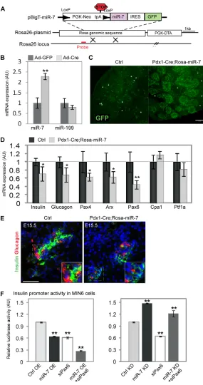
To examine the consequences of miR-7 overexpression in vivo, we generated a conditional miR-7 transgenic mouse line. miR-7a-1 genomic sequence was inserted by homologous recombination into the ubiquitously expressed Rosa26 locus (Fig. 5A) as previously described (Srinivas et al., 2001). In this knock-in model, the expression of miR-7 is coupled to the expression of enhanced green fluorescent protein (EGFP) and both are conditionally blocked by a ‘Neo-STOP’ cassette, flanked by LoxP sites. Thus, miR-7 and EGFP expression are induced only in tissues that express the Cre transgene. To validate the inducible expression of miR-7, we isolated mouse embryonic fibroblasts (MEFs) from Rosa26-miR-7 mice. Upon adenoviral introduction of Cre recombinase, the expression of miR-7 was specifically upregulated by twofold, relative to control infection, whereas the level of an unrelated miRNA, miR-199, remained unchanged (Fig. 5B).
To stimulate the production of miR-7 early in the pancreatic lineage, we crossed the Rosa26-miR-7 allele to a Pdx1-Cre transgene (Gu et al., 2002). Pdx1-Cre;Rosa26-miR-7 mice
specifically expressed GFP protein in E13.5 pancreatic cells (Fig. 5C), while littermates that did not harbor the Pdx1-Cre transgene did not express GFP (‘Ctrl’).
We next characterized E15.5 pancreata of the Pdx1-Cre;Rosa26-miR-7 mice by qPCR. Expression of the mature endocrine cell markers, insulin and glucagon, was downregulated (to 72% and 68% of control, respectively). Quantification of the expression of different transcription factors revealed a reduction in Arx (to 63%) and Pax4 (to 62%), and in the miR-7 target gene Pax6 (to 49%, Fig. 5D). Notably, Cpa1 and Ptf1a levels were unchanged, indicating that the exocrine lineage was unaffected by miR-7 mis-expression. A decrease in the expression of endocrine markers was also seen by immunostaining for insulin and glucagon at E15.5 (Fig. 5E). Therefore, when miR-7 was overexpressed in Pdx1-Cre;Rosa26-miR-7 embryos, Pax6 levels decreased and the expression of insulin and glucagon was downregulated, providing in vivo evidence for control of Pax6 by miR-7.
miR-7;Pax6 interactions upstream of insulin promoter activation
Finally, to elucidate the genetic interactions of miR-7 and Pax6, we used a luciferase reporter, driven by the insulin promoter (Melkman-Zehavi et al., 2011). As the insulin promoter is directly activated by Pax6 (Sander et al., 1997), it provides a model system for monitoring the effect of miR-7; Pax6 axis on insulin expression. Transfecting MIN6 cells with either siRNA against Pax6 (‘siPax6’) or miR-7 overexpression vector (‘miR-7 OE’), resulted in downregulation of insulin promoter activity, relative to scrambled siRNA, or empty miRvec, respectively (Fig. 5F). These results support the observed decrease in insulin mRNA expression by miR-7 OE in explants (Fig. 4G) and in miR-7 transgenic model (Fig. 5D). The combined effect of siPax6 together with miR-7 OE, significantly repressed the activity of the insulin promoter. Furthermore, transfecting MIN6 cells with knockdown oligos against miR-7 (‘miR-7 KD’) resulted in upregulation of insulin promoter activity, relative to scrambled oligos (‘Ctrl KD’), consistent with insulin mRNA upregulation in miR-7 KD-treated explants (Fig. 3D). However, when inhibited simultaneously, siPax6 reversed the effect of miR-7 KD, suggesting that Pax6 mediates miR-7 regulation of insulin expression (Fig. 5F, right panel). These results stress the epistatic relationship of miR-7 upstream of Pax6, in the control of insulin expression. Altogether, our results suggest that miR-7 acts to limit Pax6 expression levels, allowing precise endocrine cell maturation in the development of the pancreas.
DISCUSSION
The role of Pax6 in development has been extensively studied. However, how Pax6 levels are fine-tuned in tissues is not well understood (Baulmann et al., 2002; Ding et al., 2009). Here, we discover a miR-7-based mechanism that controls and refines Pax6 levels in the endocrine pancreas, through a conserved binding site in the 3⬘UTR of Pax6 mRNA. This mechanism enables accurate insulin and glucagon expression and proper - and -cell differentiation.
miR-7 is a new component in the regulation of the endocrine lineage
We show that miR-7 is expressed specifically in the endocrine lineage. miR-7 is expressed relatively early in newly born endocrine precursor cells that express Ngn3, probably coinciding with early expression of endocrine transcription factors such as NeuroD/2 (Jensen et al., 2000). The expression of miR-7 is maintained in
D
E
V
E
LO
P
M
E
N
endocrine cells during their specification to - and -cells. Intriguingly, miR-7 is not detected at E15.5 in Cpa1+exocrine cells
or in Hnf1b+duct cells, in accordance with its specific endocrine
expression. Independent evidence for the endocrine-specific expression of miR-7 emerges from genetic studies, wherein nullification of Ngn3 completely abrogates miR-7 expression. Intriguingly, miR-375 expression is only partially reduced in Ngn3 knockout embryos, suggesting differences in the control of miR-375 and miR-7 expression. In agreement with our observations, in human fetuses, miR-7 has been shown to be expressed from around the ninth week of gestation and reaches maximal expression at the peak
of pancreatic endocrine development (Correa-Medina et al., 2009). Thus, miR-7 expression and presumably its function, appear to be conserved in both mouse and human development.
Pax6 is an important target of miR-7 in the developing pancreas
The endocrine expression of miR-7 suggests that it plays important roles in differentiation of the endocrine pancreas. Bioinformatics predictions suggest that miR-7 has a battery of targets, which play a role in pancreas biology, including recently studied IRS2 (Lewis et al., 2005; Kefas et al., 2008). We focused on regulation of Pax6
Fig. 5. Overexpression of miR-7 in vivo reduces the expression of endocrine genes.(A) Schematic of the targeting construct for conditional miR-7-IRES-GFP misexpression. LoxP sites are indicated by arrowheads. (B)miR-7 upregulation is Cre dependent. qPCR analysis of miR-7 and miR-199 levels in primary embryonic fibroblasts harvested from Rosa-miR-7 transgenic mice and infected with an adenovirus that expresses either Cre-GFP (‘Ad-Cre’) or GFP alone (‘Ad-GFP’). Data are normalized to sno234 (three independent MEF lines, each in triplicate). (C)GFP is specifically expressed in E13.5 Pdx1-Cre;Rosa-miR7 pancreatic epithelium but not in littermates that do not harbor the Cre recombinase (‘Ctrl’). Asterisks depict erythrocyte autofluorescence. (D,E)miR-7 mis-expression represses endocrine genes. (D)qPCR analysis of pancreatic genes in E15.5 Pdx1-Cre;Rosa-miR-7 samples, as indicated. Data are normalized to Hprt and Gapdh mRNA, and then to the expression levels in littermate controls (n6 each genotype, three litters). Error bars represent ±s.e.m. (**P<0.05, *P<0.01). (E)Immunostaining for insulin (green) and glucagon (red) in sections of Pdx1-Cre;Rosa-miR-7 pancreas and control littermates. Blue, nuclei. Scale bar: 50m. (F)Pax6;miR-7 interaction upstream of insulin promoter activation in MIN6 cells. Insulin promoter activity is downregulated by miR-7 overexpression, relative to control (‘miR-7 OE’, ‘Ctrl OE’, respectively) and is suppressed by siRNA against Pax6 (‘siPax6). Combining miR-7 OE with siPax6 enhanced the suppressions of the insulin promoter (left panel). Insulin promoter activity is upregulated by miR-7 knockdown, relative to negative control scrambled oligo (‘miR-7 KD’, ‘Ctrl KD’, respectively). Concomitant introduction of miR-7 KD with siPax6 restores insulin promoter activity (right panel). All fireflyluciferase data is normalized to the activity of Renillaluciferase co-transfected and presented relative to the control experiment. n3 independent experiments. Error bars represent ±s.e.m. **P<0.05.
D
E
V
E
LO
P
M
E
N
[image:8.612.51.336.58.598.2]by miR-7 because of our bioinformatic analysis and because Pax6 levels are known to be tightly regulated. For example, continuous Pax6 overexpression may be harmful to endocrine cells (Yamaoka et al., 2000), whereas complete knockout results in loss of mature endocrine markers, including insulin and glucagon (Sander et al., 1997; St-Onge et al., 1997). Furthermore, loss of even a single Pax6 allele causes glucose intolerance in humans and mice (Yasuda et al., 2002; Ding et al., 2009). Plausibly, Pax6 dose sensitivity in the endocrine pancreas and in other developing organs (van Raamsdonk and Tilghman, 2000) provides a rationale for the required control by miR-7. Our experimental data suggest that, indeed, miR-7 provides another mechanism for refining Pax6 expression. Plausibly, miR-7 has additional targets in pancreas development and homeostasis that are yet to be discovered.
The Pax6-miR-7 pathway affects endocrine differentiation
We report that heterozygous expression of Pax6 is haploinsufficient and leads to defined changes in the expression of endocrine genes, including MafB, insulin and glucagon. These molecular phenotypes are also observed upon miR-7 overexpression, in both explant and transgenic models, supporting molecular interaction of miR-7 and Pax6. Furthermore, miR-7 knockdown alleviates the effect of Pax6 downregulation in the control of the insulin promoter. Taken together, these analyses provide substantiating evidence for miR-7 activity upstream of Pax6 and its effector MafB (Nishimura et al., 2008) during development. Pax6 is a negative regulator of Ghrelin-positive -cell fate (Heller et al., 2005; Dames et al., 2010). Lower numbers of ghrelin-positive -cells and higher insulin-positive and glucagon-positive cell numbers in miR-7 KD, are consistent with miR-7 function in the differentiation of endocrine cell types, upstream of Pax6. Intriguingly, - and -cells exhibit differential sensitivity to reduction in Pax6 levels, either in a heterozygous Pax6 model or when miR-7 is overexpressed. By contrast, Pax6 upregulation predominantly causes an increase in glucagon and Arx levels, consistent with observations by others (Sander et al., 1997; Heller et al., 2004; Nishimura et al., 2008). The mechanisms underlying differential Pax6 sensitivity may be related to Pax4 expression in -cells (Smith et al., 1999) or to additional molecular components that are not yet known.
Network of miR-7 and pancreatic transcription factors
The overall picture emerging from our analyses is that during development, endocrine transcription factors direct endocrine cell differentiation. Unexpectedly, miR-7 partially counteracts the activity of these transcription factors, acting as an inhibitory genetic element in endocrine cell maturation. Intriguingly, miR-7 has already been shown to act downstream of the neurogenin homolog Atonal in Drosophilaretinal development (Li et al., 2009). We therefore suggest that miR-7 may act similarly in mammals to minimize unwanted changes in the endocrine developmental program and to control Pax6 levels (see Fig. 6). In so doing, miR-7 may convey robustness to pancreas developmental, like its homolog in the fly. In future studies, it may be important to consider cases wherein pathological miR-7 expression causes abnormal Pax6 levels in humans. Another interesting direction for future studies is to explore potential Pax6 3⬘UTR isoforms, as expression may be differentially regulated in cis by such alternative mRNA variants (Jan et al., 2011). Potentially, some Pax6 3⬘UTR isoforms would not harbor binding sites for endocrine-enriched miRNAs, thus avoiding post-transcriptional regulation.
In summary, our analysis characterizes miR-7 as a novel component in the regulation of endocrine cell differentiation. Better understanding of the compound miRNA-transcription factor network in pancreas development may pave the way to more effective maturation of -cells in culture towards cell-based therapy for diabetes.
Acknowledgements
We thank Yuval Dor, Judith Magenheim, Ruth Ashery-Padan, Michael Walker and Tal Melkman-Zehavi for protocols, reagents and productive discussions. We thank Jun-ichi Miyazaki for MIN6 cells; Benjamin Purow for miR-7 reporter plasmid; and Reuven Agami for miRvec-7.
Funding
This work was supported by a grant from the Juvenile Diabetes Research Foundation [#99-2007-71 to E.H.]; the German-Israeli Foundation (GIF) Young Investigator Award; the European Foundation for the Study of Diabetes (EFSD)/D-Cure Young Investigator Award; EFSD-Lilly; and the Israel Science Foundation. Additional funding was provided by the Wolfson family charitable trust; the Celia Benattar Memorial Fund for Juvenile Diabetes; Carolito Stiftung; the Charlene Vener New Scientist Fund; the Kimmel Institute for Stem Cell Research; the estates of F. Sherr, L. Asse and N. Baltor; the Charlestein Foundation; and the Fraida Foundation. E.H. is the incumbent of the Helen and Milton A. Kimmelman Career Development Chair, and research in this lab is supported by Dr Sydney Brenner and friends. The funders had no role in study design, data collection and analysis, decision to publish or preparation of the manuscript.
Competing interests statement
[image:9.612.335.531.62.346.2]The authors declare no competing financial interests.
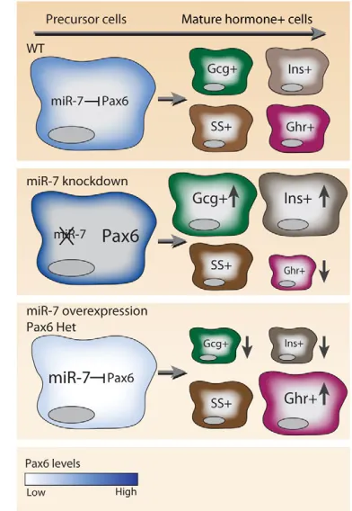
Fig. 6. A schematic model for miR-7-Pax6 interactions in pancreas development. The regulation of Pax6 levels (blue) by miR-7, regulates the differentiation of hormone-expressing endocrine cells. miR-7 knockdown de-represses Pax6 and results in reduced ghrelin (Ghr) expression and preference towards insulin and glucagon-positive cells (Ins and Gcg). Similarly, miR-7 overexpression or heterozygous (Het) expression of Pax6 results in reduced Pax6 and reciprocal changes in the expression of hormones.
D
E
V
E
LO
P
M
E
N
Author contributions
S.K.-R., A.N., A.D.M. and E.H. designed the experiments; E.H. and S.K.-R. wrote the manuscript; M.A.B. and K.A.L. designed and provided reagents; S.K.-R., A.D.M., I.A. and A.N. conducted all experiments and analyses in this work.
Supplementary material
Supplementary material available online at
http://dev.biologists.org/lookup/suppl/doi:10.1242/dev.080127/-/DC1
References
Ashery-Padan, R., Zhou, X., Marquardt, T., Herrera, P., Toube, L., Berry, A. and Gruss, P.(2004). Conditional inactivation of Pax6 in the pancreas causes early onset of diabetes. Dev. Biol. 269, 479-488.
Avnit-Sagi, T., Kantorovich, L., Kredo-Russo, S., Hornstein, E. and Walker, M. D.(2009). The promoter of the pri-miR-375 gene directs expression selectively to the endocrine pancreas. PLoS ONE4, e5033.
Bartel, D. P.(2004). MicroRNAs: genomics, biogenesis, mechanism, and function.
Cell116, 281-297.
Baulmann, D. C., Ohlmann, A., Flugel-Koch, C., Goswami, S., Cvekl, A. and Tamm, E. R.(2002). Pax6 heterozygous eyes show defects in chamber angle differentiation that are associated with a wide spectrum of other anterior eye segment abnormalities. Mech. Dev. 118, 3-17.
Berger, J., Berger, S., Tuoc, T. C., D’Amelio, M., Cecconi, F., Gorski, J. A., Jones, K. R., Gruss, P. and Stoykova, A.(2007). Conditional activation of Pax6 in the developing cortex of transgenic mice causes progenitor apoptosis.
Development134, 1311-1322.
Bonal, C. and Herrera, P. L.(2008). Genes controlling pancreas ontogeny. Int. J. Dev. Biol. 52, 823-835.
Bravo-Egana, V., Rosero, S., Molano, R. D., Pileggi, A., Ricordi, C., Dominguez-Bendala, J. and Pastori, R. L.(2008). Quantitative differential expression analysis reveals miR-7 as major islet microRNA. Biochem. Biophys. Res. Commun. 366, 922-926.
Collinson, J. M., Quinn, J. C., Buchanan, M. A., Kaufman, M. H., Wedden, S. E., West, J. D. and Hill, R. E.(2001). Primary defects in the lens underlie complex anterior segment abnormalities of the Pax6 heterozygous eye. Proc. Natl. Acad. Sci. USA98, 9688-9693.
Collombat, P., Mansouri, A., Hecksher-Sorensen, J., Serup, P., Krull, J., Gradwohl, G. and Gruss, P.(2003). Opposing actions of Arx and Pax4 in endocrine pancreas development. Genes Dev. 17, 2591-2603.
Correa-Medina, M., Bravo-Egana, V., Rosero, S., Ricordi, C., Edlund, H., Diez, J. and Pastori, R. L.(2009). MicroRNA miR-7 is preferentially expressed in endocrine cells of the developing and adult human pancreas. Gene Expr. Patterns9, 193-199.
Dames, P., Puff, R., Weise, M., Parhofer, K. G., Goke, B., Gotz, M., Graw, J., Favor, J. and Lechner, A.(2010). Relative roles of the different Pax6 domains for pancreatic alpha cell development. BMC Dev. Biol. 10, 39.
Dennis, G., Jr, Sherman, B. T., Hosack, D. A., Yang, J., Gao, W., Lane, H. C. and Lempicki, R. A.(2003). DAVID: database for annotation, visualization, and integrated discovery. Genome Biol. 4, P3.
Ding, J., Gao, Y., Zhao, J., Yan, H., Guo, S. Y., Zhang, Q. X., Li, L. S. and Gao, X.(2009). Pax6 haploinsufficiency causes abnormal metabolic homeostasis by down-regulating glucagon-like peptide 1 in mice. Endocrinology150, 2136-2144.
Gradwohl, G., Dierich, A., LeMeur, M. and Guillemot, F.(2000). neurogenin3 is required for the development of the four endocrine cell lineages of the pancreas. Proc. Natl. Acad. Sci. USA97, 1607-1611.
Gu, G., Dubauskaite, J. and Melton, D. A.(2002). Direct evidence for the pancreatic lineage: NGN3+ cells are islet progenitors and are distinct from duct progenitors. Development129, 2447-2457.
Gunawardana, S. C., Hara, M., Bell, G. I., Head, W. S., Magnuson, M. A. and Piston, D. W.(2005). Imaging Beta cell development in real-time using pancreatic explants from mice with green fluorescent protein-labeled pancreatic Beta cells. In Vitro Cell. Dev. Biol. Anim. 41, 7-11.
Heller, R. S., Stoffers, D. A., Liu, A., Schedl, A., Crenshaw, E. B., 3rd, Madsen, O. D. and Serup, P.(2004). The role of Brn4/Pou3f4 and Pax6 in forming the pancreatic glucagon cell identity. Dev. Biol. 268, 123-134.
Heller, R. S., Jenny, M., Collombat, P., Mansouri, A., Tomasetto, C., Madsen, O. D., Mellitzer, G., Gradwohl, G. and Serup, P.(2005). Genetic determinants of pancreatic epsilon-cell development. Dev. Biol. 286, 217-224.
Hornstein, E. and Shomron, N.(2006). Canalization of development by microRNAs. Nat. Genet. 38 Suppl., S20-S24.
Jan, C. H., Friedman, R. C., Ruby, J. G. and Bartel, D. P.(2011). Formation, regulation and evolution of Caenorhabditis elegans3⬘UTRs. Nature469, 97-101.
Jensen, J., Heller, R. S., Funder-Nielsen, T., Pedersen, E. E., Lindsell, C., Weinmaster, G., Madsen, O. D. and Serup, P.(2000). Independent development of pancreatic alpha- and beta-cells from neurogenin3-expressing precursors: a role for the notch pathway in repression of premature differentiation. Diabetes49, 163-176.
Kefas, B., Godlewski, J., Comeau, L., Li, Y., Abounader, R., Hawkinson, M., Lee, J., Fine, H., Chiocca, E. A., Lawler, S. et al.(2008). microRNA-7 inhibits the epidermal growth factor receptor and the Akt pathway and is down-regulated in glioblastoma. Cancer Res. 68, 3566-3572.
Kloosterman, W. P., Lagendijk, A. K., Ketting, R. F., Moulton, J. D. and Plasterk, R. H.(2007). Targeted inhibition of miRNA maturation with morpholinos reveals a role for miR-375 in pancreatic islet development. PLoS Biol.5, e203.
Kredo-Russo, S. and Hornstein, E.(2011). MicroRNA knock down by cholesterol-conjugated antisense oligos in mouse organ culture. Methods Mol. Biol. 732, 89-97.
Kroeber, M., Davis, N., Holzmann, S., Kritzenberger, M., Shelah-Goraly, M., Ofri, R., Ashery-Padan, R. and Tamm, E. R.(2010). Reduced expression of Pax6 in lens and cornea of mutant mice leads to failure of chamber angle development and juvenile glaucoma. Hum. Mol. Genet. 19, 3332-3342.
Landgraf, P., Rusu, M., Sheridan, R., Sewer, A., Iovino, N., Aravin, A., Pfeffer, S., Rice, A., Kamphorst, A. O., Landthaler, M. et al.(2007). A mammalian microRNA expression atlas based on small RNA library sequencing. Cell129, 1401-1414.
Lewis, B. P., Burge, C. B. and Bartel, D. P.(2005). Conserved seed pairing, often flanked by adenosines, indicates that thousands of human genes are microRNA targets. Cell120, 15-20.
Li, X., Cassidy, J. J., Reinke, C. A., Fischboeck, S. and Carthew, R. W.(2009). A microRNA imparts robustness against environmental fluctuation during development. Cell137, 273-282.
Lynn, F. C., Skewes-Cox, P., Kosaka, Y., McManus, M. T., Harfe, B. D. and German, M. S.(2007). MicroRNA expression is required for pancreatic islet cell genesis in the mouse. Diabetes56, 2938-2945.
Lyttle, B. M., Li, J., Krishnamurthy, M., Fellows, F., Wheeler, M. B., Goodyer, C. G. and Wang, R.(2008). Transcription factor expression in the developing human fetal endocrine pancreas. Diabetologia51, 1169-1180.
Martin, M., Hauer, V., Messmer, M., Orvain, C. and Gradwohl, G.(2007). Transcription factors in pancreatic development. Animal models. Endocr. Dev. 12, 24-32.
Melkman-Zehavi, T., Oren, R., Kredo-Russo, S., Shapira, T., Mandelbaum, A. D., Rivkin, N., Nir, T., Lennox, K. A., Behlke, M. A., Dor, Y. et al.(2011). miRNAs control insulin content in pancreatic beta-cells via downregulation of transcriptional repressors. EMBO J. 30, 835-845.
Murtaugh, L. C. and Melton, D. A.(2003). Genes, signals, and lineages in pancreas development. Annu. Rev. Cell Dev. Biol. 19, 71-89.
Nishimura, W., Rowan, S., Salameh, T., Maas, R. L., Bonner-Weir, S., Sell, S. M. and Sharma, A.(2008). Preferential reduction of beta cells derived from Pax6-MafB pathway in MafB deficient mice. Dev. Biol. 314, 443-456.
Pena, J. T., Sohn-Lee, C., Rouhanifard, S. H., Ludwig, J., Hafner, M., Mihailovic, A., Lim, C., Holoch, D., Berninger, P., Zavolan, M. et al.(2009). miRNA in situ hybridization in formaldehyde and EDC-fixed tissues. Nat. Methods6, 139-141.
Poy, M. N., Eliasson, L., Krutzfeldt, J., Kuwajima, S., Ma, X., Macdonald, P. E., Pfeffer, S., Tuschl, T., Rajewsky, N., Rorsman, P. et al.(2004). A pancreatic islet-specific microRNA regulates insulin secretion. Nature432, 226-230.
Poy, M. N., Hausser, J., Trajkovski, M., Braun, M., Collins, S., Rorsman, P., Zavolan, M. and Stoffel, M.(2009). miR-375 maintains normal pancreatic alpha- and beta-cell mass. Proc. Natl. Acad. Sci. USA106, 5813-5818.
Sander, M., Neubuser, A., Kalamaras, J., Ee, H. C., Martin, G. R. and German, M. S.(1997). Genetic analysis reveals that PAX6 is required for normal transcription of pancreatic hormone genes and islet development. Genes Dev. 11, 1662-1673.
Schedl, A., Ross, A., Lee, M., Engelkamp, D., Rashbass, P., van Heyningen, V. and Hastie, N. D.(1996). Influence of PAX6 gene dosage on development: overexpression causes severe eye abnormalities. Cell86, 71-82.
Silahtaroglu, A. N., Nolting, D., Dyrskjot, L., Berezikov, E., Moller, M., Tommerup, N. and Kauppinen, S.(2007). Detection of microRNAs in frozen tissue sections by fluorescence in situ hybridization using locked nucleic acid probes and tyramide signal amplification. Nat. Protoc. 2, 2520-2528.
Smith, S. B., Ee, H. C., Conners, J. R. and German, M. S.(1999). Paired-homeodomain transcription factor PAX4 acts as a transcriptional repressor in early pancreatic development. Mol. Cell. Biol. 19, 8272-8280.
Srinivas, S., Watanabe, T., Lin, C. S., William, C. M., Tanabe, Y., Jessell, T. M. and Costantini, F.(2001). Cre reporter strains produced by targeted insertion of EYFP and ECFP into the ROSA26 locus. BMC Dev. Biol. 1, 4.
St-Onge, L., Sosa-Pineda, B., Chowdhury, K., Mansouri, A. and Gruss, P.
(1997). Pax6 is required for differentiation of glucagon-producing alpha-cells in mouse pancreas. Nature387, 406-409.
Stark, A., Brennecke, J., Bushati, N., Russell, R. B. and Cohen, S. M.(2005). Animal MicroRNAs confer robustness to gene expression and have a significant impact on 3⬘UTR evolution. Cell123, 1133-1146.
Tkatchenko, A. V.(2006). Whole-mount BrdU staining of proliferating cells by DNase treatment: application to postnatal mammalian retina. Biotechniques40,
29-30, 32.
D
van Raamsdonk, C. D. and Tilghman, S. M.(2000). Dosage requirement and allelic expression of PAX6 during lens placode formation. Development127, 5439-5448.
Wang, S., Yan, J., Anderson, D. A., Xu, Y., Kanal, M. C., Cao, Z., Wright, C. V. and Gu, G.(2010). Neurog3 gene dosage regulates allocation of endocrine and exocrine cell fates in the developing mouse pancreas. Dev. Biol. 339, 26-37.
Wienholds, E., Kloosterman, W. P., Miska, E., Alvarez-Saavedra, E., Berezikov, E., de Bruijn, E., Horvitz, H. R., Kauppinen, S. and Plasterk, R. H.(2005). MicroRNA expression in zebrafish embryonic development. Science 309, 310-311.
Yamaoka, T., Yano, M., Yamada, T., Matsushita, T., Moritani, M., Ii, S., Yoshimoto, K., Hata, J. and Itakura, M.(2000). Diabetes and pancreatic tumours in transgenic mice expressing Pax 6. Diabetologia43, 332-339.
Yasuda, T., Kajimoto, Y., Fujitani, Y., Watada, H., Yamamoto, S., Watarai, T., Umayahara, Y., Matsuhisa, M., Gorogawa, S., Kuwayama, Y. et al.(2002). PAX6 mutation as a genetic factor common to aniridia and glucose intolerance.
Diabetes51, 224-230.
Zhou, Q., Law, A. C., Rajagopal, J., Anderson, W. J., Gray, P. A. and Melton, D. A.(2007). A multipotent progenitor domain guides pancreatic
organogenesis. Dev. Cell13, 103-114.