TEXTURE CONTENT BASED MRI IMAGE RETRIEVAL
USING GABOR WAVELET AND PROGRESSIVE
RETRIEVAL STRATEGY
1N. KUMARAN, 2R. BHAVANI 1
Assistant Professor, Department of CSE, Annamalai University, Annamalainagar 2
Professor, Department of CSE, Annamalai University, Annamalainagar
E-mail: [email protected], [email protected]
ABSTRACT
Due to the vast number of medical technologies and equipments the medical images are growing at a rapid rate. This directs to retrieve efficient medical images based on visual contents. This paper proposed the content based medical image retrieval system by means of Gabor wavelet to extract texture features of MRI scan images. Then the k-means clustering, progressive retrieval strategy and Euclidean distance are used to retrieve best MRI scan images for the query image in medical diagnosis. The experimental results demonstrate the efficiency of this system in clusters accuracy and best MRI scan image retrieval against standard Haralick’s, texture spectrum and Gabor wavelet based texture features.
Keywords: CBMIR, Euclidean Distance, Gabor Wavelet, K-Means Clustering, Progressive Retrieval
1. INTRODUCTION
Content Based Image Retrieval (CBIR) is a technique in which different visual contents have been measured to search and retrieve images from the mass amount of image databases based on the input image. The Content Based Medical Image Retrieval (CBMIR) systems are medical domain specific search engine for medical image databases, which indexing and retrieving medical images according to their visual contents like texture, shape and other information. [1-4]
Today, there are huge numbers of medical images being created in hospitals around the world. It is estimated that the amount of such images will further increase exponentially in the future. The significance of new technologies such as X-Ray radiography, Ultrasound, Computed Tomography (CT), Magnetic Resonance Imagining (MRI) and Picture Archiving and Communication Systems (PACS) have resulted in an explosive growth in the number of images stored in the database. Because these medical images are vary person to person. Medical images classifying, indexing and retrieval in manual methods are very expensive and time consuming. This will lead various systems for storage, organization, indexing and retrieval of the medical images in different fields like medical
diagnosis, surgical planning, monitoring, teaching and research.
Due to the nature of medical images, CBMIR system is still faced with challenges. Low resolution and strong noise are two common characteristics in most medical images. With these characteristics, medical images cannot be precisely extracted for the visual content of their features. One more characteristic of medical images is that many images are represented in gray level rather than colour. Even with the change of intensity, monochrome may fail to clearly display the actual circumstance of lesion area.
Generally, the medical image database contains a lot of texture based information capable for retrieval purpose. This paper, proposed the new CBMIR system in various parts of the human body MRI scan images such as spine, brain, knee and abdomen using Gabor wavelet [5] based texture features with k-means clustering [6-7], progressive retrieval strategy [8] and Euclidean distance measure [9]. The accuracy, precision and recall rate of this system is high compared with standard Haralick’s [10], texture spectrum [11] and Gabor wavelet based texture features.
In the next section of the paper, we describe related works of the system. We present brief discussion on the proposed work and Gabor wavelet transform in section 3 and 4. In sections 5 and 6, we explain about Gabor wavelet based texture features and k-means clustering. In section 7, we deal with image retrieval. Section 8 shows experiments and results. In section 9, we conclude our work with future prospects.
2. RELATED WORK
Medical images have become a key investigation tool for medical diagnosis. With the growth of medical databases, new applications committed to statistical analysis of medical data have appeared. There are many existing systems that provide different methods and algorithms for CBMIR. The most important intention of all these systems is to prove the improvement of results so as to give support to the doctors and radiologists in diagnosis of treatments.
In [12], the authors had highlighted some of the problems unique to automated retrieval from large medical image databases and presented solutions to some of them in the specific context of HRCT images of the lung and liver. Here 255 features were extracted using Sequential Forward Search (SFS) algorithm and decision-tree based approach and Expectation Maximization (EM) clustering algorithm was used to achieve 77.39% of precision rate. Tatjana Zrimec [13] presented a prototype medical image aid system that was designed to enable quick and efficient access to images, from a large data set, by content such as texture, structure and other geometric properties. They used supervised machine learning and Region of Interest (ROI) to determine a subset of features that was the most descriptive for a particular image modality. This system helped us to investigate different approaches to content-based medical
image retrieval, feature vector construction and active indexing.
D.V. Tsishkou et al. [14] constructed an approach for indexing and retrieval of large medical image database using Tree structured index in Tree Tessellation Algorithm with high logarithmic complexity and the accuracy of 74.5%. They described a medical image indexing and retrieval problem, reviewed medical image types, examined medical data content analysis and conclude that automatic systems for this problem are needed.
In [15], they described a medical image retrieval system using low level features and high level semantic features with 90% of precision and recall rate. Here medical images were segmented into several sub images using fuzzy C-mean clustering algorithm and extracting 3-gray level features using colour moments. Then the sub images were changed to binary image and seven shape features and four texture features were extracted using co-occurrence matrix. Then the genetic algorithm was used to select optimal features and the text information in the medical image was chosen for the semantic content of the report of radiologists.
Paul Miki Willy et al. [16] created an experimental design of an intelligent Content-based Medical Image Retrieval (CBMIR) for handling multiple organs of interest through three main processes. First, CBMIR identifies all such organs by comparing the images directly using the Hausdorff distance to the single, healthy organs stored in an organ database. After that, CBMIR builds image classes using neural networks and, finally, recognize the proper class for a query image using a multicriteria optimization approach resulted in more accurate retrieved images. K. Ait saadi et al. [17] demonstrated a CBMIR using the 2 x 2 Discrete Cosine Transform (DCT) and Information Dominance Strategy (IDS). In this work, the texture features were directly generated from the 2 x 2 DCT coefficients and the searching process was carried out by calculating the cosine distance measure between the signature of the query image and the database image . This system suffered from high computational complexity and high compression ratio.
from the DICOM header which was used to perform the initial search. This pre-filtering of the images reduced the number of images to be searched. Then texture features and shape features were found by Gabor filter and the fixed block resolution format. Image retrieval is performed by Euclidean distance measure. This system reduced the time taken to search the entire medical image database. Also the average precision rate of 80% is achieved. In [19], the authors extracted time-frequency coefficients from each scale and each direction of the image using Gabor wavelet. The energy was computed according to the coefficients, and dominant multi-scale and multi-direction fuzzy set was computed based on all energy computed. The standardized energy was used to measure the dominance of each element. Texture feature vector was computed according to the fuzzy set. The Euclidean distance similarity measure was carried out between the fuzzy sets. When two images were of the same kind, their similarity measure was carried out between the texture feature vectors of two images. The experiments shown that the method proposed in the paper has good retrieval performances for normal medical image retrieval only.
Horsthemke et al. [20] explained two different texture features based CBMIR systems. The first system can be used to provide context-sensitive tools for computer-aided diagnosis with pixel-level co-occurrence matrices. The second system can be used directly as a computer-aided diagnosis system for case-based and evidence-based medicine with pixel level and global level co-occurrence matrices. In [21], the authors expressed a novel scheme based on Texture Unit. This scheme reduced texture unit values from 0 to 6561 to 0 to 255 based on Lag values. The texture spectrum based texture features were extracted on both schemes and an Integrated Histogram Bin Matching (IHBM) similarity measure was used and the comparison was made. The proposed methods yield a high precision rate than the existing method.
Peiqiang Zhang et al. [22] proposed a method using co-occurrence matrix to extract texture feature and edge histogram to extract shape feature of medical images. Then Euclidean distance was used for medical image retrieval. Results of experimentation showed that the system had a recall rate about 90% and applied to medical image retrieval with promising effect. In [23], the authors presented an evaluation of the diagnosis of dementia using texture analysis of brain MRI with
Gabor wavelets and further classified by the Back Propagation Network. Here three different types of texture features were extracted. The first containing the GLCM features, the second had the Haralick’s features and the third had Gabor wavelet based Haralick’s features. From the comparison of the average efficiency, the statistical features extracted from Gabor wavelets provided better efficiency of 97% than the other two methods.
B. G. Prasad et al. [24] evaluated the performance of two statistical methods of texture features proposed by Haralick’s and Tamura for retrieving similar cases for CT scan brain images. To speed up the search process, selected features were extracted and indexed using hash structure. The Euclidean distance measure was used for similarity measurement. Both the methods were compared based on precision and recall. Tamura features were found to provide better retrieval results for CT scan brain images. In our previous work [25-26] the performance measures for spine MRI scan image retrieval proved that texture Spectrum based texture features (black-white symmetry, geometric symmetry, degree of direction, orientation features and central symmetry) were somewhat good compared to Haralick’s texture features (contrast, angular second moment, coarseness, entropy) and the combination of both features of image retrieval was the best.
Sanghavi et al. [27] focused on recent advances in CBIR system in the medical domain. It also focused on various feature extraction techniques and algorithms implemented for CBIR systems in different cases of medical domain. The authors concluded that CBIR had become an important research area in the medical domain. In another work [28], we explained the Gabor wavelet based texture features extraction and k-means clustering. Here, the Euclidean distance measure was used to retrieve related images for the query image in medical diagnosis. The experimental results demonstrated the efficiency of this system in clusters and MRI image retrieval against Haralick’s and Texture Spectrum based texture features.
direction rotational variant feature where as this method was rotational invariant. The effectiveness of this algorithm was confirmed by combining it with the Gabor transform.
3. PROPOSED WORK
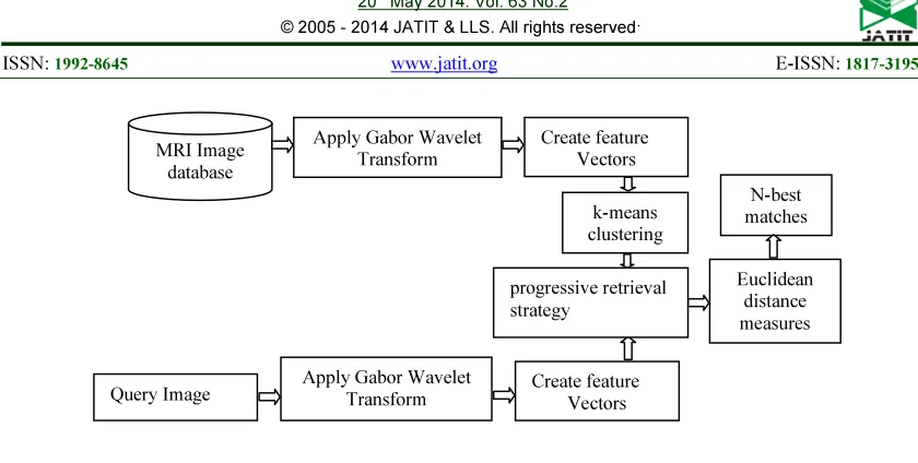
The block diagram of the proposed CBMIR system is shown in figure 1 in which various parts of the human body MRI scan images such as spine, brain, knee and abdomen are used to construct the training data set.
In our work, we apply Gabor Wavelet Transform (GWT) to each MRI scan image in the training image database. Then sixteen texture based feature vector values such as mean, standard deviation, skew and kurtosis are extracted with four directions θ = {0°, 45°, 90° and 135°} as feature components and the database are created.
After that, using k-means clustering the training images are clustered by means of the extracted texture features based database. When a testing MRI scan image is submitted, the same texture feature extraction and feature vector value construction process has been applied to obtain the feature vector values for the testing image. Next, progressive retrieval strategy is used to determine more precise MRI scan images for the testing query image from the clustered images. So, it increases the retrieval accuracy and reduces response time than the existing methods. For similarity comparison between the query MRI scan image and the more precise MRI scan images, Euclidean distance function is used. The closest Euclidean distance values for the query image are ranked and best MRI scan images are retrieved for medical diagnosis.
4. GABOR WAVELET TRANSFORM
In the spatial domain, a GWis a complex exponential modulated by a Gaussian function. In the most general the Gabor wavelet is defined as follows:
*
(1)
Where, x, y denotes the pixel position in the spatial domain, is the radial centre frequency of the complex exponential, is the orientation of the GW, and is the standard deviation of the Gaussian function. By selecting different centre frequencies and orientations, we can obtain a family of Gabor kernels, which can be used to extract features from an image.
The GW forms complex valued function like real and imaginary parts. Other wavelets are shifted, rotated and diluted versions of these two. Given an image f(x, y), GW features are extracted by convolution operation f(x, y) with
as follows:
(2) Where, denotes the convolution operator. For a neighborhood window of size W × W for W = 2t + 1, the discrete convolution operation of the image f(x, y) with GWwe get
(3)
We can write down,
(4)
With,
(5)
(6)
Where, is a complex conjugate of .
5. GABOR WAVELET BASED TEXTURE FEATURES
The feature vectors μ(x, y), STD(x, y), Skewness and Kurtosis are constructed as feature components using equation (7) to (10).
(8)
(9)
(10)
Where, (X, Y) is the image dimension. Table 1 shows the sample values of the feature vectors for an MRI spine and brain images.
6. K-MEANS CLUSTERING
Cluster analysis is the process of grouping objects into subsets that have meaning in the context of a particular problem. K-means clustering is a method of cluster analysis which aims to partition and observations into k clusters in which each observation belongs to the cluster with the nearest mean.
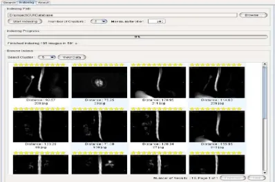
[image:5.595.88.287.506.638.2]. In this problem, the K-means clustering algorithm can be formally stated as follows: Given 1850 normal and abnormal MRI scan images as training images in a 16-dimensional metric space, determine a partition of the images into maximum 20 clusters and 100 iterations, such that the images in a cluster are more similar cases to each other than two images in different clusters.
Figure 2: Sample output of fifth cluster
This work initializes 20 clusters by arbitrarily selecting one image to represent each cluster. Each of the remaining images is assigned to a cluster and the clustering criterion is used to calculate the cluster mean. These means are used as the new cluster points and each image is reassigned to the cluster that it is most similar to. This continues until there is no longer a change when the
clusters are recalculated. The figure 2 shows the sample output of the fifth cluster out of twenty clusters.
7. IMAGE RETRIEVAL
In the clustered images, we use progressive retrieval strategies in order to balance between computational complexity and retrieval accuracy. Consider the feature vectors of the query image and the clustered image as (A, B, C) and (X, Y, Z) respectively. Then, we can roughly filter the clustered images as follows,
(11)
Where, the filtering constant β Є (0, 1) is used to adjust the sifted the clustered images. If F is false, then clustered image can be identified as far apart from Query image and therefore is discarded; else, clustered image be kept for further match. Then we use the precise filtering threshold α as similarity criteria to determine more precise images. Consider the feature vector of image A as VAF = {ΔA1, ΔA2… ΔAn} and image B as VBF = {ΔB1, ΔB2… BAn}. Define the similarity αi of ΔAi and ΔBi as,
Min(ΔAi, ΔBi)/Max(ΔAi, ΔBi), ΔAi≠0orΔBi≠0
αi=
1 where, ΔAi = 0 orΔBi = 0 (12)
and we can give the similarity criteria,
(13)
Where, and
.
Obviously, the similarity criteria are 0 ≤ α ≤ 1. If α exceeds a given threshold, it means that mismatch occurs and that image should be discarded; else, it will be kept for further match. After that the Euclidean distance is calculated between the query image and the selected precise images. If xi and yi are 2D feature vectors of selected precise images and query image respectively then the distance measure is defined as,
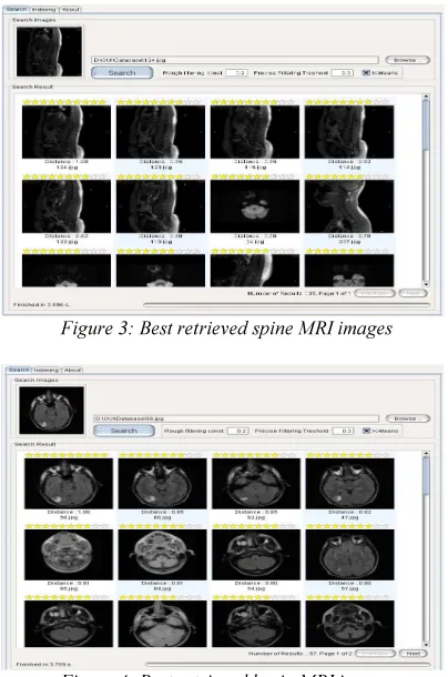
The calculated distances are sorted in increasing order and display the first N images as the best similar MRI scan images for medical treatment. The sample output screens are shown in figure 3 and figure 4.
Figure 3: Best retrieved spine MRI images
Figure 4: Best retrieved brain MRI images
8. EXPERIMENTS AND RESULTS
This method is implemented on a computer system using Java as the programming language and MySQL as the backend. In this work, we used around 1850 MRI scan images as training set and 150 MRI scan images as testing set in BMP format with the size of 256 x 256 as a database. Different parts of the human body MRI scan images such as 900 spine, 450 brain, 325 knee and 325 abdomen images are used.
The effectiveness of the k-means clustering algorithm can be measured by accuracy, sensitivity and specificity. Normally they are defined as,
Accuracy = (TP+TN) / (FN+FP+TN+TP) (15) Sensitivity = TP / (TP + FN) (16) Specificity = TN / (TN + FP) (17)
According our system, the consideration of True Positive (TP), True Negative (TN), Full Negative (FN) and Full Positive (FP) images is as follows:
TP - Correctly clustered MRI scan images TN - Wrongly clustered MRI scan images
FN - System judge correctly clustered MRI scan images but actually wrong
FP - System judge wrongly clustered MRI scan images but actually correct
The K-means clustering algorithm gave a test accuracy of 73.9% while using Haralick’s texture features alone and gave a test accuracy of 78.3% while using texture spectrum based texture features alone. Furthermore, when the both features are combined then the k-means clustering algorithm’s accuracy increases to 85.9%. The Gabor wavelet based texture features and k-means clustering algorithm gave a test accuracy of 91.4%. The proposed Gabor wavelet based texture features, k-means clustering algorithm and progressive retrieval strategy gave the test accuracy of 98.7%. Table 2 shows the accuracy, sensitivity and specificity of our method compared with other methods. The accuracy of the reporting radiologist’s diagnosis was also analysed. The proposed method yielded best recognition rates and help radiologists for the best medical diagnosis.
[image:6.595.304.512.552.667.2]Figure 5 shows the Empirical Receiver operating characteristic curves in support of various cutoff points for different texture features based MRI scan image retrieval systems with a false positive rate on the X-axis and true positive rate on the Y-axis.
Figure 5: Empirical ROC curves
different texture features based MRI scan image retrieval systems. Along with the growth of the database, the feature extraction time and response time increases proportionally. These results compared to Haralick’s and texture spectrum based texture features.
Table 3: Comparison of Feature Extraction Time and the Response Time
The texture spectrum based texture features like black-white symmetry, geometric symmetry, degree of direction, four orientation features and central symmetry are extracted with the extraction time of 321 seconds which compares very lesser to other techniques. It’s response time for best image retrieval was 4.85 seconds. The Haralick’s features are extracted with the extraction time of 378 seconds. But its response time was 5.23 seconds. The fusion of texture spectrum and Haralick’s based texture features are extracted with the extraction time of 454 seconds. Its response time for best image retrieval was 7.05 seconds. Gabor Wavelet based texture features like mean, standard deviation, Skewness and Kurtosis are extracted for four different orientations with the extraction time of 411 seconds. But its response time is 3.742 seconds which is lesser compared to other techniques. Our proposed method response time is 3.709 seconds which compare very lesser to all other techniques.
The effectiveness of the proposed method can be measured by recall and precision, which are often referred together since they measure the different aspects of the system performance. Recall is defined as the ratio between the number of retrieved relevant images and the total number of relevant images in the database. The performance measures of recall are given in figure 6 which
[image:7.595.303.513.161.251.2]proved that our proposed method has a highest recall rate than other methods.
Figure 6: Performance measures based on recall
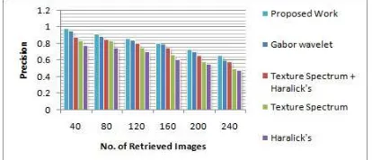
Precision measures the retrieval accuracy and is defined as the ratio between the number of retrieved relevant images and the number of total retrieved images. The performance measures of precision are given in figure 7 which proved that our proposed method has highest precision rate than other methods.
Figure 7: Performance measures based on precision
9. CONCLUSION
This paper have been proposed an efficient progressive retrieval strategy based MRI scan image retrieval method using Gabor Wavelet based texture features, K-means clustering algorithm and Euclidean distance measure. Our Experimental results demonstrate that the proposed method has the best accuracy, sensitivity, specificity, precision and recall rate than other conventional methods such as Haralick’s, texture spectrum and Gabor wavelet based MRI scan image retrieval methods. Further, a progressive retrieval strategy is suggested along with K-means clustering algorithm to reduce the response time whereas maintaining an excellent level of retrieval accuracy. We have planned to extend our work for all types of human body scan images.
REFRENCES:
[1] H. Muller, N. Michoux, D. Bandon & A. Geissbuhler, A review of content-based image retrieval systems in medical applications: clinical benefits and future directions, Methods
Number of Features
Feature Extraction
Time ( in Seconds)
Response Time ( in Seconds)
Gabor Wavelet Features
16 411 3.742
3.709 With progressive retrieval
Texture Spectrum
Features
8 321 4.85
Haralick’s
Features 14 378 5.23
Texture Spectrum
+ Haralick’s
Features
[image:7.595.304.511.366.455.2]International journal of Medical Informatics, 73, 2004, 1-23.
[2] X. S. Zhou, S. Zillner, M. Moeller, M. Sintek & et. al., Semantics and CBIR: A Medical Imaging Perspective, ACM International Conf. on Content-based Image and Video Retrieval, 2008, 571-580.
[3] Ceyhun Burak Akgül, Daniel L. Rubin, Sandy Napel, Christopher F. Beaulieu & et. al., Content-Based Image Retrieval in Radiology: Current Status and Future Directions, Journal of Digital Imaging, 24(2), 2011, 208-222. [4] Sh. Akbarpour, A Review on Content Based
Image Retrieval in Medical Diagnosis International Journal on Technical and Physical Problems of Engineering, 5(2), 2013, 148-153.
[5] B. S. Manjunath & W. Y. Ma, Texture features for browsing and retrieval of image data, IEEE Trans. Pattern Analysis and Machine Intelligence, 18(8), 1996, 837-842.
[6] Gobert N. Lee & Hiroshi Fujita, K-means clustering for classifying unlabelled MRI data, 9th IEEE Conference of the Australian Pattern Recognition Society on Digital Image Computing Techniques and Applications, 2007, 92-98.
[7] B. Ramamurthy & K.R. Chandran, Content Based Medical Image Retrieval with Texture Content Using Gray Level Co-occurrence Matrix and K-Means Clustering Algorithms, Journal of Computer Science, 8 (7), 2012, 1070-1076.
[8] Parulpreet singh & Kulvinder Singh Mann, An Approach of Image Retrieval Using Content Based Retrieval System, International Journal of Advanced Research in Computer Science and Software Engineering, 3(2), 2013, 52-59. [9] Swarnambiga Ayyachamy,Vasuki S., &
Manivannan, Distance measures for medical image retrieval, International Journal of Imaging Systems and Technology, 23(1), 2013, 9–21.
[10] R. Haralick, K. Shanmugam & I. Dinstein, Textural features for image classification, IEEE Trans. on Systems on Man and Cybernetics, 3(6), 1973, 610-621.
[11] D. He & L. Wang, Texture features based on texture spectrum, Pattern Recognition, 24(5), 1991, 391–399.
[12] Avi Kak & Christina Pavlopoulou, Content-Based Image Retrieval from Large Medical Databases, IEEE Proc. on the First
International Symposium on 3D Data Processing Visualization and Transmission, 2002, 1-10.
[13] Tatjana Zrimec, A Content-based Retrieval System for Medical Images, IEEE 7th International Conf. on Control, Automation, Robotics and Vision, IEEE, 2002, 180-185. [14] D.V. Tsishkou, E.I. Bovbel & Liventseva M.M,
Medical Images Indexing and Retrieval, IEEE 7th International symposium on Signal Processing and its Applications, 2003, 185-187. [15] Hong Shao, Wen-cbeng Cui & Hong Zhao,
Medical Image Retrieval Based on Visual Contents and Text Information, IEEE International Conf. on Systems, Man and Cybemeties, 2004, 1098-1103.
[16] Paul Miki Willy & Karl-Heinz Kufer, Content-based Medical Image Retrieval (CBMIR): An Intelligent Retrieval System for Handling Multiple Organs of Interest, IEEE Proc. of the 17th Symposium on Computer-Based Medical Systems, 2004, 103-108.
[17] K. Ait saadi, A.Zemouri, Z. Brahimi & H.Meraoubi, Indexing and Retrieval Medical images based on 2X2 DCT and IDS Compression, IEEE Proc. of 5th International Conf. on Intelligent Systems Design and Applications, 2005, 239-244.
[18] A. Grace Selvarani & Dr. S. Annadurai, Medical Image Retrieval By Combining Low Level Features and Dicom Features , IEEE International Conf. on Computational Intelligent and Multimedia Applications, 2007, 587 – 589.
[19] Gang Zhang & Zong-Min Ma, Texture Feature Extraction and Description Using Gabor Wavelet in Content-Based Medical Image Retrieval, Proc. of the International Conf. on Wavelet Analysis and Pattern Recognition, 2007, 169-173.
[20] William Horsthemke , Daniela Raicu & Jacob Furst, Task-Oriented Medical Image Retrieval, MICCAI work shop proceedings , 2007, 31-44, [21] V. Vijaya Kumar, N. Gnaneswara Rao, &
A.L.Narsimha Rao, RTL: Reduced Texture spectrum with Lag value Based Image Retrieval for Medical Images, International Journal of Future Generation Communication and Networking, 2(4), 2009, 39-48.
[23] T. R. Sivapriya, V. Saravanan & P. Ranjit Jeba Thangaiah, Texture Analysis of Brain MRI and Classification with BPN for the Diagnosis of Dementia, Engineering and Information Technology Communications in Computer and Information Science, 20(4), 2011, 553-563. [24] Prasad, B.G. & Krishna, A.N., Statistical texture
feature-based retrieval and performance evaluation of CT brain images, IEEE International Conf. on Electronics Computer Technology, 2011, 1-4.
[25] N.Kumaran & Dr.R.Bhavani, Spine MR Image Retrieval using Co-occurrence Matrix and Texture Spectrum, CiiT International Journal of Digital Image Processing, 3(12), 2011, 766-772.
[26] N.Kumaran & Dr.R.Bhavani, Spine MRI Image Retrieval using Texture Features, International Journal of Computer Applications, 46(24), 2012, 1-7.
[27] Sanghavi J.B., Bhoyar K.K. & Gawande U.H., Review of Content Based Image Retrieval Systems of Medical Domain, Advances in Medical Informatics, 2(1), 2012, 22-24.
[28] N.Kumaran & Dr.R.Bhavani, MRI Image Retrieval Using Gabor Wavelet Based Texture Features, International Journal of Advanced Research in Science and Technology, 2(1), 2013, 40-45.
Figure 1: The design of the proposed retrieval system
Table 1: Extracted Sample Values of Feature Vectors
MRI Images
Ɵ μ(x, y) SD(x,y) Skew Kurtosis μ(x, y) SD(x,y) Skew Kurtosis
0 40.221 37,203 2.062 3.554 35.132 32.262 1.221 2.455 45 13.120 16,336 3.060 12.602 12.501 10.451 3.011 16.830 90 41.010 38,251 1.303 2.422 33.435 31.547 2.004 3.508 135 26.031 19,162 1.431 4.023 14.201 12.534 2.220 11.031
Table 2: Comparison of Accuracy, Sensitivity & Specificity
Methods Accuracy (%) Sensitivity (%) Specificity (%)
Proposed work (Gabor Wavelet features + k-means
clustering + Progressive Retrieval Strategy) 98.7 95.3 99 Gabor Wavelet features + k-means clustering 91.4 60.4 97.5
Texture Spectrum + Haralick’s Features + k-means
clustering 85.9 79.4 89.5
Texture Spectrum features + k-means clustering 78.3 66 87 Haralick’s Features + k-means clustering 73.9 57.8 84.7
MRI Image database
Apply Gabor Wavelet Transform
Create feature Vectors
k-means clustering
Euclidean distance measures
N-best matches
Apply Gabor Wavelet
Transform Create feature Vectors Query Image
[image:10.595.70.526.527.624.2]