Copyright © 2009, American Society for Microbiology. All Rights Reserved.
Identification of 20 Common Human Enterovirus Serotypes by Use of
a Reverse Transcription-PCR-Based Reverse Line Blot
Hybridization Assay
䌤
Fei Zhou,
1,2Fanrong Kong,
1Kenneth McPhie,
1Mala Ratnamohan,
1Linda Donovan,
1Frank Zeng,
1Gwendolyn L. Gilbert,
1,2and Dominic E. Dwyer
1,2*
Centre for Infectious Diseases and Microbiology (CIDM), Institute of Clinical Pathology and Medical Research (ICPMR),
Westmead, New South Wales, Australia,
1and The University of Sydney, Sydney, New South Wales, Australia
2Received 24 April 2009/Returned for modification 29 May 2009/Accepted 25 June 2009
The more than 100 human enterovirus (HEV) serotypes can also be classified into four species, HEV-A to
-D, based on phylogenetic analysis of multiple gene regions. Current molecular typing methods depend largely
on reverse transcription-PCR (RT-PCR) amplification and nucleotide sequencing of the entire or 3
ⴕ
half of the
VP1 gene. An RT-PCR-based reverse line blot (RLB) hybridization assay was developed as a rapid and efficient
approach to characterize common HEVs. Twenty HEV serotypes accounted for 87.1% of all HEVs isolated at
an Australian reference virology laboratory from 1979 to 2007. VP1 sequences of all known HEV prototype
strains were aligned to design one sense primer and three antisense primers for RT-PCR. After sequencing of
the complete VP1 genes of 37 previously serotyped examples of the commonest 20 serotypes and alignment of
these VP1 sequences with GenBank sequences, four serotype-specific probes for each serotype were designed
for RLB. The RT-PCR–RLB assay was then applied to 132 HEV isolates, made up of the previously sequenced
37 isolates and another 95 serotyped clinical isolates. The RT-PCR–RLB genotypes corresponded with the
serotypes for 131/132 isolates; the one exception was confirmed by VP1 sequencing, and the genotype was
confirmed by repeat conventional serotyping. Genotyping by RT-PCR–RLB complements traditional
serotyp-ing methods and VP1 sequencserotyp-ing and has the advantages of convenience, speed, and accuracy. RT-PCR–RLB
allows detection of specific enteroviral serotypes or genotypes associated with HEV outbreaks and significant
disease.
Human enteroviruses (HEVs), members of the
Enterovirus
genus of the family
Picornaviridae
, are common human
patho-gens associated with a wide spectrum of diseases. These range
from asymptomatic infection to serious illness, especially in
children (20) and the immunocompromised. Clinical
syn-dromes include aseptic meningitis (1, 25), encephalitis (8, 26),
fever and rash, acute flaccid paralysis (29), and gastrointestinal
and respiratory illness. Some HEVs (e.g., EV71) have caused
large outbreaks in Southeast Asia (21). Polioviruses (PVs)
(including vaccine strains) have reemerged recently and have
been associated with clinical disease (30). Although Australia
is “polio-free,” there have been recent outbreaks of polio in
nearby Indonesia (5) and recent importation of a case from
Pakistan (31).
The original classification of HEVs consisted of PVs,
cox-sackieviruses A (CVA) and B (CVB), and echoviruses (E),
based on biological activity and disease. Conventional
ap-proaches for detecting and characterizing HEVs are based on
the time-consuming and labor-intensive procedures of viral
isolation in cell culture and neutralization using reference
an-tisera (7). Current classification of the over 100 HEV serotypes
(http://www.picornaviridae.com) divides the members of HEV
genera into four species (A, B, C, and
HEV-D), based on genome organization, sequence similarity, and
biological properties (23). At present, molecular typing
meth-ods largely depend on reverse transcription-PCR (RT-PCR)
amplification and nucleotide sequencing of the entire or 3
⬘
half
of the VP1 gene (23). Comparison of the VP1 sequence with
databases of VP1 sequences of HEV prototype and variant
strains (18) allows genotype assignation. Other molecular
methods that assist in serotype-specific identification include
multiplex real-time hybridization probe RT-PCR for specific
detection and differentiation of enterovirus type 71 (EV71)
and CVA16 (28), combined multiplex RT-PCR and microarray
assay to detect and differentiate EV71 and CVA16 (4),
molec-ular characterization of PV strains by RT-PCR–restriction
fragment length polymorphisms (2), and an integrated
micro-RT-PCR system for automatic detection of microorganisms,
including EV71 (11). Experience with microwell
oligonucleo-tide arrays (27) and other non-sequencing-based molecular
methods to genotype other HEV serotypes is limited.
The 20 most frequent HEV serotypes identified at the
ref-erence virology laboratory of the Centre for Infectious
Dis-eases and Microbiology (CIDM) in New South Wales,
Austra-lia, from 1979 to 2007 included HEV-A (CVA16 and EV71),
HEV-B (E3, -6, -7, -9, -11, -14, -17, -18, and -30, CVB1, -2, -3,
-4, and -5, and CVA9), and HEV-C (PV1, -2, and -3) serotypes.
These 20 serotypes accounted for 87.1% of all HEVs referred
for typing during this period.
In order to establish a quick, accurate, and cost-effective
* Corresponding author. Mailing address: Centre for Infectious
Dis-eases and Microbiology (CIDM), Institute of Clinical Pathology and
Medical Research (ICPMR), Westmead Hospital, Darcy Road,
West-mead, New South Wales, 2145 Australia. Phone: (612) 9845 6255. Fax:
(612) 9893 8659. E-mail: [email protected].
䌤
Published ahead of print on 1 July 2009.
2737
on May 16, 2020 by guest
http://jcm.asm.org/
TABLE
1.
Oligonucleotide
primers
and
probes
for
RT-PCR–RLB
assay
Primer or probe a Target Tm ( oC) GenBank accession no. Sequence (5 ⬘ –3 ⬘ ) c No. of aligned sequences d(prototype strain GenBank no.) % Covered strains (no. of covered strains/total no. of strains) VP1-ALL-Sn HEVs 59.09 AF029859 (E1) 2540NYT NMM NGC NGY NGA RAY NGG2560 86 prototype strains VP1-A-Ab HEV-A 61.42 AY421760 (CVA2) 3066AAN GWN GGR TAN CCR TCR TAR AAC CA3041 16 prototype strains VP1-B-Ab HEV-B 60.38 AF029859 (E1) 2953GGR TTN GTN GAN GWY TGC C2935 54 prototype strains VP1-C-Ab HEV-C 59.34 AF499635 (CVA1) 3069RWA NCC RTC RTA RAA RTG NGM RTA NGC3043 13 prototype strains VP1-CVA16-Sp1 Serotype CVA16 64.47 FJ868280 b 224CAC AGG AGA CAG CCA TTG G242 104 (U05876) 95.2 (99/104) VP1-CVA16-Sp2 Serotype CVA16 64.62 AB292123 63GCA GGA GAC AGC CAT TGG80 VP1-CVA16-Sp3 Serotype CVA16 61.94 AB119644 60CAC AAG AGA CAG CCA TTG G78 VP1-CVA16-Sp4 Serotype CVA16 60.85 U05876 2669CTC AAG AGA CCA CGA TTG G2687 VP1-EV71-Sp1 Serotype EV71 63.04 EF063152 3017CAT TCA TGT CAC CTG CGA G3035 622 (U22521) 70.4 (438/622) VP1-EV71-Sp2 Serotype EV71 63.03 FJ868281 b 577CCA TTT ATG TCA CCT GCG AG596 VP1-EV71-Sp3 Serotype EV71 62.14 U22521 3013TGC CAT TTA TGT CAC CTG C3031 VP1-EV71-Sp4 Serotype EV71 63.16 DQ841993 578CGT TCA TGT CAC CCG CGA G596 VP1-CVA9-Sp1 Serotype CVA9 63.45 FJ868282 b 226ATG GAG GAA TAC AAG ACC ACA GA248 20 (D00627) 95 (19/20) VP1-CVA9-Sp2 Serotype CVA9 60.26 AY466022 2673ATG GAG GAG TAT AAA ACC ACA GA2695 VP1-CVA9-Sp3 Serotype CVA9 61.51 D00627 2673ATG GAA GAG TAC AAG ACC ACT GA2695 VP1-CVA9-Sp4 Serotype CVA9 63.22 AB167978 91ATG GAG GAT TAC AAA ACC ACA GA113 VP1-CVB1-Sp1 Serotype CVB1 63.62 FJ868283, b FJ868284 b 358ATA ACG AGT GCC CAA CAA CC377 18 (M16560) 88.9 (16/18) VP1-CVB1-Sp2 Serotype CVB1 63.99 AB167989 234ATA ACG AGC GCT CAA CAA CC253 VP1-CVB1-Sp3 Serotype CVB1 65.10 M16560 2809ATA ACA AGT GCC CAG GAG CC2828 VP1-CVB1-Sp4 Serotype CVB1 62.67 AY186745 2790ATA ACG AGT GCC CAA CAG C2808 VP1-CVB2-Sp1 Serotype CVB2 62.14 AB234339 132TGG AAA GTG AGT GTT AGA CAA GC154 11 (AF085363) 100 (11/11) VP1-CVB2-Sp2 Serotype CVB2 64.27 FJ868285 b 280TGG AAG GTG AGT GTT AGA CAA GC302 VP1-CVB2-Sp3 Serotype CVB2 63.03 AF225468 280TGG AAA GTG AGT GTT AGG CAA G301 VP1-CVB2-Sp4 Serotype CVB2 61.82 AM711056 280TGG AAA GTC AGT GTT AGG CAG300 VP1-CVB3-Sp1 Serotype CVB3 62.61 AM711026 256CGG TAT GCT GAA TGG GTG273 63 (M16572) 88.9 (56/63) VP1-CVB3-Sp2 Serotype CVB3 63.40 M16572 2705GCG GTA TGC TGA ATG GGT A2723 VP1-CVB3-Sp3 Serotype CVB3 67.29 EU144042 2708CGG TAT GCC GAA TGG GTG2725 VP1-CVB3-Sp4 Serotype CVB3 65.24 FJ868286, b FJ868287 b 256CGG TAC GCT GAA TGG GTG273 VP1-CVB4-Sp1 Serotype CVB4 58.24 FJ868288, b FJ868289 b 222GAT TTA CAT CAA ATA CTC AAG TGC245 93 (X05690) 71 (66/93) VP1-CVB4-Sp2 Serotype CVB4 58.98 X05690 2666CGT AAT TTA TAT AAA ATA CTC CAG TGC2692 VP1-CVB4-Sp3 Serotype CVB4 60.10 AF160047 254ATC TAC ATC AAA TAC TCA AGT GCT G278 VP1-CVB4-Sp4 Serotype CVB4 58.89 AF160069 244TGC ATG TGT GAT TTA TAT AAA ATA CTC270 VP1-CVB5-Sp1 Serotype CVB5 60.56 AY695109 310CGC AAA CTT GAG ATG TTT ACA TA332 36 (AF114383) 77.8 (28/36) VP1-CVB5-Sp2 Serotype CVB5 64.76 FJ868290, b FJ868291 b 316TTG GAA ATG TTC ACA TAT GCC A337 VP1-CVB5-Sp3 Serotype CVB5 61.99 AF114383 2756CGA AAG CTT GAG ATG TTT ACG TA2778 VP1-CVB5-Sp4 Serotype CVB5 60.56 AM711080 315CGC AAA CTA GAA ATG TTC ACA TA337 VP1-E3-Sp1 Serotype E3 60.22 FJ868292, b FJ868293 b 157GTC AAG AAT TAT CAT TCC AGG AC179 24 (AY302553) 83.3 (20/24) VP1-E3-Sp2 Serotype E3 59.75 AJ241446 156TGT TAA GAA TTA TCA TTC CAG GAC179 VP1-E3-Sp3 Serotype E3 60.07 AY302553 2603TGT TAA GAA CTA TCA CTC CAG GAC2626 VP1-E3-Sp4 Serotype E3 62.55 AY919580 157GTC AAG AAC TAC CAT TCC AGG AC179 VP1-E6-Sp1 Serotype E6 60.57 FJ868294 b 367ATA ACC AGT GTG CAG GAT GA386 63 (AY302558) 82.5 (52/63) VP1-E6-Sp2 Serotype E6 60.87 FJ868295 b 363TGT CAT TAC AAG TGT ACA GGA TGA386 VP1-E6-Sp3 Serotype E6 63.18 AY302558 2813ATA ACC AGC GTG CAA GAT GA2832 VP1-E6-Sp4 Serotype E6 59.57 AB198867 252ATA ACC AGC GTA CAG GAT GA271 VP1-E7-Sp1 Serotype E7 62.17 FJ868296, b FJ868297 b 271ATG TCG TGG ACC ATA AAT GC290 17 (AY302559) 100 (17/17) VP1-E7-Sp2 Serotype E7 63.38 AY302559 2717ATG TCG TGG ACA ATA AAT GCC2737 VP1-E7-Sp3 Serotype E7 63.14 DQ227458 1975ATG TCG TGG GCC ATA AAT G1993 VP1-E7-Sp4 Serotype E7 62.55 AJ241447 270CAT GTC ATG GAC CAT AAA TGC290 VP1-E9-Sp1 Serotype E9 62.53 FJ868298, b FJ868299 b 271TTT GAT GCG TGG GAG ATA AG290 23 (X84981) 91.3 (21/23) VP1-E9-Sp2 Serotype E9 63.79 X84981 2718ACG CAT GGG AGA TAA GCG2735 VP1-E9-Sp3 Serotype E9 61.01 X92886 2717CTT TGA TGC ATG GGA GAT AAG2737 VP1-E9-Sp4 Serotype E9 64.92 AB167995 110TTT GAC GCG TGG GAG ATA AG129 VP1-E11-Sp1 Serotype E11 59.53 FJ868300, b FJ868301 b 307ATG AGA AGG AAA CTT GAA ATG TT329 92 (X80059) 81.5 (75/92) VP1-E11-Sp2 Serotype E11 62.27 X80059 2765ATG AGA CGC AAG CTA GAG ATC TT2787 VP1-E11-Sp3 Serotype E11 66.75 AY121418 307ATG AGG CGC AAG CTG GAG324 VP1-E11-Sp4 Serotype E11 66.47 AY121423 297CAT GGT GCA AAT GAG GCG314 VP1-E14-Sp1 Serotype E14 62.63 AB268217 169GAC AGC TGG GAC ATC AAC AT188 25 (AY302540) 80 (20/25) VP1-E14-Sp2 Serotype E14 58.94 FJ868302 b 274GAC AGC TGG GAT ATC AAC AT293 VP1-E14-Sp3 Serotype E14 58.99 FJ868303 b 274GAC AGC TGG GAC ATA AAC AT293 VP1-E14-Sp4 Serotype E14 61.20 AB268225 151CGA TAG TTG GGA CAT CAA CAT171on May 16, 2020 by guest
http://jcm.asm.org/
[image:2.585.53.540.77.619.2]method to characterize different isolates belonging to these
common serotypes, we developed an HEV RT-PCR–reverse
line blot (RLB) assay, using modifications from previously
published RLB assays (9, 10, 32, 33).
MATERIALS AND METHODS
Viruses. Our laboratory database contained 6,383 HEVs isolated between 1979 and 2007. Isolates were obtained by inoculation of clinical specimens, including specimens where HEV had been detected by real-time fluorescence PCR (24), into cultures of primary monkey kidney cells and a variety of contin-uous cell lines, including A549 (human lung carcinoma), MRC-5 (human em-bryonic lung fibroblast), and RD (human rhabdomyosarcoma) cells. The sero-type of each isolate was identified by neutralization with serosero-type-specific horse antisera, originally purchased from the National Institute of Allergy and Infec-tious Diseases (Bethesda, MD). Virus isolates were stored as unpurified cell
culture supernatants at⫺70°C.
In this study, 132 HEV isolates collected from 1990 to 2007 and belonging to the commonest 20 serotypes were randomly selected from 6,383 HEV isolates for testing by RT-PCR–RLB. Of these 132 isolates, 37 (one or more examples of the 20 commonest serotypes) were also used for sequencing of the VP1 gene.
Primer design for VP1.To amplify the VP1 region, the VP1 sequences of all 85 known prototype HEV strains (23) plus EV94 (GenBank accession no. EF107097) were aligned using the ClustalW tool provided in Biomanager (Syd-ney Bioinformatics; http://www.angis.org.au/). This allowed the design, in highly conserved regions of VP1, of one sense primer (VP1-All-Sn) and three antisense primers (VP1-A-Ab, VP1-B-Ab, and VP1-C-Ab) to cover three different species (HEV-A, -B, and -C) (Table 1). Primer sequences were evaluated using the Sigma DNA calculator (Sigma; http://www.sigma-genosys.com/calc/dnacalc.asp) and synthesized by Sigma-Aldrich (Sydney, Australia).
RT-PCR, sequencing, and analysis of complete VP1 genes.Viral RNA was
extracted from 200l of virus-infected cell culture supernatant by use of a High
Pure viral RNA kit (Roche Applied Science, Mannheim, Germany) according to
the manufacturer’s procedures and eluted with 50l elution buffer. RT was
performed under the following conditions: 22°C for 5 min, 50°C for 40 min, 72°C
for 10 min, and 4°C for 5 min. Each RT mix contained 10l RNA, 1l
deoxynucleoside triphosphates (dNTPs) (10 mM concentration of each dNTP),
4l 5⫻first-strand buffer, 1l dithiothreitol (0.1 M), 0.5l SuperScript III
reverse transcriptase (Invitrogen, CA), 0.5 l RNasin Plus RNase inhibitor
(Promega, Madison, WI), 1l random primers (hexamers) (200 ngl⫺1
) (Roche
Applied Science, Mannheim, Germany), and water to 20l. The cDNA was
stored at⫺70°C.
The complete VP1 genes of 37 isolates, providing one or two examples of each of our commonest 20 serotypes, were sequenced in two fragments by use of published primers (17). These were primer pairs 486-488 and 487-489 for HEV-A, primer pairs 490-492 and 491-493 for HEV-B, and primer pairs 494-496
and 495-497 for HEV-C. Primer 492 was modified to GGR TTI GTN GAI GWY
TGC CA (R is A or G, I is deoxyinosine, N is A, C, G, or T, W is A or T, and Y is C or T) (modifications are shown in italics). PCR was performed using an AmpliTaq DNA polymerase kit (Appled Biosystems, Foster City, CA) and the following cycling conditions: 95°C for 10 min; 35 cycles of 94°C for 30 s, 48°C for 30 s, and 72°C for 90 s; 72°C for 10 min; and a hold at 22°C. The PCR mixture
contained the following: 2l template cDNA, 0.4l each of forward (100 pmol
l⫺1
) and reverse (100 pmoll⫺1
) primers, 5l dNTPs (2 mM concentration of
each dNTP), 5l 10⫻PCR buffer, 4l MgCl2(25 mM), 0.3l AmpliTaq DNA
polymerase (5 unitsl⫺1
), and water to 50l.
PCR products (10 l) were analyzed by electrophoresis on 2% ethidium
bromide-stained agarose gels. For sequencing, PCR products of the appropriate size that produced visible bands upon UV illumination were further purified using a PCR product presequencing kit (USB Corporation, Cleveland, OH) according to the manufacturer’s procedures. These products were sequenced using a BigDye Terminator (version 3.1) cycle sequencing kit and an ABI Prism 3100 genetic analyzer (Applied Biosystems, Foster City, CA). Sequences of the two fragments of each isolate were assembled, and the previously tested serotype was confirmed by pairwise comparison of the complete VP1 sequence with a database containing VP1 sequences for the prototype and variant strains of all known HEV serotypes (18).
Serotype-specific probe design for VP1.After sequencing of the complete VP1 regions of 37 isolates and alignment of these VP1 sequences with at least 600 bases of VP1 sequence from GenBank for each of the 20 serotypes, using the ClustalW tool in Biomanager (Sydney Bioinformatics), four serotype-specific probes per serotype were designed in relatively conservative regions of VP1
VP1-E17-Sp1 Serotype E17 63.77 FJ868304, b FJ868305 b 223GTG GTC AAA ACA TAC AAA ATG GG245 11 (AY302543) 90.9 (10/11) VP1-E17-Sp2 Serotype E17 60.70 AY302543 2667GTG GTC AAA ACA TAT AAA ATG GG2689 VP1-E17-Sp3 Serotype E17 60.87 AY919430 223GTG GTT AAG ACA TAC AAA ATG GG245 VP1-E17-Sp4 Serotype E17 58.82 AF295482 223GTA GTC AAA ACA TAC AAA ATG GG245 VP1-E18-Sp1 Serotype E18 61.79 FJ868306, b FJ868307 b 329TTG ACA TTG AGA TGA CAA TGG T350 50 (AF317694) 96 (48/50) VP1-E18-Sp2 Serotype E18 60.76 AF317694 2773TTG ATA TCG AGA TGA CAA TGG T2794 VP1-E18-Sp3 Serotype E18 61.60 AB167997 201TTG ACA TTG AAA TGA CAA TGG T222 VP1-E18-Sp4 Serotype E18 58.48 AM711103 329TTG ACA TAG AGA TGA CAA TGG T350 VP1-E30-Sp1 Serotype E30 64.63 AM711074 328ATG TTC ACA TAC ATG AGG TTT GAC C352 614 (AF162711) 75.1 (461/614) VP1-E30-Sp2 Serotype E30 61.59 FJ868308, b FJ868309 b 328ATG TTT ACA TAC ATG AGG TTT GAC C352 VP1-E30-Sp3 Serotype E30 59.64 AF162711 2782ATG TTT ACG TAT ATG AGA TTT GAC CT2807 VP1-E30-Sp4 Serotype E30 64.32 AM711084 328ATG TTC ACA TAC ATG AGA TTT GAC CTT354 VP1-PV1-Sp1 Serotype PV1 62.94 AY923837 578GCA TTT CAG TAC CAT ACG TTG G599 652 (AY560657 and PV Sabin 1 strain V01150) 73 (476/652) VP1-PV1-Sp2 Serotype PV1 63.01 AY560657 3061GGA TCT CGG TAC CGT ATG TTG3081 VP1-PV1-Sp3 Serotype PV1 63.16 AY255678 580ATC TCC GTG CCG TAC GTT597 VP1-PV1-Sp4 Serotype PV1 62.90 AJ237885 578GAA TTT CGG TTC CAT ACG TTG598 VP1-PV2-Sp1 Serotype PV2 64.54 FJ868310 b 204GAG ACG AAC GCG ATC AGA GT223 78 (AY082680 and PV Sabin 2 strain X00595) 78.2 (61/78) VP1-PV2-Sp2 Serotype PV2 62.74 M12197 2696GGT CGG AGT CTA CGG TTG A2714 VP1-PV2-Sp3 Serotype PV2 65.03 AY082680 205AAG CGA ACG CGA TCA GAG T223 VP1-PV2-Sp4 Serotype PV2 66.31 AM158276 2492GGC GAA CGC GAT CAG AAT2509 VP1-PV3-Sp1 Serotype PV3 63.51 FJ868311, b FJ868312 b 430AAT AAT GGG CAT GCA CTC AAC450 67 (K01392 and PV Sabin 3 strain X00596) 79.1 (53/67) VP1-PV3-Sp2 Serotype PV3 64.95 AY221227 430AAT AAT GGG CAC GCT CTC AAC450 VP1-PV3-Sp3 Serotype PV3 66.39 AY278864 430AAC AAT GGG CAT GCA CTC AA449 VP1-PV3-Sp4 Serotype PV3 65.17 POLVP4D 2164AAT AAC GGG CAT GCA CTG AAT2184 aS, sense; A, antisense; b, biotin-labeled primer (primers were biotin labeled at the 5 ⬘ end); p, probe (probes were 5 ⬘ end labeled with a C-6 amine); n, non-biotin-labeled primer. bOne of 33 GenBank accession numbers for new complete VP1 sequences for 37 isolates. cM, A or C; R, A or G; W, A or T; S, C or G; Y, C or T; K, G or T; N, A, C, G, or T. Numbers represent the base positions at which primer/probe sequences start and finish (starting at position 1 of the corresponding GenBank sequence). dThe database of prototype strains is based on previously published sequences (23), except for EV94 (GenBank accession no. EF107097). The sequence le ngths for alignment were at least 600 bases.
on May 16, 2020 by guest
http://jcm.asm.org/
(Table 1) for use in RLB. Probe sequences were evaluated using the Sigma DNA calculator (Sigma), and the probe specificity was confirmed by a BLASTn search in Biomanager. All probes were synthesized by Sigma-Aldrich (Syd-ney, Australia).
RT-PCR for RLB.PCR (using All-Sn, A-Ab, B-Ab, and VP1-C-Ab) was performed for all 132 isolates (the 95 isolates not sequenced in VP1
were tested blinded to prior neutralization results), using a Qiagen HotstarTaq
polymerase kit (Qiagen, Clifton Hill, Victoria, Australia) and the following cycling conditions: 95°C for 15 min; 35 cycles of 94°C for 30 s, 42°C for 30 s, and 72°C for 1 min; 72°C for 10 min; and a hold at 22°C. The PCR mixture contained
the following: 2l template cDNA, 1l VP1-All-Sn (100 pmoll⫺1), 0.5l
(each) of VP1-A-Ab, VP1-B-Ab, and VP1-C-Ab (100 pmoll⫺1
), 2.4l dNTPs
(2.5 mM concentration of each dNTP), 3l 10⫻PCR buffer, 4.2l MgCl2(25
mM), 0.2l Qiagen HotstarTaqpolymerase (5 unitsl⫺1
), and water to 30l.
RLB hybridization assay.The RLB assay was performed as previously
de-scribed, with minor modifications (10). The PCR product (30l) was halved for
hybridization over the two membranes, each of which was bound to 40 probes (4 probes for each of 10 serotypes) (Fig. 1). Briefly, the PCR product was hybridized with membrane-bound probes at 58°C for 1 h, and the two membranes were
washed separately twice (10 min each time) at 58°C with prewarmed (58°C) 2⫻
SSPE (1⫻SSPE is 0.18 M NaCl, 10 mM NaH2PO4, and 1 mM EDTA [pH
7.7])–0.5% sodium dodecyl sulfate (SDS) and incubated in 15 ml of
streptavidin-peroxidase conjugate (Roche Diagnostics, Mannheim, Germany) diluted 1:4,000
in 2⫻SSPE–0.5% SDS for 60 min at 42°C. These two membranes were further
washed with 2⫻SSPE–0.5% SDS at 42°C and then with 2⫻SSPE at 25°C. If
present, bound PCR products were detected by chemiluminescence, using ECL detection liquid (GE Healthcare Limited, Buckinghamshire, United Kingdom), and visualized by exposure for 20 min to an X-ray film (Hyperfilm; GE Health-care Limited, Buckinghamshire, United Kingdom).
Identification of isolates negative by RLB.For isolates that were negative using the RLB assay, a modified species-specific RT-PCR amplification method (17) was applied. First, to determine the species (HEV-A, HEV-B, HEV-C, or HEV-D) of each isolate, four individual PCRs for each isolate were performed using an AmpliTaq DNA polymerase kit (Appled Biosystems, Foster City, CA) and the following cycling conditions: 95°C for 10 min; 35 cycles of 94°C for 30 s, 42°C for 30 s, and 72°C for 30 s; 72°C for 10 min; and a hold at 22°C. Four separate PCR mixtures for each isolate, using four different primer pairs (480-423 for HEV-A, 481-424 for HEV-B, 482-425 for HEV-C, and 483-426 for
HEV-D), were prepared to contain the following: 2l template cDNA, 0.4l
each of forward (100 pmoll⫺1
) and reverse (100 pmoll⫺1
) primers, 5l
dNTPs (2 mM concentration of each dNTP), 5l 10⫻PCR buffer, 4l MgCl2
(25 mM), 0.3l AmpliTaq DNA polymerase (5 unitsl⫺1
), and water to 50l.
Ten microliters of each of the four PCR products for each isolate was analyzed by electrophoresis on 2% ethidium bromide-stained agarose gels. After
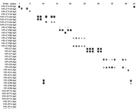
deter-FIG. 1. Representative RT-PCR–RLB results for 43 clinical isolates (see Table 1 for description and targets of probes listed on the left). The
first membrane (shown here) was bound with 40 probes (4 probes for each of 10 common serotypes), while the second (not shown) was bound with
40 probes for the other 10 common serotypes. Conventional serotype–RT-PCR–RLB results for the isolates shown, from left to right, were CVA9
(positive control), CVA9, E6*, CVA9, CVA9, E6*, E3*, CVA16, CVA16, E30, CVA16, CVB1*, CVA16, CVA16, CVB1*, CVB2, CVB2, CVB2,
CVB2, CVB2, CVB3, CVB3, CVB3, CVB3, CVB3, E7, E7, E7, CVB4*, E7, E7, E9, E9, E9, E9, E9, E17*, E3*, E9, E9, E11, E30, and CVA9
(positive control). Isolates marked with asterisks belong to one of the 10 common serotypes and were identified by the second membrane (data
not shown).
on May 16, 2020 by guest
http://jcm.asm.org/
[image:4.585.63.522.61.429.2]mining the species of each isolate, the complete VP1 gene was amplified, se-quenced, and analyzed using the above methods.
Nucleotide sequence accession numbers.Thirty-three new complete VP1 se-quences for 37 isolates have been deposited in GenBank (Table 1).
RESULTS AND DISCUSSION
Analysis of HEV database.
A total of 6,383 HEV isolates
were obtained from 1979 to 2007 from a variety of clinical
samples, including cerebrospinal fluid, stools, vesicular skin
lesions, throat swabs, and nasopharyngeal aspirates. These
in-cluded isolates of PV (1,360 isolates, all vaccine strains), CVA
(508 isolates), CVB (1,357 isolates), E (2,837 isolates), EV68
(2 isolates), EV69 (1 isolates), EV71 (91 isolates), and
untype-able HEVs (227 isolates). Of these isolates, the commonest 10
serotypes were E11, PV1, CVB3, PV2, E9, CVA9, E30, CVB5,
PV3, and E7, which accounted for 65.1% of isolates. The next
10 common serotypes were CVB4, CVB2, E6, CVA16, E14,
E17, E18, CVB1, E3, and EV71 (Table 2). The commonest 20
serotypes accounted for 87.1% (5,558/6,383 isolates) of all
iso-lates over approximately 30 years.
Analysis of complete VP1 sequences.
The original
classifica-tion of HEVs (PV, CVA, CVB, and E) was based on biological
activity and disease, even though HEVs are sometimes difficult
to culture in vitro and serological typing using reference
anti-sera is time-consuming and labor-intensive. The current
mo-lecular classification scheme divides HEVs into four species
(HEV-A to -D): in this newer system, members of an EV
species share
⬎
70% amino acid (aa) identity in P1,
share
⬎
70% aa identity in the nonstructural proteins 2C and
3CD, share a limited range of host cell receptors, share a
limited natural host range, have a genome base composition
(C
⫹
G) which varies by no more than 2.5%, and share a
sig-nificant degree of compatibility in proteolytic processing,
rep-lication, encapsidation, and genetic recombination (6).
Re-combination has been observed to occur in nature only among
members of the same species, except in the 5
⬘
-nontranslated
region (3, 12–14, 16, 19, 22).
Molecular methods with analysis of the VP1 gene can
func-tion as an excellent surrogate for serotyping by neutralizafunc-tion.
The principles include the following: (i) a partial or complete
VP1 nucleotide sequence identity of
ⱖ
75% (
⬎
85% aa
se-quence identity) between a clinical EV isolate and a prototype
serotype strain may be used to confirm the serotype of the
clinical isolate, with the proviso that the second highest score
is
⬍
70%; (ii) a best-match nucleotide sequence identity
of
⬍
70% may indicate that the isolate represents an unknown
(that is, new) serotype; and (iii) a sequence identity of 70 to
75% indicates that further characterization is required before
the isolate can be identified firmly (18). Using these criteria,
strains of homologous serotypes can easily be distinguished
from heterologous serotypes and new serotypes can be
identi-fied. Recently, 17 isolates were classified by this approach as
members of 13 new HEV types, namely, EVs 79 to 88, 97, 100,
and 101 (15). Following sequencing of VP1, we applied these
guidelines to identify 37 isolates before then designing
sero-type-specific probes; these showed that the
genotypes/sero-types of these isolates obtained by sequencing corresponded
with serotypes determined by neutralization.
VP1 primer and probe design.
The VP1 sequences of all 86
known prototype strains, including EV94, were aligned to
al-low design of one sense primer (VP1-All-Sn). Three antisense
primers (VP1-A-Ab, VP1-B-Ab, and VP1-C-Ab) were also
designed to differentiate the three different HEV species
(HEV-A, -B, and -C) (Table 1). To design each of the
species-specific VP1-A-Ab, VP1-B-Ab, and VP1-C-Ab primers, VP1
sequences of 16 HEV-A, 54 HEV-B, and 13 HEV-C prototype
strains were aligned (Table 1). The primer lengths were 19 to
27 bases, and melting temperatures (
T
m) were 59 to 62°C.
To construct probes for different common serotypes, the
complete VP1 genes of 37 isolates were sequenced and
ana-lyzed (see above). After alignment of at least 600 bases of
GenBank VP1 sequence for each of these 20 serotypes,
to-gether with the 37 complete VP1 genes sequenced in this
project, four serotype-specific probes for each serotype were
designed (Table 1). The main principles for probe design were
as follows: (i) a conservative region of one specific serotype was
selected after verifying that the probes would cover as many
strains within that serotype as possible; (ii) within the
conser-vative region, the four probes should be homologous with the
sequenced isolates, the prototype strain, and other convergent
strains; (iii) the lengths of probes should be 18 to 27 bases, and
the
T
mshould be 58 to 67°C; and (iv) the specificity of all
probes was confirmed by BLASTn search in Biomanager. The
numbers of VP1 sequences aligned for probe design for each
serotype are shown in Table 1. For the four probes per
sero-type, the proportions of covered strains among all strains
within the serotype ranged from 70.4 to 100% (Table 1). These
data demonstrate that four probes per serotype are capable of
capturing the majority of serotype strains.
Validation of primers and serotype-specific probes by
[image:5.585.44.284.89.317.2]RT-PCR–RLB.
PCR was performed using primers VP1-All-Sn,
TABLE 2. RT-PCR–RLB and serotype identification results for
132 clinical isolates
Serotype(s)
No. of isolates at CIDM (% of total no. of
isolates)
No. of isolates tested by RLB
RT-PCR–RLB result
CVA16
177 (2.8)
7
CVA16
EV71
91 (1.4)
6
EV71
CVA9
323 (5.1)
6
CVA9
CVB1
97 (1.5)
5
CVB1
CVB2
200 (3.1)
6
CVB2
CVB3
521 (8.2)
5
CVB3
CVB4
226 (3.5)
5
CVB4
CVB5
307 (4.8)
5
CVB5
E3
92 (1.4)
5
E3
E6
177 (2.8)
4
E6
E7
247 (3.9)
7
E7
E9
418 (6.6)
9
E9
E11
751 (11.8)
7
E11
E14
129 (2.0)
7
E14
E17
109 (1.7)
5
E17
E18
104 (1.6)
11
E18
E30
312 (4.9)
20
E30
PV1
546 (8.6)
3
PV1
PV2
463 (7.3)
4
PV2
PV3
268 (4.2)
4
PV3
E25
a1
Negative
aThis isolate, previously identified as E14 by serotyping but negative by RLB,
was confirmed as E25 by a modified species-specific RT-PCR amplification method (17) showing it as HEV-B and by VP1 sequencing (GenBank accession no. FJ868313). Repeat conventional serotyping confirmed the E25 serotype, consistent with the genotype.
on May 16, 2020 by guest
http://jcm.asm.org/
VP1-A-Ab, VP1-B-Ab, and VP1-C-Ab to amplify the VP1
gene. For the first membrane, bound with 40 probes for 10
common serotypes, one sequenced CVA9 isolate (GenBank
accession no. FJ868282) was used as the positive control to
determine the location of the first probe (VP1-CVA9-Sp1) and
the locations of the first and last samples of the RLB
read-out on the film (Fig. 1). Similarly, one sequenced E6 isolate
(GenBank accession no. FJ868294) was chosen as the positive
control for the second membrane, bound with 40 probes of the
other 10 common serotypes (not shown).
After hybridization with serotype-specific probes for each of
the commonest 20 serotypes, the RLB assay results were
con-cordant with the serological identification for 131 isolates,
which meant that at least one probe among the four
serotype-specific probes detected each isolate (Table 2; Fig. 1). One
isolate, previously identified as E14 by serotyping but negative
by RLB, was confirmed as E25 by a modified species-specific
RT-PCR amplification method (17) showing it as HEV-B and
by VP1 sequencing (GenBank accession no. FJ868313).
Re-peat conventional serotyping showed that the isolate was E25,
confirming the genotype.
Since the RLB assay cannot distinguish between sequences
with a one-base difference, the probes for some serotypes that
contain sequences with only one base difference from each
other showed at least one positive result with PCR products
(Fig. 1). However, given the specificity of the probes, any one
among four probes per serotype can confirm the specific
sero-type/genotype of each isolate.
Most recently, Susi et al. (27) developed a straightforward
assay for the rapid typing of HEVs by use of oligonucleotide
arrays in microtiter wells. In their studies, 10 probes for 10
different HEV reference strains were designed and employed.
Furthermore, they used serotype consensus oligonucleotide
probes for CVA9 to detect 25 CVA9 isolates. Both this and our
studies show that such methods could potentially be extended
to cover all known HEV serotypes. Theoretically, this strategy
can extend to other RNA virus detection and identification
procedures.
Conclusion.
In this study, we successfully used RT-PCR–
RLB to identify 131/132 HEV isolates belonging to the
com-monest 20 serotypes detected by neutralization at our
institu-tion; the exception was an E25 isolate, a relatively uncommon
serotype. Genotyping by RT-PCR–RLB complements
tradi-tional serotyping methods and VP1 sequencing and has the
advantages of convenience, speed, and accuracy. This will
al-low examination of specific HEV serotypes or genotypes
asso-ciated with outbreaks and significant disease. The ease and low
cost of this RT-PCR–RLB strategy will improve the diagnosis
and epidemiological investigation of enteroviral infections and
outbreaks where other typing methods are unavailable.
ACKNOWLEDGMENTS
Fei Zhou is supported by an Australian National Health and
Med-ical Research Council (NHMRC) postgraduate scholarship, a CIDM
Public Health postgraduate scholarship, and a Westmead Millennium
Foundation postgraduate scholarship.
REFERENCES
1.Archimbaud, C., M. Chambon, J. L. Bailly, I. Petit, C. Henquell, A. Mirand, B. Aublet-Cuvelier, S. Ughetto, J. Beytout, P. Clavelou, A. Labbe, P. Phil-ippe, J. Schmidt, C. Regagnon, O. Traore, and H. Peigue-Lafeuille.2009.
Impact of rapid enterovirus molecular diagnosis on the management of
infants, children, and adults with aseptic meningitis. J. Med. Virol.81:42–48.
2.Baicus, A., M. Combiescu, A. Persu, G. Oprisan, A. Aubert-Combiescu, and F. Delpeyroux.2006. The molecular characterization of poliovirus strains by the RT-PCR-RFLP assay and its use in the active surveillance for acute flaccid paralysis cases in Romania between 2001–2006. Roum. Arch.
Micro-biol. Immunol.65:120–130.
3.Brown, B., M. S. Oberste, K. Maher, and M. A. Pallansch.2003. Complete genomic sequencing shows that polioviruses and members of human entero-virus species C are closely related in the noncapsid coding region. J. Virol.
77:8973–8984.
4.Chen, T. C., G. W. Chen, C. A. Hsiung, J. Y. Yang, S. R. Shih, Y. K. Lai, and J. L. Juang.2006. Combining multiplex reverse transcription-PCR and a diagnostic microarray to detect and differentiate enterovirus 71 and
coxsack-ievirus A16. J. Clin. Microbiol.44:2212–2219.
5.Estivariz, C. F., M. A. Watkins, D. Handoko, R. Rusipah, J. Deshpande, B. J. Rana, E. Irawan, D. Widhiastuti, M. A. Pallansch, A. Thapa, and S. Imari.
2008. A large vaccine-derived poliovirus outbreak on Madura
Island—Indo-nesia, 2005. J. Infect. Dis.197:347–354.
6.Fauquet, C. M., M. A. Mayo, and J. Maniloff.2005. Virus taxonomy: classi-fication and nomenclature of viruses. Eighth report of the International Committee on the Taxonomy of Viruses. Elsevier Academic Press, San Diego, CA.
7.Grandien, M., M. Forsgren, and A. Ehrnst.1989. Enteroviruses and
reovi-ruses, p. 513–569.InN. J. Schmidt and R. W. Emmons (ed.), Diagnostic
procedures for viral, rickettsial and chlamydial infections, 6th ed. American Public Health Association, Washington, DC.
8.Huang, C., N. K. Chatterjee, and L. J. Grady. 1999. Diagnosis of viral
infections of the central nervous system. N. Engl. J. Med.340:483–484.
9.Kong, F., M. Brown, A. Sabananthan, X. Zeng, and G. L. Gilbert.2006. Multiplex PCR-based reverse line blot hybridization assay to identify 23
Streptococcus pneumoniaepolysaccharide vaccine serotypes. J. Clin.
Micro-biol.44:1887–1891.
10.Kong, F., and G. L. Gilbert.2006. Multiplex PCR-based reverse line blot hybridization assay (mPCR/RLB)—a practical epidemiological and
diagnos-tic tool. Nat. Protoc.1:2668–2680.
11.Lien, K. Y., W. C. Lee, H. Y. Lei, and G. B. Lee.2007. Integrated reverse transcription polymerase chain reaction systems for virus detection. Biosens.
Bioelectron.22:1739–1748.
12.Lindberg, A. M., P. Andersson, C. Savolainen, M. N. Mulders, and T. Hovi.
2003. Evolution of the genome of human enterovirus B: incongruence be-tween phylogenies of the VP1 and 3CD regions indicates frequent
recom-bination within the species. J. Gen. Virol.84:1223–1235.
13.Lukashev, A. N., V. A. Lashkevich, O. E. Ivanova, G. A. Koroleva, A. E. Hinkkanen, and J. Ilonen.2003. Recombination in circulating enteroviruses.
J. Virol.77:10423–10431.
14.Lukashev, A. N., V. A. Lashkevich, O. E. Ivanova, G. A. Koroleva, A. E. Hinkkanen, and J. Ilonen.2005. Recombination in circulating human en-terovirus B: independent evolution of structural and non-structural genome
regions. J. Gen. Virol.86:3281–3290.
15.Oberste, M. S., K. Maher, W. A. Nix, S. M. Michele, M. Uddin, D. Schnurr, S. al Busaidy, C. Akoua-Koffi, and M. A. Pallansch.2007. Molecular iden-tification of 13 new enterovirus types, EV79-88, EV97, and EV100-101,
members of the species human enterovirus B. Virus Res.128:34–42.
16.Oberste, M. S., K. Maher, and M. A. Pallansch.2004. Evidence for frequent recombination within species human enterovirus B based on complete
genomic sequences of all thirty-seven serotypes. J. Virol.78:855–867.
17.Oberste, M. S., K. Maher, A. J. Williams, N. Dybdahl-Sissoko, B. A. Brown, M. S. Gookin, S. Penaranda, N. Mishrik, M. Uddin, and M. A. Pallansch.
2006. Species-specific RT-PCR amplification of human enteroviruses: a tool for rapid species identification of uncharacterized enteroviruses. J. Gen.
Virol.87:119–128.
18.Oberste, M. S., S. M. Michele, K. Maher, D. Schnurr, D. Cisterna, N. Junttila, M. Uddin, J. J. Chomel, C. S. Lau, W. Ridha, S. al Busaidy, H. Norder, L. O. Magnius, and M. A. Pallansch.2004. Molecular identification and characterization of two proposed new enterovirus serotypes, EV74 and
EV75. J. Gen. Virol.85:3205–3212.
19.Oberste, M. S., S. Penaranda, K. Maher, and M. A. Pallansch.2004. Com-plete genome sequences of all members of the species human enterovirus A.
J. Gen. Virol.85:1597–1607.
20.Ooi, M. H., S. C. Wong, A. Mohan, Y. Podin, D. Perera, D. Clear, S. del Sel, C. H. Chieng, P. H. Tio, M. J. Cardosa, and T. Solomon.2009. Identification and validation of clinical predictors for the risk of neurological involvement in children with hand, foot, and mouth disease in Sarawak. BMC Infect. Dis.
9:3–14.
21.Ooi, M. H., S. C. Wong, Y. Podin, W. Akin, S. del Sel, A. Mohan, C. H. Chieng, D. Perera, D. Clear, D. Wong, E. Blake, J. Cardosa, and T. Solomon.
2007. Human enterovirus 71 disease in Sarawak, Malaysia: a prospective clinical, virological, and molecular epidemiological study. Clin. Infect. Dis.
44:646–656.
22.Oprisan, G., M. Combiescu, S. Guillot, V. Caro, A. Combiescu, F.
on May 16, 2020 by guest
http://jcm.asm.org/
roux, and R. Crainic.2002. Natural genetic recombination between
co-circulating heterotypic enteroviruses. J. Gen. Virol.83:2193–2200.
23.Pallansch, M., and R. Roos.2007. Enteroviruses: polioviruses,
coxsackievi-ruses, echovicoxsackievi-ruses, and newer enterovicoxsackievi-ruses, p. 839–894.InD. M. Knipe,
P. M. Howley, D. E. Griffin, R. A. Lamb, M. A. Martin, B. Roizman, and S. E. Straus (ed.), Fields virology, 5th ed. Lippincott Williams & Wilkins, Philadelphia, PA.
24.Rabenau, H. F., A. M. Clarici, G. Muhlbauer, A. Berger, A. Vince, S. Muller, E. Daghofer, B. I. Santner, E. Marth, and H. H. Kessler.2002. Rapid detection of enterovirus infection by automated RNA extraction and
real-time fluorescence PCR. J. Clin. Virol.25:155–164.
25.Rotbart, H. A., P. J. Brennan, K. H. Fife, J. R. Romero, J. A. Griffin, M. A. McKinlay, and F. G. Hayden.1998. Enterovirus meningitis in adults. Clin.
Infect. Dis.27:896–898.
26.Sapkal, G. N., V. P. Bondre, P. V. Fulmali, P. Patil, V. Gopalkrishna, V. Dadhania, V. M. Ayachit, D. Gangale, K. P. Kushwaha, A. K. Rathi, S. D. Chitambar, A. C. Mishra, and M. M. Gore.2009. Enteroviruses in patients
with acute encephalitis, Uttar Pradesh, India. Emerg. Infect. Dis.15:295–298.
27.Susi, P., L. Hattara, M. Waris, T. Luoma-Aho, H. Siitari, T. Hyypia, and P. Saviranta.2009. Typing of enteroviruses by use of microwell oligonucleotide
arrays. J. Clin. Microbiol.47:1863–1870.
28.Tan, E. L., V. T. Chow, S. H. Quak, W. C. Yeo, and C. L. Poh.2008. Development of multiplex real-time hybridization probe reverse transcrip-tase polymerase chain reaction for specific detection and differentiation of
enterovirus 71 and coxsackievirus A16. Diagn. Microbiol. Infect. Dis.61:
294–301.
29.Victoria, J. G., A. Kapoor, L. Li, O. Blinkova, B. Slikas, C. Wang, A. Naeem, S. Zaidi, and E. Delwart.2009. Metagenomic analyses of viruses in the stool
of children with acute flaccid paralysis. J. Virol.83:4642–4651.
30.WHO.2006. Resurgence of wild poliovirus type 1 transmission and effect of
importation into polio-free countries, 2002–2005. Wkly. Epidemiol. Rec.
81:63–68.
31.Wilder-Smith, A., K. Leder, and P. A. Tambyah.2008. Importation of
po-liomyelitis by travelers. Emerg. Infect. Dis.14:351–352.
32.Zeng, X., F. Kong, C. Halliday, S. Chen, A. Lau, G. Playford, and T. C. Sorrell. 2007. Reverse line blot hybridization assay for identification of medically important fungi from culture and clinical specimens. J. Clin.
Mi-crobiol.45:2872–2880.
33.Zhou, F., F. Kong, Z. Tong, and G. L. Gilbert.2007. Identification of
less-commonStreptococcus pneumoniaeserotypes by a multiplex PCR-based
re-verse line blot hybridization assay. J. Clin. Microbiol.45:3411–3415.