0095-1137/09/$08.00
⫹
0
doi:10.1128/JCM.00120-09
Copyright © 2009, American Society for Microbiology. All Rights Reserved.
Simultaneous Identification of 14 Genital Microorganisms in Urine by
Use of a Multiplex PCR-Based Reverse Line Blot Assay
䌤
†
Michelle L. Mckechnie,
1,2Richard Hillman,
1Deborah Couldwell,
3Fanrong Kong,
2Eleanor Freedman,
4Hui Wang,
2,5and Gwendolyn L. Gilbert
2*
Sexually Transmitted Infections Research Centre (STIRC), University of Sydney, Marian Villa, Westmead Hospital, New South Wales 2145,
Australia
1; Centre for Infectious Diseases and Microbiology, Institute of Clinical Pathology and Medical Research (ICPMR), Westmead,
New South Wales 2145, Australia
2; Parramatta Sexual Health Clinic, Level 2, Jeffery House, 158 Marsden St., Parramatta,
New South Wales 2145, Australia
3; Sydney Sexual Health Centre, Sydney Hospital and Eye Hospital, Macquarie Street,
Sydney, New South Wales 2000, Australia
4; and Research Laboratory for Infectious Skin Diseases, Department of
Dermatology, Wuhan First Hospital, Wuhan 430022, People’s Republic of China
5Received 21 January 2009/Returned for modification 27 February 2009/Accepted 31 March 2009
The aim of this study was to develop and evaluate a sensitive method for the simultaneous identification of
14 urogenital potential pathogens. A multiplex PCR-based reverse line blot (mPCR/RLB) assay was developed
to detect 14 urogenital pathogens or putative pathogens, namely
Trichomonas vaginalis
,
Streptococcus
pneu-moniae
,
Neisseria gonorrhoeae
,
Chlamydia trachomatis
,
Ureaplasma parvum
,
U. urealyticum
,
Gardnerella vaginalis
,
Haemophilus influenzae
, herpes simplex virus type 1 (HSV1) and HSV2,
N. meningitidis
,
Mycoplasma hominis
,
M.
genitalium
, and adenovirus, using two species-specific primer pairs and probes for each. The method was
validated using a reference strain or a well-characterized clinical isolate of each target organism and was found
to be both sensitive and specific. The limits of detection for the mPCR/RLB assay varied among the 14 target
organisms from 4.2
ⴛ
10
ⴚ1to 7.0
ⴛ
10
ⴚ11ng/
l of genomic DNA. There were no cross-reactions among any
of the probes. This method was used to test 529 first-voided urine specimens from male patients with and
without urethritis attending two Sydney sexual health clinics. One or more target species were detected in 193
(36%) subjects. Of 233 positive results, overall 216 (93%) were concordant between mPCR/RLB and a
com-parator method (culture and/or species-specific PCR), 9 were positive only by mPCR/RLB, and 8 were positive
only by the comparator method. The mPCR/RLB method was an accurate, convenient, and inexpensive method
for the detection of multiple potential pathogens in first-voided urine specimens from men.
Sexually transmitted infections (STIs) are a major global
health problem. Worldwide, an estimated 340 million cases
of curable STIs, including chlamydial infection, gonorrhea,
trichomoniasis, and syphilis, occur annually, and their
inci-dence is increasing in many parts of the world. In developing
countries, their complications rank in the top five disease
categories for which adults seek health care (www.who.int
/mediacentre/factsheets/fs110/en/). Many STIs cause
asymp-tomatic infection; for example, up to 70% of men and women
with gonococcal and/or chlamydial infections are symptom free
(www.who.int/mediacentre/factsheets/fs110/en/), which creates
the potential for unrecognized transmission with significant
implications for both individual and population health.
Urethritis is characterized by discharge and dysuria (37) and
is broadly classified as nongonococcal (NGU) or gonococcal. It
occurs in both men and women but often is unrecognized in
women. Acute NGU is one of the commonest STIs affecting
heterosexual men, yet a specific pathogen, most commonly
Chlamydia trachomatis
, is identified in only 50 to 70% of cases
(7). Pelvic inflammatory disease is an important complication
of STI in women;
C. trachomatis
and
N. gonorrhoeae
commonly
are implicated, but often the cause is unknown. Bacterial
vagi-nosis is the commonest cause of vaginal discharge and is
asso-ciated both with recognized STIs and other genital syndromes
(3, 18). Additional epidemiological studies are needed to
de-termine the significance of organisms other than recognized
genital pathogens in urethral and vaginal syndromes (7, 13–
15). In particular, the pathogenic roles, if any, of the two
recently defined human
Ureaplasma
species (10),
U.
urealyti-cum
(previously
U. urealyticum
biovar 2) and
U. parvum
(pre-viously
U. urealyticum
biovar 1), and several other genital (32,
43, 44) and respiratory pathogens (20, 30, 33, 42) in NGU are
unclear.
The high level of sensitivity of nucleic acid amplification
tests, such as PCR, allows the use of less invasive specimen
types, including first-voided urine specimens or self-collected
vaginal swabs that are unsuitable for less sensitive methods,
such as culture and antigen tests (8). This paper describes the
development and evaluation of a multiplex PCR-based reverse
line blot (mPCR/RLB) assay (19) that can detect any of 14
recognized and potential genital pathogens in urine specimens
for use in clinical and epidemiological studies of genital
infec-tions.
MATERIALS AND METHODS
Reference strains.Previously well-characterized clinical isolates ofC. tracho-matis,N. gonorrhoeae,M. genitalium, herpes simplex virus type 1 (HSV1) and HSV2, adenovirus,T. vaginalis,M. hominis,G. vaginalis, andN. meningitidis,
* Corresponding author. Mailing address: Centre for Infectious
Dis-eases and Microbiology, Institute of Clinical Pathology and Medical
Research, Westmead Hospital, Darcy Road, Westmead, New South
Wales 2145, Australia. Phone: (612) 9845 6255. Fax: (612) 9893 8659.
E-mail: [email protected].
† Supplemental material for this article may be found at http://jcm
.asm.org/.
䌤
Published ahead of print on 8 April 2009.
1871
on May 16, 2020 by guest
http://jcm.asm.org/
provided by the Centre for Infectious Disease and Microbiology (CIDM) diag-nostic laboratory, were used as positive controls. All isolates had been identified according to routine methods (27). In addition, the following organisms were purchased from the American Type Culture Collection (ATCC; Manassas, VA):
U. urealyticum(ATCC 27813 and ATCC 27814),U. parvum(ATCC 27818),
Haemophilus influenzae(ATCC 10211), andStreptococcus pneumoniae(ATCC 27336).
Clinical specimens.Five hundred twenty-nine male patients with and without urethral symptoms were enrolled in a study of NGU at Parramatta Sexual Health Clinic (PSHC) and Sydney Sexual Health Centre (SSHC) from November 2006 to September 2007. Men with characteristic gonococcal urethritis, in whom Gram stains of urethral discharge showed gram-negative diplococci, and men who had been treated with antibiotics in the previous 6 weeks were excluded from the study. First-voided urine specimens were collected. Specimens from the SSHC were split, and one portion was sent to the routine diagnostic laboratory serving the clinic forC. trachomatisPCR (Roche COBAS Amplicor). Specimens were stored at 4°C at the clinic and transported in weekly batches, in a cool box, to the Centre for Infectious Diseases and Microbiology (CIDM), where they were stored at 4°C until DNA extraction was performed within 24 h of receipt. Specimens from PSHC were stored under the same conditions until being tested forC. trachomatisusing a Roche COBAS Amplicor at the CIDM diagnostic laboratory.
In addition, to assist in the validation of mPCR/RLB results, urethral swabs were collected from all subjects. A Gram-stained smear was examined at the clinic. The swabs were placed in Stuart’s transport medium and transported to the CIDM diagnostic laboratory, where cultures forN. gonorrhoeae, aerobic/ facultative bacteria (includingS. pneumoniae,H. influenzae,N. meningitidis, and
G. vaginalis),M. hominis, andUreaplasmaspp. were performed. Swabs were plated on New York City medium, 5% horse blood, chocolate (both in Columbia agar base; Oxoid, Basingstoke, United Kingdom), and A8 mycoplasma (Oxoid, Basingstoke, United Kingdom) agars, incubated for 24 to 48 h in CO2, and
identified by microscopic and colony morphology and biochemical and antigen tests (27).
DNA extraction.The Roche COBAS Amplicor extraction kit (Roche Diag-nostics Australia Pty Limited Systems, Australia) was used per the manufactur-er’s instructions. Briefly, the urine specimens were vortexed thoroughly for 10 s before 500l of each specimen was transferred to a tube containing 500l of wash buffer. The specimens then were incubated at 37°C for 15 min and centri-fuged at 13,000⫻gfor 5 min. The supernatant was discarded, 250l of lysis buffer was added, and after incubation at room temperature for 15 min, 250l of specimen diluent was added to the lysate. The tubes then were vortexed and centrifuged for 10 min at 13,000⫻gand stored at⫺70°C.
Primer and probe design.Two sets of species-specific primers and probes, targeting highly conserved regions, were designed for each organism. The prim-ers and probes used for this assay are shown in Table 1 and in the supplemental material. Primers and probes were designed to have similar physical character-istics to allow simultaneous amplification and hybridization in a multiplex reac-tion without the loss of sensitivity as follows: melting temperature (Tm), 58 to 65°C; length, 18 to 30 bp; moderate, weak, or no secondary structure; no dimer formation; and amplicon sizes, 80 to 400 bp (19). Some primers were selected from published papers and modified to match the desired characteristics. All probes and primers were checked for specificity against all sequences in GenBank using SeqSearch in the Australian National Genomic Information Ser-vices (ANGIS) programs (http://www.angis.org.au). The adenovirus primers used were designed to allow annealing to all 51 known adenovirus types by introducing degenerative base positions (1). Oligonucleotide primers were biotinylated at the 5⬘end, and probes had a 5⬘amine group and were synthesized by Sigma Aldrich (Sydney, Australia).
mPCR amplification.mPCR amplification was performed using a 25-l reac-tion mixture containing 10l template DNA, 0.075l of each forward (100 pmol
l⫺1) and reverse (100 pmoll⫺1) primer, 1.25l deoxynucleoside
triphos-phates (0.125 mM of each deoxynucleoside triphosphate), 2.5l 10⫻buffer (Qiagen), 3.0l 25 mM MgCl2(final concentration, 3.0 mM), 0.2l Qiagen
HotStarTaqpolymerase (5 Ul⫺1
), and water to 25l. The thermal profile involved initial denaturation for 15 min at 95°C, 40 cycles of 30 s at 94°C, 55°C for 30 s, and 72°C for 90 s, and a final extension for 10 min at 72°C, followed by a hold at 22°C. Inhibition controls were not included in the assay.
RLB assay.The RLB assay was performed as previously described (19). Briefly, probes were labeled and fixed to the membrane in various concentrations (0.6 to 10.8 pmol/l) to determine the optimal conditions. Each PCR product was denatured and immediately chilled on ice. Hybridization was performed at 60°C for 60 min. The washed membrane was incubated in peroxidase-labeled streptavidin conjugate (Roche, Germany) at 42°C for 60 min. The membrane
then was incubated in chemiluminescence blotting substrate (ECL direct system; Roche) for 2 min and covered with Hyperfilm X-ray film (Amersham). The film was exposed for 5 min.
sPCR.Single PCRs (sPCRs), using different targets from those used in the mPCR/RLB assay, were used as comparator methods forT. vaginalis, HSV1 and HSV2,M. genitalium, and adenovirus to confirm the specificity of the mPCR targets. The oligonucleotide primers used for sPCR are shown in Table 2. sPCRs for the other nine pathogens, using the same primers as those used in the mPCR, were performed when culture and mPCR/RLB results were discrepant. The same primers were used to confirm results, as sPCR generally is more sensitive than mPCR and the possibility of cross-reactions, which can occur in mPCR, is avoided.
sPCRs were performed using a 25-l reaction mixture containing 10l tem-plate DNA, 0.25l of each forward (100 pmoll⫺1
) and reverse (100 pmoll⫺1
) primer, 1.25l deoxynucleoside triphosphates (0.125 mM of each), 2.5l 10⫻
buffer (Qiagen), 3.0l 25 mM MgCl2 (final concentration, 3.0 mM), 0.2l
Qiagen HotStarTaqpolymerase (5 Ul⫺1), and water to 25l. The thermal
profile involved initial denaturation for 15 min at 95°C, 40 cycles of 30s at 94°C, 55°C for 30s, and 72°C for 90 s, and a final extension for 10 min at 72°C, followed by a hold at 22°C.
Plasmid construction.Reference strains of all species were amplified using primers (Table 1; also see the supplemental material) targeting species-specific genes. Amplified products were inserted into a pGEM-T Easy vector system (Promega) and transformed into competentEscherichia colicells JM109 accord-ing to the manufacturer’s instructions. The Luria-Bertani–ampicillin–5-bromo-4-chloro-3-indolyl-aˆ-D-galactopyranoside–isopropyl--D-thiogalactopyranoside plates were screened for positive clones and subcultured. The clones then were extracted. DNA concentrations were determined using UV spectrometry.
Analytical sensitivity.The analytical sensitivity of the mPCR/RLB assay was estimated using a series of 10-fold dilutions of plasmid templates of all species to determine the lowest limit of detection, which was expressed as nanograms per microliter of DNA in the last sample positive in the dilution series for each organism.
The results of mPCR/RLB assays were compared to those of the Roche COBAS Amplicor PCR forC. trachomatisin the same specimens (performed by routine diagnostic laboratories serving both clinics) and with those of urethral swab cultures forN. gonorrhoeae,Ureaplasmaspp.,M. hominis, and aerobic/ facultative bacteria, includingS. pneumoniae,H. influenzae, andG. vaginalis.
Final results, after repeating any tests that were discrepant, were accepted as true positives if (i) both RLB probes were positive or one was positive and the result was confirmed by sPCR; (ii) the culture was positive; and/or (iii) sPCR using an alternative target was positive.
Data analysis.Data analysis was carried out using SPSS software (version 15.0; SPSS Inc., Chicago, IL). Univariate analysis was performed using Fisher’s exact test and the chi-squared test for categorical variables. A 95% confidence interval was used.
RESULTS
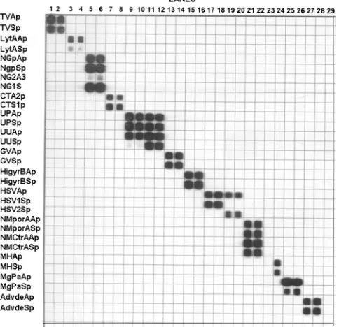
Testing and validation with clinical isolates and reference
strains of target organisms showed that each target species was
amplified, hybridized, and correctly identified by the mPCR/
RLB assay (Fig. 1). Initially nonspecific binding during
hybrid-ization was observed, which was eliminated by adjusting
reac-tion condireac-tions and components. Final probe concentrareac-tions
used to label the RLB membrane were 10.8 pmol/
l for HSV2,
5.4 pmol/
l for
S. pneumoniae
and adenovirus, 0.6 pmol/
l for
N. gonorrhoeae
, and 1.8 pmol/
l for all other species. In all
cases both probes gave positive results in the RLB if target
DNA was present in the sample.
Analytical sensitivity of mPCR/RLB and comparison to
al-ternative methods.
The limit of detection by mPCR/RLB
ranged from 4.2
⫻
10
⫺1to 7.0
⫻
10
⫺11ng/
l for different
species. Results for mPCR/RLB and the comparator method
for each target species (except HSV2, which was not detected
in any specimens) are shown in Table 3. Of a total of 233
positive results, 211 (90%) were concordant in mPCR/RLB
and comparator methods on initial testing; 14 were positive in
on May 16, 2020 by guest
http://jcm.asm.org/
mPCR/RLB only (of which 10 were resolved by repeating the
comparator method); and 7 were positive in the comparator
method only.
Of the 55 specimens in which
C. trachomatis
was detected
using the mPCR/RLB method, 5 (10%) initially were negative
using the Roche COBAS Amplicor PCR. However, on
retest-ing,
C. trachomatis
was detected in all five specimens by the
Amplicor PCR. All specimens that initially were positive with
the Amplicor PCR also were positive with mPCR/RLB.
Al-though subjects with clinical or microscopic evidence of
gonor-rhea on presentation were excluded, seven had positive tests
for gonorrhea in the mPCR/RLB test and were culture
posi-tive. Only two of these specimens were positive for
N.
gonor-rhoeae
using the Roche COBAS Amplicor PCR when tested
initially, but all were positive on retesting.
Ureaplasma
spp. were identified in 86 urethral swab
speci-mens by culture but in only 84 urine specispeci-mens by mPCR/RLB
(
U. parvum
, 31;
U. urealyticum
, 53). All mPCR/RLB results
were confirmed by species-specific sPCR.
[image:3.585.45.542.80.510.2]Clinical specimens.
The age of the 529 subjects ranged from
19 to 76 years (mean, 37 years; median, 35 years). One or more
pathogens or putative pathogens were identified in 193 (36%)
men, including 136 of 277 (49%) men with urethritis symptoms
and 57 of 252 (23%) asymptomatic men. Two or more target
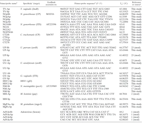
TABLE 1. Oligonucleotide primers and probes developed or modified for the mPCR/RLB assay used in this study
aPrimer/probe nameb Specificityc(target) GenBank
accession no. Primer-probe sequence
d(5⬘–3⬘) T
me(°C)
Reference or sourceg
TV-Ap
T. vaginalis
(
btuB
)
904TGT TGT GAG CTT GAG TGT ACG G883
65.1084
This study
TV-Sp
916CGA TCT TAA CCA CCT TGT TTC C945
63.31923
14 (mod)
NGpSb
N. gonorrhoeae
(CP)
M10316
3249TGC TGT TTC AAG TCG TCC AG3268
64.06359
This study
NGpAp
3317GAT AGT CAT AGC AGG GCT GTT C3296
61.5549
This study
NGpSp
3452CCG TAA CGT CTC TAA GTC TGC TT3474
62.51156
This study
NGpAb
3503CGA AGC CGC CAG CAT AGA GC3484
71.22094
This study
NG16Sb
N. gonorrhoeae
(ITS)
AF223396
404CCA AAA CTT AAC AAA TGA AAG CAA G428
63.41
This study
NG1S
453TGA TTT GCG AAG TAG AAT AAC G474
60.64
This study
NG2A2
456ATC AAA ATA AGC TGC TAA AAA CAG433
59.41
This study
NGITSAb
490TGT TAA AGA TCG ATG CGT CGT472
64.33
This study
CT24b
C. trachomatis
(CP)
X06707
840GGG ATT CCT GTA ACA ACA AGT CAG G864
67.33045
This study
CTS1p
865TTG CGC ATA ATT TTA GGC TTG885
63.59178
This study
CTA2p
1021ACA CTT TGT CTC GAT GAA AGA CA999
62.57137
This study
CT27b
1047CCT CTT CCC CAG AAC AAT AAG AAC
AC1022
67.37814
This study
UP-Sb
U. parvum
(
ureB
)
AF085731
637GAT CAC ATT TTC ACT TGT TTG AAG TG662
64.37767
23 (mod)
UP-Ap
702CTT CAT TTC CTT TTT CAT CAA AAA ATA
C675
63.43446
This study
UP-Sp
688AAA AAG GAA ATG AAG ATA AAG AAC
G712
61.40322
23 (mod)
UP-Ab
735AAC GTC GTC CAT AAG CAA CTT TG713
65.8875
23 (mod)
UU-Ap
U. urealyticum
(
ureB
)
705CTT CAT TTC CTT TTT CAT CAA AAA ATA
C678
63.43446
This study
UU-Sp
691AAA AAG GAA ATG AAG ATA AAG AAC
G715
61.40322
23 (mod)
UU-Ab
739AAA CGA CGT CCA TAA GCA ACT TTA716
64.34317
23 (mod)
GV-Ap
G. vaginalis
(ITS)
416TCC TGT CTA CCA AGG CAT CC397
63.97078
This study
GV-Sp
632CGT GTG ATA ACC GTC AGG TG651
64.06355
This study
HSV1-Sp
HSV1 (gD)
545CGT TTG AGA CCG CCG GCA562
73.04224
This study
HSV2-Sp
572CCT TCG AGA CCG CGG GTA589
68.53278
This study
NM-Sb
N. meningiditis
(
porA
)
AY319969
929GCT TCG GTA ATG CAG TTC CA948
64.94242
17 (mod)
NM-Ap
1010CTG GTA TTT TCG CCT TTT TTA C989
17 (mod)
NM-Sp
953TCA GCT ATG CCC ATG GTT970
This study
MH-Ap
M. hominis
(
gap
)
770CTG AAT AAA CAA CTG TTT TAA CAC CTT
CGCT740
68.7016
2 (mod)
MH-Sp
702CAG GTG CTA AAA AGG TGT TTA TTA CTG
CT730
66.55416
This study
MgPAa-Ap
M. genitalium
(
mgpA
)
1463TAT CAT ACC TTC TGA TTG CAA AGT1445
60.39573
This study
MgPa-Sp
1473CGG TAG AGC TTT ATA TGA TAT TAA CTT
AGC1502
61.46476
This study
AdVdeSb
Adenovirus (hexon)
GCC SCA RTG GKC WTA CAT GCA CAT C
f69.2
1 (mod)
AdVdeAp
CCY ACR GCC AGI GTR WAI CGM RCY TTG TA
68.16371
1 (mod)
AdVdeSp
GCC CGY GCM ACI GAI ACS TAC TTC
63.76665
1 (mod)
AdVdeAb
CAG CAC SCC ICG RAT GTC AAA
62.80243
1 (mod)
aIn addition to primers specifically designed or modified for this study, published primers were used without modification for several targets. Details are shown in the supplemental material.
bThe suffix b indicates a biotin-labeled primer, and p indicates an amine-labeled probe. An A indicates antisense, and S indicates sense. cAbbreviations: CP, cryptic plasmid; ITS region, intergenic spacer region; gD, glycoprotein D.
dNumbers represent the base positions at which the primer/probe sequence starts and finishes (starting at point 1 of the corresponding gene sequence in GenBank). eMelting temperatures were provided by the primer synthesizer (Sigma-Aldrich).
fS⫽G⫹C; R⫽A⫹G; K⫽G⫹T; W⫽A⫹T; Y⫽C⫹T; M⫽A⫹C; I⫽inosine. gSome primers were modified (mod) from the published primers.
on May 16, 2020 by guest
http://jcm.asm.org/
organisms were identified in 30 men (16 symptomatic and 14
asymptomatic) (Table 4). Figure 2 shows the numbers and
percentages of specimens in which each organism was detected
by symptom status. A simple comparison of results in
symp-tomatic and asympsymp-tomatic men showed that
C. trachomatis
(48/277 and 7/252, respectively;
P
⬍
0.001),
N. gonorrhoeae
(7/277 and 0/252, respectively;
P
⫽
0.01), and
M. genitalium
(12/277 and 3/252, respectively;
P
⫽
0.03) were detected
sig-nificantly more frequently in men with symptoms. There were
no significant differences in detection rates between
symptom-atic and asymptomsymptom-atic men for any other pathogens. A
de-tailed analysis of clinical and epidemiological data will be
re-ported separately (D. Couldwell, unpublished data).
DISCUSSION
We have developed an mPCR/RLB hybridization assay that
permits the reliable, simultaneous detection of 14 known or
potential urogenital pathogens, many of which are difficult to
identify by other methods. We have used mPCR/RLB
previ-ously to identify multiple pathogens in respiratory specimens
and blood cultures (45, 46). Others (40) have used gel-based
mPCR to identify
Ureaplasma
spp.,
M. genitalium
, and
M.
hominis
in first-voided urine samples, and there is a recent
report of an mPCR to detect 16 pathogens using a microplate
assay (25). mPCR/RLB potentially is applicable to routine
diagnosis, can be modified to add or delete targets, and is
particularly suitable for epidemiological studies to examine the
roles of putative pathogens in genital syndromes. It is more
practicable and less expensive than microarray technology.
Overall, 75 target organisms were detected in 57 of 252
asymptomatic men, and 158 were detected in 136 of 277 men
with symptoms; multiple organisms were identified in
approx-imately equal numbers of men with and without symptoms.
They included two mixed infections with
N
.
gonorrhoeae
and
C.
trachomatis
, which is not uncommon and probably results from
simultaneous transmission (25, 31). Both are well-established
genital pathogens, whether or not they cause symptoms, and
M. genitalium
also has been implicated in NGU (7, 24). In this
study, all three were significantly associated with the presence
of urethral symptoms (
P
⬍
0.05). The rate of the detection of
M. genitalium
was similar to that reported by others (5, 25, 40),
and more widespread testing for it in patients with NGU has
been advocated (6).
The roles of the other organisms or combinations of
organ-isms targeted in this study are uncertain, since many commonly
are found among the normal genital flora. Providing further
evidence for their roles in urethritis was the aim of the clinical
component of this study (of which the results will be reported
separately). There were differences between symptomatic and
asymptomatic men in the rates of the detection of HSV1,
adenovirus, and
U. urealyticum
as in other studies (7, 50), but
the numbers were small and overall differences did not reach
statistical significance.
[image:4.585.43.285.434.668.2]The comparison of mPCR/RLB results to those of
alterna-tive detection methods showed very good correlation. Several
TABLE 2. Oligonucleotide primers used for sPCR
Primer name Specificity (target) GenBank
accession no. Primer sequence
a(5⬘-3⬘) T
mb(°C) Reference
Tv1
T. vaginalis
18S rRNA gene
U17510
874TAA TGG CAG AAT CTT TGG AG894
59.2
25
Tv2
1185GAA CTT TAA CCG AAG GAC TTC1165
58.3
HSVPolA1
HSV DNA polymerase
ATC ATC TAC CGC GAC ACG GACT
68.8
49
HSVPolA2
TCC ACG CCC TTG ATG AGC ATC T
72.0
MG16-45F
M. genitalium
16S rRNA gene
X77334
45TAC ATG CAA GTC GAT CGG AAG TAG C69
68.8
16
MG16-447R
469AAA CTC CAG CCA TTG CCT GCT AG447
69.2
AD1
Adenovirus hexon gene
U20821
CTG ATG TAC TAC AAC AGC ACT GGC AAC ATG GG
76.1
36
AD2
GCG TTG CGG TGG TGG TTA AAT GGG TTT ACG
TTG TCC AT
83.4
GV1
G. vaginalis
LO8167
364TTA CTG GTG TAT CAC TGT AAG G385
55.8
51
16S-23S rRNA gene
GV2
23S
695CCG TCA CAG GCT GAA CAG T677
64.1
aNumbers represent the base positions at which the primer/probe sequence starts and finishes (starting at point 1 of the corresponding gene sequence in GenBank). bMelting temperatures were provided by the primer synthesizer (Sigma-Aldrich).
FIG. 1. mPCR/RLB results using reference strains. Lanes: 1 and 2,
T. vaginalis
; 3 and 4,
S. pneumoniae
; 5 and 6,
N. gonorrhoeae
; 7 and 8,
C. trachomatis
; 9 and 10,
U. parvum
; 11 and 12,
U. urealyticum
; 13 and
14,
G. vaginalis
; 15 and 16,
H. influenzae
; 17 and 18, HSV1; 19 and 20,
HSV2; 21 and 22,
N. meningitidis
; 23, blank; 24,
M. hominis
; 25 and 26,
M. genitalium
; and 27 and 28, adenovirus type 1.
on May 16, 2020 by guest
http://jcm.asm.org/
organisms were detected in very few (
⬍
5) specimens (
T.
vagi-nalis
,
S. pneumoniae
,
G. vaginalis
,
N. meningitidis
, and
adeno-virus), but results agreed in all but one (one culture negative
and mPCR/RLB positive for
N. meningitidis
). mPCR/RLB
identified
N. gonorrhoeae
, HSV1, and
M. genitalium
in all
spec-imens that were positive by comparator methods and HSV1 in
three additional specimens. It did not detect
M. hominis
in one
and
Ureaplasma
spp. in two urine specimens from men whose
urethral swabs were culture positive. These specimens were
from men attending SSHC, where urine specimens were stored
at 4°C for several days before being processed, which may have
reduced the sensitivity of mPCR/RLB compared to that of the
culture of urethral swabs, which were stored at room
temper-ature in Stuart’s transport medium. The refrigeration of
spec-imens for several days had no apparent effect on the detection
of other pathogens. Stellrecht et al. (40), using urine and swabs
for PCR, recorded sensitivities similar to those of culture for
Ureaplasma
spp.,
M. genitalium
, and
M. hominis
, and similar
sensitivities have been reported by others for other species (2,
12, 23, 40, 50). Nevertheless, these results suggest that
speci-mens should be processed for PCR as soon as possible after
collection and, if they cannot be tested immediately, stored as
DNA extracts.
False-negative mPCR/RLB results also may have resulted
from the prolonged storage of DNA extracts (up to 18 months
at
⫺
20°C) prior to testing, which can affect DNA quality and
PCR efficiency (28), or from PCR inhibitors in urine (9, 34).
Initial false-negative results for Roche COBAS Amplicor
PCR for
N. gonorrhoeae
and
C. trachomatis
(some from both
diagnostic laboratories performing routine testing for the two
clinics) were positive on retesting. These results reflect the
real-life pitfalls of diagnostic testing, even by reputable
labo-ratories using generally reliable assays.
A significant limitation of this study, in common with other
studies of new, potentially more sensitive tests, was that there
was no single gold standard for the analysis of mPCR/RLB. We
chose to culture, where practicable, a different specimen
(ure-thral swab) for target bacteria, for which urine would have
been inappropriate, to identify men with urogenital
coloniza-tion/infection. To optimize the reliability of the mPCR/RLB,
we measured limits of detection, quantitatively, using cloned
targets; confirmed all positive results using culture or sPCRs;
designed primers and probes based on targets used in
well-TABLE 3. Comparison of results of mPCR/RLB and comparator methods in detection of genital infection/colonization with 14 recognized or
putative genital pathogens
Organism Limit of detectiona(ng/l) No. positively detected by
mPCR/RLBb Comparator methodc(target)
No. positively detected by comparator method
T. vaginalis
1.5
⫻
10
⫺91
sPCR (18S rRNA gene)
1
S. pneumoniae
7.4
⫻
10
⫺11
Culture
1
N. gonorrhoeae
4.3
⫻
10
⫺27
Culture and Amplicor PCR
2 (
⫹
5)
dC. trachomatis
7.0
⫻
10
⫺1155
Amplicor PCR
50 (
⫹
5)
dUreaplasma
spp.
e7.8
⫻
10
⫺984
Culture
86
G. vaginalis
7.8
⫻
10
⫺33
sPCR (16-23S rRNA gene)
3
H. influenzae
7.0
⫻
10
⫺831
Culture
35
HSV1
4.3
⫻
10
⫺88
sPCR (
pol
)
5
N. meningitides
8.2
⫻
10
⫺82
Culture
1
M. hominis
5.5
⫻
10
⫺115
Culture
16
M. genitalium
2.1
⫻
10
⫺915
sPCR (16S rRNA gene)
15
Adenovirus
6.3
⫻
10
⫺74
sPCR (hexon gene)
4
a
For mPCR/RLB. b
sPCR, using the same primers as those for mPCR, was performed on specimens with discrepant mPCR/RLB and culture results. In all cases the mPCR/RLB and sPCR results were concordant.
c
Comparator methods were either the culture of urethral swab collected at the same time as first-voided urine specimen or sPCR on the same urine DNA extract as that used for mPCR/RLB, using a different, species-specific target (except for adenoviruses, for which the same hexon gene target was used).
d
Of the 7 and 55 specimens positive by mPCR/RLB forN. gonorrhoeaeandC. trachomatis, respectively, only 2 and 50 were positive initially in the Roche Amplicor PCR; all were positive on retesting.
e
[image:5.585.42.543.90.228.2]Urethral specimens were cultured for ureaplasmas, but isolates were not speciated.Ureaplasmaspp. identified in the mPCR/RLB-positive specimens are shown in Fig. 2.
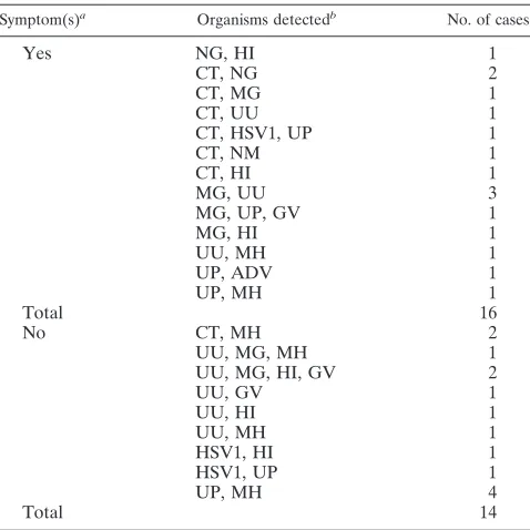
TABLE 4. Mixed genital infections/colonization with target
organisms detected by mPCR/RLB assay from 30 men
with and without urethral symptoms
Symptom(s)a Organisms detectedb No. of cases
Yes
NG, HI
1
CT, NG
2
CT, MG
1
CT, UU
1
CT, HSV1, UP
1
CT, NM
1
CT, HI
1
MG, UU
3
MG, UP, GV
1
MG, HI
1
UU, MH
1
UP, ADV
1
UP, MH
1
Total
16
No
CT, MH
2
UU, MG, MH
1
UU, MG, HI, GV
2
UU, GV
1
UU, HI
1
UU, MH
1
HSV1, HI
1
HSV1, UP
1
UP, MH
4
Total
14
aA symptomatic man was a patient who had urethral symptoms (dysuria, urethral discomfort, or urethral discharge), and an asymptomatic man was a patient who presented with no symptoms.
bAbbreviations: CT,C. trachomatis; NG,N. gonorrhoeae; HI,H. influenzae; UU,U. urealyticum; GV,G. vaginalis; UP,U. parvum; MH,M. hominis; MG,M. genitalium; ADV, adenovirus; and NM,N. meningitidis.
on May 16, 2020 by guest
http://jcm.asm.org/
[image:5.585.44.283.436.675.2]established sPCR methods, which had been shown to be
spe-cific; and avoided contamination by the use of appropriate
negative and no-DNA controls. There were relatively few
dis-crepancies between methods, and we believe that the decision
to regard any confirmed positive result as a true positive for the
purposes of comparison was justified. Nevertheless, we cannot
exclude the possibility that a small number of mPCR/RLB
results were false positives. Even in this relatively large number
of subjects, most target organisms were identified too
infre-quently to calculate accurate sensitivities and predictive values.
This study confirmed the sensitivity and specificity of the
mPCR/RLB assay for the detection of a wide range of
poten-tial urogenital pathogens in first-voided urine specimens.
How-ever, cross-reactivity can occur if primers and probes are not
designed correctly, and the optimization of reaction
compo-nents and conditions is required to produce a stable system
without nonspecific reactions. The advantages of mPCR/RLB
are that it can simultaneously test up to
⬃
40 specimens for up
to
⬃
40 target genes in a single reaction, and it could be used
for a variety of specimens other than urine, including cervical
smears collected for cytology (48) or self-collected vaginal
swabs. Given the high frequency of the multiple species
iden-tified, we believe that such an approach could contribute to an
effective public health response to STIs.
ACKNOWLEDGMENTS
We thank Vitali Sintchenko and Heather Gidding for assistance in
planning the study. Victor Weixiong provided some of the primer/
probe designs. We also thank the virology staff at CIDM for
perform-ing all DNA extractions, Bin Wang from the Westmead Millennium
Institute for assistance with cloning experiments, and clinical research
teams at SSHC and PSHC for the collection of urine samples and
clinical data.
REFERENCES
1.Allard, A., B. Albinsson, and G. Wadell. 2001. Rapid typing of human adenovirus by general PCR combined with restriction endonuclease analysis. J. Clin. Microbiol.39:498–505.
2.Baczynska, A., H. F. Svenstrup, J. Fedder, S. Birkelund, and G.
Chris-tiansen.2004. Development of real-time PCR for detection ofMycoplasma hominis. BMC Microbiol.35:1–13.
3.Bailey, J. V., C. Farquhar, C. Owen, and P. Mangtani.2004. Sexually trans-mitted infections in women who have sex with women. Sex. Transm. Infect.
80:244–246. 4. Reference deleted.
5.Bjornelius, E., P. Lidbrink, and J. J. Jensen.2000.Mycoplasma genitaliumin nongonococcal urethritis—a study in Swedish male STD patients. Int. J. STD AIDS11:292–296.
6.Bowden, F. J., S. N. Tabrizi, S. M. Garland, and C. K. Fairley.2002. Sexually transmitted infections: new diagnostic approaches and treatments. Med. J. Aust.176:551–557.
7.Bradshaw, C. S., S. N. Tabrizi, T. R. H. Read, S. M. Garland, C. A. Hopkins, L. M. Moss, and C. K. Fairley.2006. Etiologies of non-gonococcal urethritis: bacteria, viruses, and the association with orogenital exposure. J. Infect. Dis.
193:336–345.
8.Chen, M. Y.2005. Changes in testing methods for genitalChlamydia tracho-matisin New South Wales, Australia. Sex. Health2:251–253.
9.Chong, S., D. Jang, X. I. Song, J. Mahony, A. Petrich, P. Barriga, and M. Chernesky.2003. Specimen processing and concentration ofChlamydia tra-chomatisadded can influence false negative rates in the LCx assay but not in the APTIMA Combo 2 assay when testing for inhibitors. J. Clin. Microbiol.
41:778–782.
10.Deguchi, T., T. Yoshida, T. Miyazawa, M. Yasuda, H. Tamaki, H. Ishiko, and S. Maeda.2004. Association ofUreaplasma urealyticum(biovar 2) with non-gonoccocal urethritis. Sex Trans. Dis.31:192–195.
11. Reference deleted.
12.Falk, L., and J. S. Jensen.2005. Signs and symptoms of urethritis and cervicitis among women with or withoutMycoplasma genitaliumor Chla-mydia trachomatisinfection. Sex Trans. Infect.81:73–78.
13.Hagman, M., L. Forslin, H. Moi, and D. Danielsson.1991.Neisseria menin-gitidisin specimens from urogenital sites. Sex Trans. Dis.18:228–232. 14.Hardick, J., S. Yang, S. Lin, D. Duncan, and C. Gaydos.2003. Use of the
Roche LightCycler instrument in a real-time PCR forTrichomonas vaginalis
in urine samples from females and males. J. Clin. Microbiol.12:5619–5622. 15.Hartmann, A. A., and P. Elsner.1988. Urethritis caused byNeisseria
men-ingitidisgroup B: a case report. Sex. Trans. Dis.15:150–151.
16.Jensen, J. S., B. E. D. Birthe, and P. Lidbrink.2004. Use of TaqMan 5⬘
nuclease real-time PCR for quantitative detection ofMycoplasma genitalium
DNA in males with and without urethritis who were attendees at a sexually transmitted disease clinic. J. Clin. Microbiol.42:683–692.
17.Jordens, Z. J., and J. E. Heckels.2005. A novel porA-based real time PCR for detection of meningococcal carriage. J. Med. Microbiol.54:463–466. 18.Keane, F. E. A., B. J. Thomas, L. Whitaker, A. Renton, and D.
Taylor-Robinson.1997. An association between non-gonococcal urethritis and bac-terial vaginosis and the implications for patients and their sexual partners. Genitourin. Med.73:373–377.
[image:6.585.134.451.69.253.2]19.Kong, F., and G. L. Gilbert.2006. Multiplex PCR-based reverse line blot hybridisation assay (mPCR/RLB)—a practical epidemiological and diagnos-tic tool. Nat. Protoc.1:2668–2680.
FIG. 2. Results of mPCR/RLB on first-voided urine specimens for 14 target urogenital organisms from men with and without symptoms of
urethritis. Numbers on bars indicate the percentages of subjects with positive results for each target organism. Denominators are 277 for
symptomatic men and 252 for asymptomatic men; the
y
axis shows the number of positive specimens. The arrows indicate organisms detected
significantly more frequently in men with urethral symptoms than in men without symptoms (
P
ⱕ
0.05).
on May 16, 2020 by guest
http://jcm.asm.org/
20.Koroglu, M., Y. Yakupogullari, and F. Aydogan.2007. A case of urethritis due toStreptococcus pneumoniae. Sex. Trans. Dis.34:1040.
21. Reference deleted. 22. Reference deleted.
23.Mallard, K., K. Schopfer, and T. Bodmer.2005. Development of real-time PCR for the differential detection and quantification ofUreaplasma urealyti-cumandUreaplasma parvum. J. Clin. Microbiol.60:13–19.
24.Manhart, L. E., K. K. Holmes, and J. P. Hughes.2007.Mycoplasma geni-taliumamong young adults in the United States: an emerging sexually trans-mitted infection. Am. J. Public Health97:1118–1125.
25.Masue, N., T. Deguchi, S. Yokoi, T. Yamada, K. Ohkusu, and T. Ezaki.2007. System for simultaneous detection of 16 pathogens related to urethritis to diagnose mixed infection. Int. J. Urol.14:39–42.
26. Reference deleted.
27.Murray, P. R., E. J. Baron, J. Jorgensen, M. A. Pfaller, and M. L. Landry (eds.).2007. Manual of clinical microbiology, 9th ed. ASM Press, Wash-ington, DC.
28.Ng, D. P. K., D. Koh, S. G. L. Choo, V. Ng, and Q. Fu.2004. Effect of storage conditions on the extraction of PCR-quality genomic DNA from saliva. Clin. Chim. Acta343:191–194.
29. Reference deleted.
30.Noble, R. C. 1985. Colonisation of the urethra withStreptococcus pneu-moniae: a case report. Genitourin. Med.61:345–346.
31.Oriel, J. D.1975. Infection with chlamydia group A in men with urethritis due toN. gonorrhoeae. J. Infect. Dis.131:376–382.
32.Povlsen, K., E. Bjornelius, P. Lidbrink, and I. Lind.2002. Relationship of
Ureaplasma urealyticumbiovar 2 to non-gonococcal urethritis. Eur. J. Clin. Microbiol. Infect. Dis.21:97–101.
33.Quentin, R., J. M. Mellovet, and P. Y. Sizaret.1989. Typing of urogenital, maternal and neonatal isolates ofHaemophilus influenzaeandHaemophilus parainfluenzaein correlation with clinical source of isolation and evidence for a genital specificity ofH. influenzaebiotype IV. J. Clin. Microbiol.27:2286– 2294.
34.Rauter, C., M. Mueller, I. Diterich, S. Zeller, D. Hassler, T. Meergans, and T. Hartung.2005. Critical evaluation of urine-based PCR assay for diagnosis of Lyme borreliosis. Clin. Diagn. Lab. Immun.12:910–917.
35. Reference deleted.
36.Sarantis, H. J. G., M. Brown, M. Petric, and R. Tellier.2004. Comprehensive detection and serotyping of human adenovirus by PCR and sequencing. J. Clin. Microbiol.42:3963–3969.
37.Scholes, D., A. Stergachis, F. E. Heidrich, H. Andrilla, K. K. Holmes, and W. E. Stamm.1996. Prevention of pelvic inflammatory disease by screening for cervical chlamydial infection. N. Engl. J. Med.334:1362–1366. 38. Reference deleted.
39. Reference deleted.
40.Stellrecht, K. A., A. M. Woron, N. G. Mishrik, and R. A. Venezia.2004. Comparison of multiplex PCR assay with culture for detection of genital mycoplasmas. J. Clin. Microbiol.42:1528–1533.
41. Reference deleted.
42.Sturm, S. A. W.1986.Haemophilus influenzaeandHaemophilus parainflu-enzaein nongonococcal urethritis. J. Infect. Dis.153:165–167.
43.Taylor-Robinson, D.2007. The role of mycoplasmas in pregnancy outcomes. Best Pract. Res. Clin. Obstet. Gynaecol.21:425–438.
44.Taylor-Robinson, D., and W. M. McCormack.1980. The genital mycoplas-mas (the first of two parts). N. Engl. J. Med.302:1003–1010.
45.Wang, H., F. Kong, P. Jelfs, G. James, and G. L. Gilbert.2004. Simultaneous detection and identification of common cell culture contaminant and patho-genic mollicutes strains by reverse line blot hybridization. Appl. Environ. Microbiol.70:1483–1486.
46.Wang, Y., F. Kong, G. L. Gilbert, M. Brown, W. Gao, S. Yu, and Y. Yang.
2008. Use of a multiplex PCR-based reverse line blot (mPCR/RLB) hybri-disation assay for the rapid identification of bacterial pathogens. Clin. Mi-crobiol. Infect.14:155–160.
47. Reference deleted.
48.Xiong, L., F. Kong, H. Zhou, and G. L. Gilbert.2006. Use of PCR and reverse line blot hybridization assay for simultaneous detection and serovar identification ofChlamydia trachomatis. J. Clin. Microbiol.44:1413–1418. 49.Yamamoto, L. J., D. G. Tedder, R. Ashley, and M. J. Levin.1991. Herpes
simplex virus DNA in cerebrospinal fluid of a patient with mollaret’s men-ingitis. N. Engl. J. Med.325:1082–1085.
50.Yoshida, T., T. Deguchi, S.-T. Meda, Y. Kubota, M. Tamaki, S. Yokoi, M. Yasuda, and H. Ishiko.2007. Quantitative detection ofUreaplasma parvum
(biovar 1) andUreaplasma urealyticum(biovar 2) in urine specimens from men with and without urethritis by real-time polymerase chain reaction. Sex. Trans. Dis.34:416–419.
51.Zariffard, M. R., M. Saifuddin, B. E. Sha, and G. T. Spear.2002. Detection of bacterial vaginosis-related organisms by real-time PCR for Lactobacilli,
Gardnerella vaginalisandMycoplasma hominis. FEMS Immun. Med. Micro-biol.34:277–281.