N A N O E X P R E S S
Open Access
An injectable hybrid nanoparticle-in-oil-in-water
submicron emulsion for improved delivery of
poorly soluble drugs
Shuo Wang
1, Hua Wang
1, Wenquan Liang
1*and Yongzhuo Huang
2,3*Abstract
Poor drugability problems are commonly seen in a class of chemical entities with poor solubility in water and oil, and moreover, physicochemical instability of these compounds poses extra challenges in design of dosage forms. Such problems contribute a significant high failure rate in new drug development. A hybrid nanoparicle-in-oil-in-water (N/O/W) submicron emulsion was proposed for improved delivery of poorly soluble and unstable drugs (e.g., dihydroartemisinin (DHA)). DHA is known for its potent antimalarial effect and antitumor activity. However, its insolubility and instability impose big challenges for formulations, and so far, no injectable dosage forms are clinically available yet. Therefore, an injectable DHA N/O/W system was developed. Unlike other widely-explored systems (e.g., liposomes, micelles, and emulsions), in which low drug load and only short-term storage are often found, the hybrid submicron emulsion possesses three-fold higher drug-loading capacity than the conventional O/W emulsion. Of note, it can be manufactured into a freeze-drying form and can render its storage up to 6 months even in room temperature. The in vivo studies demonstrated that the PK profiles were significantly improved, and this injectable system was effective in suppressing tumor growth. The strategy provides a useful solution to effective delivery of such a class of drugs.
Keywords:Poor solubility, Drug delivery, Nano emulsion, Nanoparticle, Dihydroartemisinin
Background
Poor solubility of a chemical entity is a major obstacle affecting its drugability [1]. It is estimated that more than 40% of the new chemical entities generated in drug discovery programs are poorly soluble [2]. With growing interest in the role of modern pharmaceutics in early stage of drug discovery and development, rationale de-sign of drug delivery systems for poorly soluble drugs has been widely explored, such as emulsions [3,4], solid lipid nanoparticles [5,6], micelles, and liposomes [7,8]. Emulsions are one of the most commonly used drug car-riers for poorly water-soluble drugs due to the unique advantage in mass manufacturing, easy sterilization, and excellent physicochemical stability. However, there still remain two problems waiting to be solved, i.e., low drug-loading capacity and quick drug release. Moreover, for
the drugs that remain insoluble in both water and oil, there is no satisfactory solution yet. As a case in point, dihydroartemisinin (DHA), a potent antimalarial drug [9,10], is poorly soluble and unstable, and its oral bioavail-ability is low due to degradation in the gastrointestinal tract and poor intestinal absorption. Moreover, because of its unfavorable solubility property, no injectable dosage form is clinically available yet, thus, severely limiting its use in medical emergency in severe malaria. According to WHO, an estimated 781,000 deaths from malaria in 2009, and most victims are children under five [11]. Further-more, DHA recently has been demonstrated for its extra-ordinary antitumor activity and its promising development into anticancer drug [12-14]. Therefore, to develop an in-jectable drug delivery system for DHA is not only clinically beneficial for these diseases, but also such exploration is meaningful for rationale design of drug delivery for the class of water/oil insoluble drugs.
Herein, we propose a novel hybrid system of the nano-particle-in-oil-in-water (N/O/W) submicron emulsion. Considering the oil compatibility, solid lipid nanoparticles * Correspondence:[email protected];[email protected]
1
Zhejiang University College of Pharmaceutical Sciences, 388 Yuhangtang Road, Hangzhou 310058, China
2
Shanghai Institute of Materia Medica, Chinese Academy of Sciences, 501 Hai-ke Road, Shanghai 201203, China
Full list of author information is available at the end of the article
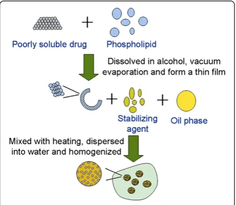
(SLN) were selected. Drugs were incorporated into SLN and dispersed in oil as oily phase, and the oily phase was used to dispersed in aqueous solution with homogenization for prep-aration of the N/O/W submicron emulsion (Figure 1).
Materials and methods Materials
Dihydroartemisinin (purity>99%) was kindly provided by Beijing Zhongyan Tongrentang Medicine R&D Co., Ltd. (Beijing, China). Poloxamer 188 was purchased from BASF Corporation (Ludwigshafen, Germany), cholesterol from Pharmacia Biothech (Piscataway, NJ, USA), and soya phosphatidylcholine from Lipoid GmbH (Ludwighafen, Germany). Mannitol, sucrose, and glyceryl monostearate were purchased from Sinopharm Chemical Reagent Co., Ltd. (Shanghai, China). Hepatic H22 cell line was kindly
provided by Zhejiang University of Traditional Chinese Medicine. All other chemicals were of analytical grade.
Preparation of DHA N/O/W emulsion
DHA (150 mg) and lecithin (900 mg) were dissolved in 30-ml alcohol and then mixed with soybean oil (5,000 mg) and glyceryl monostearate (150 mg). The mixture was used as oily phase and warmed up to 60°C and then dropwise added to the aqueous phase containing glycerol (1,100 mg) and poloxamer 188 (900 mg) with agitation at high speed and temperature (8,000 to 10,000 rpm and 60°C). After adjusting the pH from 6 to 7, the primary emulsion was homogenized by microfluid technology at a pressure of 200 to 1,000 bar (8 to 10 cycles) to obtain the DHA N/O/W emulsion with DHA concentration about 3.0 mg/ml. In order to maintain DHA stability, the freeze-dried emulsion was prepared with a complex of sucrose and mannitol (4:1) as lyoprotectant.
Meanwhile, the DHA O/W emulsion was prepared as follows. DHA (49 mg) and lecithin (1,000 mg) were dis-solved in alcohol and attached to a rotary evaporator to remove the organic solvent. Soybean oil (5,000 mg) was then added to dissolve the dried film as oil phase. Aque-ous phase contained glycerol (1,100 mg); poloxamer, 188 (1,000 mg). The DHA O/W emulsion was prepared as described above.
Characterization of the DHA N/O/W emulsion
Size and zeta potential of the DHA N/O/W emulsion were determined by laser scattering technique using Malvern ZS90 (Malvern Instruments Ltd., Malvern, Worcestershire, UK) after appropriate dilution with double distilled water.
The primary N/O/W emulsion with a size of about 10
μm (before microfluid process), through light
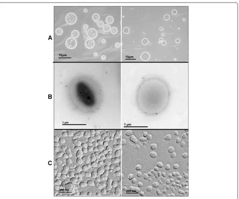
micros-copy (MPS-30, Leica Microsystems, Wetzlar, Hesse, Germany), was employed to observe the encapsulation of particles in O/W emulsion. Furthermore, morphology of the mid-product of DHA N/O/W emulsion (about 1
μm) was studied with transmission electron microscopy
(TEM) (JEM-1200EX, JEOL Ltd., Akishima, Tokyo, Japan). The sample for TEM was dropped onto a 400-mesh copper grid coated with carbon film. The sample was dried in air before TEM observation. The sample for freeze-fracture transmission electron microscopy (FF-TEM) was prepared as follows. A droplet of microemul-sion was sandwiched between two copper electron mi-croscopy grids, snapfrozen by immersion in liquid ethane, and then loaded into a double-replica device and
immersed in liquid nitrogen (−196°C). The double
rep-lica device was mounted on the rotary sample stage of a Balzers BAF-400D freeze etch device (Balzers AG,
Bal-zers, Liechtenstein) cooled to −110°C. Fracturing was
carried out at a vacuum of 3 × 10⁻³mbar. The fractured surfaces were shadowed with platinum (45°) and carbon (90°). The replicas were then washed in chloroform, methanol, and finally, distilled water. Dried replicas were examined with TEM operating at an accelerating voltage of 80 kV.
Drug loading capacity
The DHA N/O/W emulsion was dissolved in methanol and then injected to the high-performance liquid chro-matography (HPLC) system. Chromatographic
separ-ation was achieved using a C18column (150 × 4.6 mm, 5
μm, Agilent Technologies, Palo Alto, CA, USA) with
[image:2.595.57.292.498.702.2]column temperature at 40°C. The mobile phase con-sisted of 40:60 double-distilled water containing 0.1% acetic acid (Millipore Corporation, Milford, MA, USA)/ acetonitrile (HPLC grade, Merck & Co., Inc., White-house Station, NJ, USA). The flow rate was set to 1.0 ml/min, and UV detection wavelength was 210 nm. Figure 1Schematic illustration of the preparation of
Pharmacokinetics study
The experimental protocols were approved by the Animal Ethics Review Committee of Zhejiang University. The male rabbits (2.0 ± 0.2 kg) were divided into three groups (6 per group). DHA solution, DHA O/W emulsion or DHA N/O/W emulsion was injected via ear vein at a dose of 6.2 mg/kg. Blood samples were collected from the mar-ginal vein on another ear before administration and at pre-determined time points after intravenous administration. Plasma sample (100μl) was spiked with 10μl of the IS so-lution (artemisinin, 1 μg/ml), and the mixture was vor-texed for 10 s. The sample was extracted with 2-ml ethyl acetate, vortexed for 3 min, and centrifuged for 15 min at 3,500 rpm. The upper layer was transferred into another test tube and evaporated to dryness under a gentle stream of N2gas at 37°C. The residue was reconstituted in 100μl
[image:3.595.57.290.88.249.2]of methanol/water (60:40). The concentration of DHA in Figure 2The changes of DHA content and size of the
reconstituted N/O/W emulsion at different storage periods.
[image:3.595.59.541.302.702.2]plasma samples was determined by LC-MS method (ESI, positive mode, Agilent 1100 LC-MSD, Agilent Technolo-gies, Palo Alto, CA, USA). Pharmacokinetic analysis was conducted using Kinetica Software (Version 4.4, Thermo Electron Corporation, Waltham, MA, USA).
In vivo antitumor efficacy of DHA N/O/W emulsion Hepatic H22 tumor cells (5 to 6 × 10⁶ cells per mouse)
were inoculated subcutaneously to the Institute for Can-cer Research (ICR) mice (6 to 8 weeks, 22 to 26 g) at the right axilla. Tumors were measured by a vernier caliper, and its volume (V) was calculated as follows:
V¼d2D=2;
wheredand D are the shortest and the longest diameter
of the tumor in mm, repectively.
When the tumor grew to a mean size of approxi-mately 300 mm3, mice were treated intravenously (i.v.) with DHA solution, DHA O/W emulsion, or N/O/W emulsion.
Hemolysis test and vascular irritation test
Blood sample was collected from New Zealand white rabbit (2.5 kg) ear vein. A 10-ml aliquot of defibrinated blood and 10 ml of saline were mixed and centrifuged. The supernatant was discarded, and the wash step was repeated three times until no red color was observed in the supernatant. Erythrocytes were diluted with saline to
2% (v/v). The DHA N/O/W emulsion at varying
concen-trations was mixed with 2% erythrocyte solution in equal volume. The morphology of the erythrocytes was observed under the microscope, and the color change of the solution was monitored with spectrophotometry
(λ= 414 nm) for hemolysis evaluation. Hemolysis effect
was calculated as follows:
Hemolysisð Þ ¼% Asample−Anegative
Apositive−Anegative 100%;
where Asample is the absorbance of the tested sample;
Anegative, the absorbance of the negative control (i.e.,
mixture of equal volumes of 2% erythrocytes and saline); and Apositive, the absorbance of the positive control (i.e.,
mixture of equal volumes of 2% erythrocytes and dis-tilled water).
For vascular irritation test, DHA N/O/W emulsion with a DHA dose of 6.2 mg/kg was injected to the mar-ginal ear vein once per day for 7 days. At the eighth day, the rabbits were killed by aortic bleeding under pento-barbital anesthesia. The vascular irritation was investi-gated by histological examination of the injection site.
Statistical analysis
Each experiment was performed in triplicate, and the values were expressed as mean ± S.D. Statistical analysis was performed using Student's t test. Significant differ-ences are demonstrated by symbols (*, **).
Results and discussion
Preparation and characterization of DHA N/O/W emulsion DHA N/O/W emulsion was prepared by a modified
solvent evaporation and membrane emulsification
[image:4.595.56.291.89.258.2]method. The drug-loading capacity was improved by the DHA N/O/W emulsion, in which DHA concentration Figure 4Plasma concentration-versus-time profiles of DHA
after intravenous administration of 6.2 mg/kg DHA N/O/W emulsion to rabbits, DHA solution and DHA O/W emulsion as controls.
Table 1 Pharmacokinetic parameters of DHA
Parameters DHA N/O/W emulsion DHA O/W emulsion DHA solution
t1/2α(min) 9.40 ± 0.28*,** 3.97 ± 0.34 3.10 ± 0.09
t1/2β(min) 160.91 ± 47.21*,** 73.31 ± 1.61* 20.20 ± 6.56
Cl (mlmin−1kg) 0.0023 ± 0.0002*,** 0.0038 ± 0.0002* 0.0069 ± 0.0013
AUC (minngml−1) 219,759.00 ± 14,782.77*,** 131,564.5 ± 8,396.89* 73,025.35 ± 14,149.00
MRT (min) 97.65 ± 20.27*,** 32.22 ± 4.03* 9.40 ± 2.44
Parameters of DHA after intravenous administration of DHA N/O/W emulsion to rabbits, DHA solution, and DHA O/W emulsion as controls. The results are
[image:4.595.57.540.635.716.2]was 3.0 mg/ml, over three times higher than that of the DHA O/W emulsion (0.98 mg/ml).
The freeze-dried DHA N/O/W emulsion was able to reconstitute in distilled water within 1 min. The average
particle size and zeta potential were 175 nm and −48.1
mV. The storage stabilities of DHA N/O/W emulsion indicated that DHA N/O/W emulsion was stable at 4°C for 1 month. The stability was further improved in freeze-dry form, and the reconstituted emulsion showed merely slight changes in the average particle size and content of DHA at 25°C up to 6 months (Figure 2).
The representative light micrographs of the DHA N/ O/W and DHA O/W emulsions are presented in Fig-ure 3A. Particles were observed in the oil droplets of pri-mary N/O/W emulsion, clearly showing the successful encapsulation of NP in oil droplets (magnification, ×400). As a control, no particles were seen in primary O/W emulsion. Such NP encapsulation in N/O/W
emulsion (1 μm) was also found in TEM micrographs
(Figure 3B). FF-TEM is a powerful tool to provide a dir-ect image of the inside structures of nanomaterials and
their morphology in a liquid sample, including
emulsions [15] and nanoparticles [16]. Thus, FF-TEM was used to observe the internal structure of DHA N/O/ W emulsion (Figure 3C). However, with the decrease in particle size (nanoscale), the difference between DHA N/O/W and DHA O/W emulsions becomes less appar-ent. It primarily contributed to the fact that the imaging resolution is not high enough to distinguish the internal cores (nanoparticulate core and nanodroplet oil core) under the freeze-fracture condition. Furthermore, the shell structure of the N/O/W becomes thinner with the decreasing particle size, and thus, the double layer of the shell is less apparent. However, a minor distinction can be seen that ‘wrinkles’ existed in the oil droplets of N/ O/W submicron emulsion while not in O/W emulsion.
Pharmacokinetics study
The plasma concentration-versus-time profiles are
shown in Figure 4. The half-life of DHA was signifi-cantly prolonged in the group treated with DHA N/O/ W emulsion compared with the group of the DHA O/W emulsion or the DHA solution. The pharmacokinetic parameters are listed in Table 1. In the animals treated with DHA solution, the blood DHA was rapidly cleared and hardly detected after 0.75 h, with a t1/2β of 20 min.
However, a very significant improvement was seen in the group of DHA N/O/W emulsion, showing eight-fold in-crease int1/2β(160 min). As a result, the area under the
plasma concentration-versus-time curve (AUC) of DHA N/O/W emulsion group displayed nearly three times higher than that of the DHA solution group and 1.7 times higher than that of the DHA O/W emulsion. It demonstrated the advantages of improved PK profiles and the potential superior treatment outcomes of DHA N/O/W emulsion.
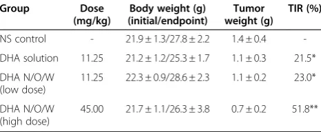
In vivo antitumor efficacy of DHA N/O/W emulsion Tumor-bearing mice transplanted with murine hepatic H22cells were used to investigate the antitumor effect of
DHA. During the treatment regime, tumor volume was monitored at a daily basis. Figure 5 shows the variation
of tumor volume after H22bearing mice were treated by
i.v. injection of saline, DHA solution, and DHA N/O/W emulsion, respectively. The group treated with DHA N/ O/W emulsion (45 mg/kg eq) displayed the strongest
tumor growth inhibition and smallest tumor volumes (P
[image:5.595.57.292.88.255.2]<0.01). In contrast, severe side effects (e.g., animal death) were seen in the group treated with DHA Figure 5Mean relative tumor volume in DHA N/O/W emulsion
(45 mg/kg), DHA N/O/W emulsion (11.25 mg/kg), DHA solution (11.25 mg/kg), and N.S. control. (n = 10).
Table 2 In vivo anti-tumor effects of DHA and DHA N/O/ W emulsion on growth of H22tumor in ICR mice (n = 10)
Group Dose
(mg/kg)
Body weight (g) (initial/endpoint)
Tumor weight (g)
TIR (%)
NS control - 21.9 ± 1.3/27.8 ± 2.2 1.4 ± 0.4
-DHA solution 11.25 21.2 ± 1.2/25.3 ± 1.7 1.1 ± 0.3 21.5*
DHA N/O/W (low dose)
11.25 22.3 ± 0.9/28.6 ± 2.3 1.1 ± 0.2 23.0*
DHA N/O/W (high dose)
45.00 21.7 ± 1.1/26.3 ± 3.8 0.7 ± 0.2 51.8**
Note:*
P<0.05 and**
P<0.01 vs. N.S. control.
#
[image:5.595.303.540.112.158.2]P<0.05 vs. DHA solution or low-dose group.
Table 3 Hemolysis test of DHA N/O/W dry emulsion by spectrophotometry
Emulsion volume/total volume (%)
Negative control
Positive control
13.3 10.0 6.7 3.3
[image:5.595.57.290.620.716.2]solution (45 mg/kg eq), indicating the over-tolerance dose. The tumor inhibition rate of DHA N/O/W emul-sion was 51.8% (Table 2). Thus, it demonstrated the enhanced antitumor efficacy and the reduced toxicity of DHA with the N/O/W emulsion formulation.
Elementary safety assay
Hemolysis and vascular irritation evaluations are import-ant for parenteral preparations before in vivo investiga-tion because vessels and erythrocytes are the initial parts of the body that encounter and interact with i.v. injected preparations. Thus, the tests provide preliminary safety evaluation for DHA N/O/W emulsion. The DHA N/O/ W emulsion displayed excellent biocompatibility. There were neither broken nor agglutinated erythrocytes observed under the microscope when exposed to the DHA N/O/W emulsion in the tested concentration range, and all samples showed less than 5% of hemolytic degree (Table 3).
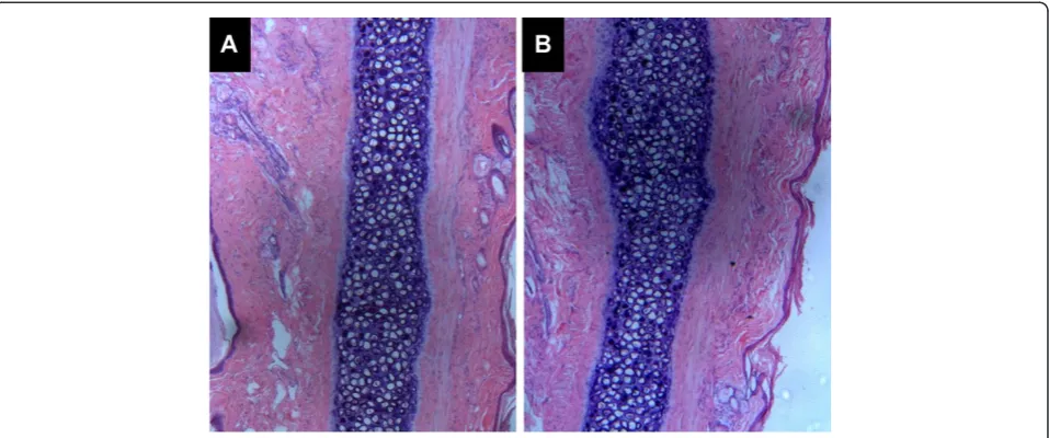
The vascular irritation test also demonstrated the safety of the DHA N/O/W emulsion in rabbits as shown in Figure 6. There was no swelling, thrombus, degener-ation, or inflammatory cell infiltration observed in the histological examination after i.v. administration of the DHA N/O/W emulsion at the dose of 2.5 mg/kg. Our results indicated the biocompatibility of the injectable DHA N/O/W emulsion.
Conclusion
A novel N/O/W submicron emulsion with high drug-loading capacity was prepared for intravenous adminis-tration of poorly soluble and unstable drugs. Our strat-egy is able to not only enhance the drug solubility in the
emulsion but also improve its storage stability by freeze-drying. The pharmacokinetics of N/O/W emulsion are characterized by the significantly prolonged half-life and slow plasma clearance compared with conventional O/ W emulsion and DHA solution. Presumably due to the improved PK profiles, the toxic effect of DHA was greatly reduced. Furthermore, the antitumor efficacy was
demonstrated in vivo studies using a hepatic H22 tumor
transplanted mouse model. More importantly, with the advantages of the high drug-loading capacity, easy manufacturing process, and excellent biocompatibility, the N/O/W emulsion provides a promising solution for developing injectable drug delivery systems for a class of drugs of poor water solubility and stability.
Competing interests
The authors declare that they have no competing interests.
Authors’contributions
SW performed the experimental laboratory work. HW carried out the TEM and FF-TEM analyses. WL and YH conceived and designed the experiments. SW and YH prepared the manuscript. All authors read and approved the final manuscript.
Acknowledgements
This study was funded by the National Natural Science Foundation of China (NSFC nos. 30873176 and 81172996) as well as partially supported by Shanghai Pujiang Scholarship and School of Pharmacy, Fudan University & The Open Project Program of Key Lab of Smart Drug Delivery (Fudan University), MOE & PLA, China.
Author details
1Zhejiang University College of Pharmaceutical Sciences, 388 Yuhangtang
Road, Hangzhou 310058, China.2Shanghai Institute of Materia Medica, Chinese Academy of Sciences, 501 Hai-ke Road, Shanghai 201203, China. 3
[image:6.595.60.539.89.289.2]Key Lab of Smart Drug Delivery (Fudan University), MOE & PLA, 826 Zhangheng Rd, Shanghai 201203, China.
Received: 19 February 2012 Accepted: 25 March 2012 Published: 13 April 2012
References
1. Fahr A, Liu X:Drug delivery strategies for poorly water-soluble drugs.
Expert Opin Drug Deliv2007,4:403–416.
2. Lipinski CA, Lombardo F, Dominy BW, Feeney PJ:Experimental and computational approaches to estimate solubility and permeability in drug discovery and development settings.Adv Drug Deliv Rev2001, 46:3–26.
3. Bansal T, Mustafa G, Khan ZI, Ahmad FJ, Khar RK, Talegaonkar S:
Solid self-nanoemulsifying delivery systems as a platform technology for formulation of poorly soluble drugs.Crit Rev Ther Drug Carrier Syst2008, 25:63–116.
4. Chen Y, Li G, Wu X, Chen Z, Hang J, Qin B, Chen S, Wang R: Self-microemulsifying drug delivery system (SMEDDS) of vinpocetine: formulation development and in vivo assessment.Biol Pharm Bull2008, 31:118–125.
5. Hanafy A, Spahn-Langguth H, Vergnault G, Grenier P:Tubic Grozdanis M, Lenhardt T, Langguth P: Pharmacokinetic evaluation of oral fenofibrate nanosuspensions and SLN in comparison to conventional suspensions of micronized drug.Adv Drug Deliv Rev2007,59:419–426.
6. Ye J, Wang Q, Zhou X, Zhang N:Injectable actarit-loaded solid lipid nanoparticles as passive targeting therapeutic agents for rheumatoid arthritis.Int J Pharm2008,352:273–279.
7. Pastorino F, Brignole C, Marimpietri D, Cilli M, Gambini C, Ribatti D, Longhi R, Allen TM, Corti A, Ponzoni M:Vascular damage and anti-angiogenic effects of tumor vessel-targeted liposomal chemotherapy.Cancer Res 2003,63:7400–7409.
8. Harvey RD, Barlow DJ, Drake AF, Kudsiova L, Lawrence MJ, Brain AP, Heenan RK:The effect of electrolyte on the encapsulation efficiency of vesicles formed by the nonionic surfactant, 2C18E12.J Colloid Interface Sci2007, 315:648–661.
9. Klayman DL:Qinghaosu (artemisinin): an antimalarial drug from China.
Science1985,228:1049–1055.
10. Grace JM, Aguilar AJ, Trotman KM, Peggins JO, Brewer TG:Metabolism of beta-arteether to dihydroqinghaosu by human liver microsomes and recombinant cytochrome P450.Drug Metab Dispos1998,26:313–317. 11. WHO:WHO World Malaria Report 2010. Geneva; 2010.
12. Lee J, Zhou HJ, Wu XH:Dihydroartemisinin downregulates vascular endothelial growth factor expression and induces apoptosis in chronic myeloid leukemia K562 cells.Cancer Chemother Pharmacol2006, 57:213–220.
13. Disbrow GL, Baege AC, Kierpiec KA, Yuan H, Centeno JA, Thibodeaux CA, Hartmann D, Schlegel R:Dihydroartemisinin is cytotoxic to
papillomavirus-expressing epithelial cellsin vitroandin vivo.Cancer Res 2005,65:10854–10861.
14. Singh NP, Lai HC:Synergistic cytotoxicity of artemisinin and sodium butyrate on human cancer cells.Anticancer Res2005,25:4325–4331. 15. Gao Y, Li N, Hilfert L, Zhang S, Zheng L, Yu L:Temperature-induced
microstructural changes in ionic liquid-based microemulsions.Langmuir 2009,25:1360–1365.
16. Weber CH, Chiche A, Krausch G, Rosenfeldt S, Ballauff M, Harnau L, Gottker-Schnetmann I, Tong Q, Mecking S:Single lamella nanoparticles of polyethylene.Nano Lett2007,7:2024–2029.
doi:10.1186/1556-276X-7-219
Cite this article as:Wanget al.:An injectable hybrid nanoparticle-in-oil-in-water submicron emulsion for improved delivery of poorly soluble drugs.Nanoscale Research Letters20127:219.
Submit your manuscript to a
journal and benefi t from:
7Convenient online submission 7Rigorous peer review
7Immediate publication on acceptance 7Open access: articles freely available online 7High visibility within the fi eld
7Retaining the copyright to your article