in immunoregulation
Hong Jiang, Leonard Chess
J Clin Invest.
2004;
114(9)
:1198-1208.
https://doi.org/10.1172/JCI23411
.
The immune system evolved to protect organisms from a virtually infinite variety of
disease-causing agents but to avoid harmful responses to self. Because immune protective
mechanisms include the elaboration of potent inflammatory molecules, antibodies, and killer
cell activation — which together can not only destroy invading microorganisms, pathogenic
autoreactive cells, and tumors, but also mortally injure normal cells — the immune system is
inherently a “double-edged sword” and must be tightly regulated. Immune response
regulation includes homeostatic mechanisms intrinsic to the activation and differentiation of
antigen-triggered immunocompetent cells and extrinsic mechanisms mediated by
suppressor cells. This review series will focus on recent advances indicating that distinct
subsets of regulatory CD4
+and CD8
+T cells as well as NK T cells control the outgrowth of
potentially pathogenic antigen-reactive T cells and will highlight the evidence that these
suppressor T cells may play potentially important clinical roles in preventing and treating
immune-mediated disease. Here we provide a historical overview of suppressor cells and
the experimental basis for the existence of functionally and phenotypically distinct
suppressor subsets. Finally, we will speculate on how the distinct suppressor cell subsets
may function in concert to regulate immune responses.
Review Series Introduction
Find the latest version:
An integrated view of suppressor T cell subsets
in immunoregulation
Hong Jiang and Leonard Chess
Department of Medicine and Pathology, Columbia University College of Physicians and Surgeons, New York, New York, USA.
The immune system evolved to protect organisms from a virtually infinite variety of disease-causing agents but
to avoid harmful responses to self. Because immune protective mechanisms include the elaboration of potent
inflammatory molecules, antibodies, and killer cell activation — which together can not only destroy invading
microorganisms, pathogenic autoreactive cells, and tumors, but also mortally injure normal cells — the immune
system is inherently a “double-edged sword” and must be tightly regulated. Immune response regulation includes
homeostatic mechanisms intrinsic to the activation and differentiation of antigen-triggered immunocompetent
cells and extrinsic mechanisms mediated by suppressor cells. This review series will focus on recent advances
indi-cating that distinct subsets of regulatory CD4
+and CD8
+T cells as well as NK T cells control the outgrowth of
potentially pathogenic antigen-reactive T cells and will highlight the evidence that these suppressor T cells may
play potentially important clinical roles in preventing and treating immune-mediated disease. Here we provide a
historical overview of suppressor cells and the experimental basis for the existence of functionally and
phenotypi-cally distinct suppressor subsets. Finally, we will speculate on how the distinct suppressor cell subsets may function
in concert to regulate immune responses.
The potential capacity for the immune response to induce or acti-vate disease was clearly recognized at the turn of the 20th century by Paul Ehrlich, who emphasized that the immune system must carefully distinguish between self and non-self in order to avoid autoimmunity. Ehrlich envisioned that during the ontogeny and outgrowth of the immunocompetent clones responsive to foreign antigens, there had to be mechanisms to control the outgrowth of clones reactive with self (1). Moreover, the failure to control the outgrowth of autoreactive cells would lead to a state of “horror autotoxicus,” or autoimmunity. Ehrlich’s ideas were amplified and developed with the elaboration of the clonal selection hypoth-esis (2–4). This hypothhypoth-esis was further refined with identification of the antigen receptors on T and B cells and discovery that the antigen specificity of these receptors is a consequence of random recombination of the many V, D, and J genes encoding the antigen-binding sites of these receptors, a process that could generate more than 109 distinct receptors (5).
According to a modern interpretation of the clonal selection hypothesis, multiple clones of immunocompetent cells displaying unique antigen-specific receptors exist prior to interaction with antigens and in the case of T cells get selected on the basis of inter-action with self-peptides bound to MHC molecules in the thymus. The majority of thymocytes bearing high-affinity receptors for self-antigens are eliminated centrally during thymic differentia-tion by an apoptotic mechanism termed negative selecdifferentia-tion. How-ever, many self-reactive T cells with low to intermediate affinity for self-antigen escape thymic negative selection and are released into the periphery, where they are capable of
autoantigen-driv-en activation, proliferation, and differautoantigen-driv-entiation into potautoantigen-driv-entially pathogenic effector cells (6–9). Thus, mechanisms that normally regulate the outgrowth or function of these self-reactive T cells ultimately control the initiation and progression of autoimmune disease. A corollary of these notions is that autoimmune diseases arise from either the failure to eliminate or inactivate high-affinity immunocompetent cells during their ontogeny and/or the failure of the immune system to control the outgrowth or function of intermediate self-reactive clones that escape into the periphery.
In addition to direct autoimmune attack, the immune system can also induce disease, because the very protective immunologic mechanisms that are employed to limit the outgrowth of invading foreign pathogens or tumor cells can induce “collateral damage” on normal, uninfected cells in the vicinity of immune attack. This col-lateral damage is mediated in large part by inflammatory cytokines and is thought to account for the destruction of normal tissues clinically observed during the physiologic immune attack aimed at the elimination of foreign pathogens or tumors. To prevent immune disease either induced directly by autoimmune attack or to control collateral damage occurring during all immune respons-es, a complex network of interacting regulatory peripheral mecha-nisms has coevolved to prevent or dampen immune-mediated dis-eases. These regulatory systems include mechanisms intrinsic to the antigen activation and differentiation of T cells as well as those mediated by regulatory “suppressor” T cells. This JCI review series on regulatory T cells will focus on recent advances indicating that distinct subsets of regulatory CD4+ (10–12) and CD8+ T cells (refs. 13, 14 and the present article) as well as NK T cells (NKT cells) (15) function to suppress the outgrowth of potentially pathogenic antigen-reactive T cells. The series will highlight the resurrection of the idea that suppressor T cells play potentially important clini-cal roles in the prevention and treatment of immune-mediated disease. The articles will review evidence that suppressor cells are essential for the control of autoimmunity and are also involved in the control of the immune response to transplanted allografts (12), allergens (16), and infectious pathogens. The series will also
Nonstandard abbreviations used: AICD, activation-induced cell death; CD40L, CD40 ligand; EAE, experimental autoimmune encephalomyelitis; α-GalCer, α -galac-tosylceramide; ILT3, Ig-like transcript 3; IPEX syndrome, immunodysregulation, poly-endocrinopathy, enteropathy, X-linked syndrome; MBP, myelin basic protein; NKT cells, NK T cells; PLP, proteolipid protein; T1D, type 1 diabetes.
Conflict of interest: The authors have declared that no conflict of interest exists.
include a review of T cell vaccination, which may be mediated by some or all of the Tregs (17).
In this series introductory article, we will emphasize that the regulation mediated by these suppressor T cell subsets is superim-posed on intrinsic regulatory mechanisms induced by antigen acti-vation alone. Thus, we will first describe the intrinsic homeostatic mechanisms that control immune responses independent of sup-pressor cells. We will then briefly describe the experimental histori-cal basis for the existence of the CD4+, CD8+, and NKT suppressor subsets and attempt to give an overview of their unique functions. Finally, as a prelude to the rest of the articles in the series, we will speculate on how the distinct subsets of suppressor cells function in concert to regulate immune responses.
Homeostatic regulatory mechanisms intrinsic to antigen activation and differentiation that function independently of suppressor cells
TCR affinity, apoptosis, and antigen-induced cell death. Immune responses are initiated when the antigen-specific TCRs expressed by resting CD4+ T cells are triggered by MHC-peptide complexes in concert with costimulatory molecules expressed by APCs (18– 20). This triggering induces CD4+ T cells to proliferate, secrete cytokines, and express cell-surface molecules including the IL-2 receptor (CD25), CTLA-4, and CD40 ligand (CD40L) criti-cal for the subsequent growth and functional differentiation of T cells. To regulate the immune response, the immune system has also evolved several homeostatic feedback mechanisms to downregulate and control the outgrowth, differentiation, and function of peripheral antigen-activated CD4+ T cells (Figure 1). One level of control resides at the initial clonal activation of the T cell receptor itself by MHC-peptide complexes. The functional consequences of T cell signaling are dependent on the affinity and duration of binding of the TCRs with MHC-peptide complexes (21, 22). For example, there is a threshold of affinity necessary for optimal activation and differentiation of T cells, whereas trig-gering of very high-affinity TCRs can induce apoptotic pathways leading to activation-induced cell death (AICD) (23).
The homeostatic role of costimulatory molecules in immunoregulation. During the initial T cell-MHC-peptide interaction, other recep-tor ligand interactions also become pivotal in ultimately dictat-ing the functional fate of T cells. For example, one of the earliest
antigen activation–induced cell-surface molecules expressed by T cells is CD40L (24). CD40L interacts with CD40 expressed on B cells to induce antibody formation and with CD40 expressed on antigen-presenting DCs to induce cellular immune responses (25, 26). Another critical consequence of the interaction of CD40L with CD40 expressed on APCs is the upregulation of other key costimulatory molecules, including CD80 (B71) and CD86 (B72) (27, 28). These molecules interact with CD28 expressed on naive T cells to provide the costimulatory signal required for optimal activation of T cells. However, following activation, a structurally related molecule termed CD152 or CTLA-4 is expressed that also binds CD80 and CD86 but delivers inhibitory signals to T cells. The consequence of CTLA-4 negative signaling is abrogation of functional activation. The resulting nonfunctional T cells are termed anergic or tolerant. Thus, blockade of either the CD40L/ CD40 pathway or the CD28/B7 pathway can lead to inhibition of immune responses and tolerance induction (20, 28–30).
The differentiation of CD4+ T cells into subsets expressing different arrays
of cytokines. A third general set of regulatory mechanisms, also a con-sequence of the initial triggering of CD4+ T cells by MHC-peptide complexes, is the further differentiation of the CD4+ T cells into the functionally distinct Th1 and Th2 subsets phenotypically dis-tinguished, in part, by the elaboration of distinct sets of cytokines (31–33). In this regard, IFN-γ secreted by Th1 cells is known to downregulate the differentiation and function of TH2 cells and, conversely, IL-4. TGF-β and IL-10 inhibit Th1 cell differentiation (33–35). In addition, other cytokine-secreting Th subsets capable of secreting the immunosuppressive cytokines IL-10 and/or TGF-β
but not IL-4 (termed Tr1 or Th3 cells) (36–38) have been observed and will be reviewed in detail in several articles in this review series. A widely prevalent view is that the balance between the emergence of Th1, Th2, as well as the other Th cytokine–secreting CD4+ Tregs following antigen activation plays a major role in the outgrowth and functions of self-reactive and foreign reactive clones (39–41).
[image:3.585.42.341.80.275.2]Homeostatic regulatory mechanisms mediated by dominant suppression of the immune response The origin of the idea of suppressor T cells and its initial demise. Superim-posed on the intrinsic mechanisms of homeostatic regulation are extrinsic mechanisms mediated by suppressor T cells that control the induction and/or outgrowth of antigen-activated T cells. In
Figure 1
Homeostatic control of the outgrowth of antigen-acti-vated CD4+ T cells. Control of the peripheral
immu-nity is accomplished by mechanisms intrinsic to anti-gen activation of CD4+ T cells; including apoptosis,
induction of anergy, and differentiation into Th sub-sets that are independent of other types of regulato-ry cells. In addition, superimposed on these intrinsic mechanisms are control mechanisms mediated by distinct subsets of NKT, CD4+, and CD8+ regulatory
this regard, the idea that suppressor T cells may be critical to the control of virtually all immune responses arose in the laboratory of Richard Gershon at Yale University in the late 1960s in studies of immune tolerance to foreign antigens. Gershon showed initially that adoptive transfer of T cells from animals made tolerant to for-eign antigen “X” could specifically suppress the production of anti-X antibodies in recipient animals (42, 43). These studies were rap-idly extended in numerous laboratories, which documented that suppressor cells were not only involved in the peripheral regulation of cells responding to foreign antigens but also participated in the regulation of self-reactive cells and in the control of autoimmu-nity (44, 45). The first models of the pathways by which suppres-sor T cells might function in the specific regulation of immunity arose from the seminal studies by Cantor and Boyse, who showed that genetically well-defined alloantisera could be used to identify phenotypically stable and functionally distinct subsets of T cells. These studies ultimately led to the discovery of CD4+ and CD8+ T cell subsets (46–50). Implicit in these discoveries was the idea that the CD4+ and CD8+ T cell subsets were genetically programmed during ontogeny, prior to interaction with antigen, to mediate distinct functional programs. The CD4+ T cells were functionally programmed to induce both antibody responses and cell-mediated immune reactions. In contrast, the CD8+ T cells were not geneti-cally programmed as inducer cells but instead were programmed to differentiate into killer cells capable of destroying tumor cells or cells infected with intracellular pathogens. However, following interaction with antigen-activated CD4+ T cells, CD8+ T cells could be induced in vitro to differentiate into suppressor cells, which in turn downregulate the activity of the CD4+ T cell population (51, 52). Experiments employing allosera to the MHC molecule Qa-1 suggested that Qa-1 was expressed on the suppressor-inducer subset of CD4+ T cells (51, 52). This is of interest because eventu-ally Qa-1 was cloned and found to be an MHC class Ib molecule capable of presenting endogenous as well as exogenous peptides (53) to CD8+ T cells. However, the potential significance of Qa-1 expression on suppressor-inducer cells in vivo was not delineated until the last several years, with experiments employing mono-clonal antibodies to Qa-1 and Qa-1–knockout mice (54–56) (see below). Precise characterization of the specificity and functional phenotype of suppressor cells generated in these complex cellular experiments was impossible, largely because there were no pheno-typic markers at the time that definitively distinguished the CD8 suppressor cells from the more conventional CD8 or CD4 T cells. Moreover, the biological significance of these in vitro findings was not placed on firm in vivo footing because of the lack of mono-clonal antibodies to the murine CD4 and CD8 populations and the lack of molecular genetics–based approaches to study CD4- or CD8-deficient animals.
In this regard, it is important to emphasize that the original sup-pressor cell circuits were initially conceived and/or deduced at a time when molecular immunology was in its infancy. For example, the nature of the TCR was unknown, as was the precise structure and function of MHC molecules in restricting T cell activity. In addition, the great majority of the cytokines that are now known to regulate immune functions had not yet been identified. It was also unknown that antigen activation of the TCRs induced intrin-sic homeostatic mechanisms, including the differentiation of CD4+ T cells into Th1 and Th2 subsets which elaborated distinct sets of regulatory cytokines. Clearly, understanding the precise role of these lymphokines in suppression would have significantly
influenced the interpretation of data suggesting that an array of antigen-specific factors was uniquely secreted by suppressor cells. Lack of this information, however, caused interest in the models of T cell suppression mediated by CD8+ T cells to wane by the mid-1980s (45, 57, 58), and skepticism about the importance of T cell suppression dominated the field until the 1990s. Although some of the skepticism concerning T cell suppression mediated by CD8+ T cells in the mid-1980s was justified, it is clear now that the proverbial baby had been thrown out with the bath water. Indeed, many of the ideas and models of immunoregulation that have been dismissed were essentially correct and have far reaching bio-logical and clinical significance.
The resurrection of CD8+ suppressor T cells. The resurrection of the
concept that CD8+ T cells mediate T cell suppression was initiated, in part, by the publication of 2 articles in Science in 1992. The stud-ies showed that CD8+ T cells participate in vivo in the resistance to disease induced during the natural history of experimental autoim-mune encephalomyelitis (EAE), a well-studied model of the human disease multiple sclerosis (59, 60). For example, EAE is induced by myelin proteins such myelin basic protein (MBP), which activates encephalitogenic CD4+ Th1 cells. Mice of the B10PL strain com-pletely recover from the first episode of EAE and become highly resistant to the reinitiation of EAE by secondary immunization. If these protected mice are then depleted of CD8+ T cells through the use of monoclonal anti-CD8, the protection is reversed, and the mice develop clinical EAE upon reimmunization with MBP (59). Furthermore, mice depleted of CD8+ T cells during the initial induction of EAE and allowed to recover normal levels of CD8+ T cells are not resistant and develop EAE again upon rechallenge with MBP. Thus, CD8+ T cells require priming during the first episode of EAE to regulate CD4+ T cells triggered by secondary MBP stimula-tion in vivo. Moreover, when CD8–/– mice are bred with EAE-suscep-tible PL/J mice, the progeny of the CD8–/– and PL/J mating develop more chronic EAE than the wild-type PL/J mice, as reflected by a higher frequency of relapses (60). These experiments provide evi-dence that CD8+ T cells play a key role in both inducing resistance to autoimmune EAE and in abrogating or suppressing recurrent relapsing episodes of pathogenic autoimmunity in vivo.
intriguing and suggested that perhaps the nonclassical MHC class 1b molecules may play a role in vivo in restricting the suppression mediated by CD8+ T cells. Indeed, subsequent experiments in vivo showed that the suppression mediated by CD8+ suppressor T cells is blocked by antibodies to the MHC class 1b Qa-1 molecule (9, 54, 61). Importantly, the Qa-1+ CD4+ T cells could be employed as vac-cine T cells and used to induce regulatory CD8+ T cells in vivo. This T cell vaccination procedure protected animals from developing EAE, and this protection was abrogated by depletion of the Qa-1– restricted CD8+ T cells in vivo (61, 62).
In very recent experiments, the Cantor laboratory has created Qa-1–deficient mice and used them to directly demonstrate, in vivo, the important role that Qa-1 plays in the regulatory pathway mediated by CD8+ T cells in the control of autoimmunity (56). The authors convincingly demonstrated that the Qa-1–deficient mice develop severe EAE when exposed to the myelin-associated self–pro-teolipid protein (self-PLP) peptide and fail to develop the resistance to EAE that normally develops in wild-type mice after immuniza-tion with PLP peptide. Furthermore, the failure of resistance to EAE is associated with the escape of Qa-1–deficient CD4 cells from CD8+ T cell suppression, which could be restored by lentiviral-based expression of the syngeneic Qa-1 allele. These results form the heart of the functional data on Qa-1–deficient mice, demonstrating the in vivo relevance of Qa-1 in the control of autoimmunity. Further-more, the authors also showed that Qa-1– deficient mice fail to con-trol expansion of herpes simplex virus 1–induced (HSV-1–induced)
CD4+ Th1 cells that induce ocular keratitis and blindness. These Th1 CD4+ T cells are specific for HSV peptides cross-reactive with self-corneal tissues and thus confirm the findings in another rele-vant autoimmune disease model. Moreover, these studies also show that the effect of Qa-1 on regulatory CD8+ T cells in the control of autoimmune disease in vivo is only observed during the secondary, but not the primary, immune response.
A number of biologic features of the Qa-1 molecule make it par-ticularly interesting with respect to its role as a restricting element in immunoregulation. First, Qa-1 is preferentially expressed on activated, but not resting, T cells. Moreover, the fact that surface expression of Qa-1 on activated T cells is short lived may exclude resting T cells from downregulation by Qa-1–dependent CD8+ T cells. Second, Qa-1 is of limited polymorphism, with the potential to present self- and foreign peptides to CD8+ T cells. The predomi-nant self-peptide presented by Qa-1 is Qdm, a hydrophobic peptide (AMAPRTLLL) derived from the leader sequence of certain MHC class Ia molecules (63). However, during T cell activation, Qa-1 can also bind other hydrophobic self-peptides that may serve as target antigens for the CD8+ T cells, as described above. Because the Qa-1– self-peptide complexes can interact with the CD94-NKG2 receptors expressed on NK or classical CD8+ T cells to either positively or negatively regulate function, Qa-1 may not only serve as the target of CD8 suppression, but also regulate the function of CD8+ sup-pressor T cells via the CD94-NKG2 receptors.
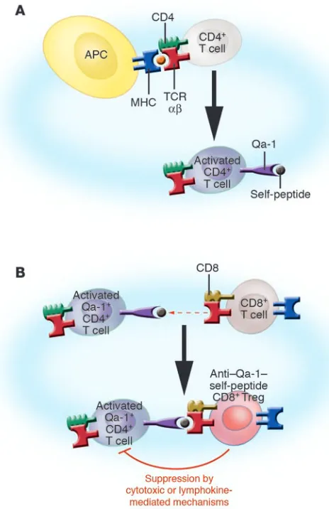
[image:5.585.45.277.82.440.2]In summary, we envision that the CD8 T cell regulatory pathway comprises a series of sequential cellular events (Figure 2). It is ini-tiated by the activation of naive CD4+ T cells during the primary immune response, in which the TCRs on CD4+ T cells interact with MHC class II–peptide complexes presented by conventional APCs. This initial interaction induces the surface expression of activa-tion-dependent Qa-1–self-peptide complexes which is not observed in resting T cells. These activation-dependent Qa-1–self-peptide complex serve as both the inducer and target structures recognized by TCR αβ on regulatory CD8+ T cells. Thus, the Qa-1–self-peptide complex expressed by activated CD4+ T cells triggers TCR αβ on regulatory CD8+ T cells. These CD8+ T cells then differentiate into effector cells, which in turn suppress CD4+ T cells expressing the same target Qa-1–self-peptide complex. This suppression is linked to specific antigen activation of CD4+ T cells, and as a consequence the peptides bound by Qa-1 will likely be those induced by antigen activation. Whether they represent specific peptides derived from the CD4 T cell TCR (TCR Vβ or Vα peptides) or peptides derived from activation molecules induced by antigen triggering is not known. The mechanism of effecting suppression of the CD4+ T cells by the Qa-1–restricted CD8+ T cells has not been definitively
Figure 2
Model of cognate interactions in the induction and function of Qa-1– restricted regulatory CD8+ T cells. (A) Initial activation of CD4+ T cell
TCRs with peptide–MHC complexes induces the expression of Qa-1 bound with a variety of self-peptides on the surface of the CD4+ T
cells. (B) Anti–Qa-1–self-peptide CD8+ precursor T cells are activated
by Qa-1–expressing CD4+ T cells. The Qa-1–restricted CD8+ Tregs
selectively downregulate certain but not all antigen-activated CD4+ T
cells based on the specific recognition of Qa-1–self-peptide complexes expressed on certain CD4+ T cells by TCR αβ on the CD8+ T cells. In
this regard, we have demonstrated in the EAE model that self-reactive CD4+ T cells, which are selectively downregulated by the CD8+ T cells,
delineated; however, the consequences of TCR triggering of CD8+ T cell precursors may be conventional and involve differentiation into specific CTLs that lyse target cells and/or secrete lymphokines that downregulate the target cells.
Furthermore, it has also been shown that a distinct population of antigen-specific, non–Qa-1–restricted CD8+CD28– cells can suppress immune responses by directly interacting with antigen-presenting DCs and rendering these cells tolerogenic (64–66). The suppression involves the upregulation of inhibitory Ig-like tran-script 3 (ILT3) and ILT4 receptors expressed on the DCs. APCs tolerized by CD8+ T cells show reduced expression of costimulatory molecules and induce antigen-specific unresponsiveness in CD4+ T helper cells. The precise function of these cells in vivo is not clear, and it is not known whether they interact with the Qa-1–restricted CD8+ suppressor cells or, alternatively, whether Qa-1–restricted T cells induce tolerogenic DCs.
Finally, the Qa-1–dependent regulatory CD8 pathway has begun to be translated from mice to humans with the in vitro findings that human CD8+ T cells can be induced to differentiate into regu-latory cells whose function is dependent on HLA-E, the human homolog of Qa-1 (67, 68). These data support the idea that clini-cally relevant methods to induce and/or enhance this suppressive pathway in humans may prove useful in the prevention and treat-ment of human autoimmune disease. The details of several of the points in this section will be expanded upon in the series reviews from the Cantor and Kumar laboratories (13, 14).
The rapid rise of CD4+ suppressor T cells. Although suppressor cells
were initially identified within the CD8+ T cell population (49, 50, 69), it was later found that suppressor cell function could be mediated by CD4+ T cells independent of CD8+ T cells (50, 70–72). For example, in the early 1980s, it was shown that coculture of graded numbers of polyclonally activated human CD4+ T cells to autologous resting CD4+ T cells inhibited the capacity of the resting CD4+ T cells to induce B cell differentiation and Ig syn-thesis in vitro (73, 74). The in vivo significance of these findings in humans were unknown. However, in vivo experiments in mice in the mid-1980s placed the idea of CD4+ suppressor cells on a firmer biological foundation. In particular, studies by Sakaguchi and colleagues on the organ-specific autoimmune disease induced in mice following neonatal thymectomy were quite revealing. These thymectomized mice were shown to have reduced numbers of CD4+ as well as CD8+ T cells. Furthermore, reconstitution of thymectomized mice by highly enriched populations of CD4+ but not CD8+ T cells from syngeneic normal mice completely inhibited disease development (70).
A decade later in 1995, Sakaguchi showed for the first time that the suppression mediated by CD4+ T cells is a function of the small subset of CD4+CD25+ cells (71). The transfer of T cells depleted in the CD4+CD25+ population into nude (athymic) mice led to a variety of autoimmune diseases, which could be prevented by injection of purified CD4+CD25+ T cells but not CD4+CD25– T cells. The field of CD4+CD25+ regulator cells, termed “CD4+CD25+ Tregs,” was thus born and has experienced an explosive growth over the past few years (75, 76). Thus, the general experimental protocols employed to characterize these suppressor cells in vivo took advantage of the observation that mice deficient in T cells (neonatally thymectomized, nu/nu mice or Rag–/– mice) do not develop autoimmune disease following
adoptive transfer of normal syngeneic spleen cells unless the spleen cells are depleted of CD4+CD25+ T cells. Thus, when
CD4+ splenic T cells prepared from normal mice were depleted of CD25+ cells and the remaining CD4+ T cells were transferred to syngeneic T cell–deficient mice, the recipients spontaneously developed various organ-specific autoimmune diseases (includ-ing type 1 diabetes [T1D], thyroiditis, and gastritis) and systemic wasting disease. Reconstitution of the CD4+CD25+ population inhibited the autoimmune development (72, 76). However, it is becoming increasingly clear that in many situations, CD4+CD25– T cells are as effective as CD4+CD25+ T cells in controlling T cell– mediated disease (77, 78).
In this regard, seminal studies of the potency of suppressor T cells by Lafaille and colleagues unequivocally showed that very small numbers of CD4+ suppressor cells can regulate immune responses in vivo independent of CD8+ T cells. This set of studies evaluated TCR-transgenic mice expressing the MBP specific for the TCR expressed on pathogenic clones and known to induce EAE (T/R+ mice). Lafaille found that these T/R+ mice rarely develop spontane-ous EAE. However, when the T/R+ mice are crossed with Rag1–/–
mice to obtain mice that have only T cells expressing the transgenic MBP-specific TCR (T/R– mice), almost all mice develop spontane-ous EAE (79). Because both T/R+ and T/R– mice have large numbers of the potentially encephalitogenic CD4+ anti-MBP T cells, these results suggest that a very small number of nontransgenic lym-phocytes, which are present in T/R+ but absent in T/R– mice, can potently suppress the in situ activation of CD4+ anti-MBP T cells mediating EAE. To identify the cellular regulatory mechanisms important in this suppression, T/R+ mice were crossed into mice deficient in either B cells, CD8+ T cells, NKT cells, TCR γδ cells, or TCR αβ cells (80, 81). Only mice that were deficient in CD4+αβ T cells developed EAE. Moreover, T/R– mice were protected from EAE by the early (that is, prior to the onset of EAE) adoptive transfer of purified CD4+ T cells from normal donors. These results support the view that under certain experimental conditions CD4+ cells alone can suppress the initiation of autoimmunity. Moreover, it was subsequently shown that both CD4+CD25+ and CD4+CD25– cells could mediate this suppression (78, 82).
follow-ing antigen triggerfollow-ing of the TCR. The CTLA-4 may be of special interest with respect to the function of CD4+CD25+ regulatory cells because, like CD25, CTLA-4 is thought to be constitutively expressed on the CD4+CD25+ suppressor populations (85), and blockade of the CTLA-4 interactions with their CD80 or CD86 receptors in vivo can abrogate suppression. Moreover, T cells trig-gered via CTLA-4 predominantly secrete TGF-β, a cytokine with suppressive functions (86). On the other hand, in vitro experiments have not been able to document a blocking effect of anti–CTLA-4 on suppressor cell function (87). The search is still on to define specific markers that distinguish the subsets of CD4+ suppressor subsets (78, 83, 84). This general issue will be further discussed in several review articles in this series (particularly refs. 10–12).
With respect to the general role of cytokines in the suppres-sion mediated by CD4+ Tregs, the cytokine profiles found in the various populations of CD4+ suppressor cells include a vari-ety of combinations of the already-known immunoregulatory cytokines (i.e., IL-10, TGF-β, IL-4, IFN-γ). For example, there are CD4+CD45Rblow activated suppressor cells that secrete large quantities of either IL-10 and IL-4 (termed Tr1 cells) and other CD4+CD45Rblow suppressor T cells that secrete large quantities of
[image:7.585.60.531.82.411.2]TGF-β (termed Th3 cells) (38). (The role of IL-10 cytokine expres-sion will be discussed in detail in the series review by O’Garra et al., ref. 11) The functional significance of these cytokine-secret-ing CD4+ T cells is supported by the findings that TGF-β –defi-cient mice develop autoimmune disease (88) and that adminis-tration of neutralizing antibodies to IL-4 or TGF-β abrogates the in vivo prevention of autoimmunity or tolerance-inducing activity of CD4+ T cells in some models (89, 90). The relation-ship of these cells to the CD4+CD25+CTLA-4+ cells is unclear. For example, the in vitro capacity of CD4+CD25+ T cells to suppress immune responses is known to be contact dependent and not due to the IL-10, TGF-β, and IL-4 cytokines alone. It is possible the Th3 and Tr1 populations arise from “conventional” resting CD4+CD25– T cells, which, following antigen activation, express CD25. Taken together, these data may suggest that CD4+CD25– T cells, which function to suppress immune responses in vivo, may not represent a lineage-specific suppressor population. In contrast, the naturally occurring CD4+CD25+ T cells responsible for preventing autoimmunity in neonatal immune-deficient mice are thought to represent a lineage-specific suppressor population arising directly from the thymus (76).
Figure 3
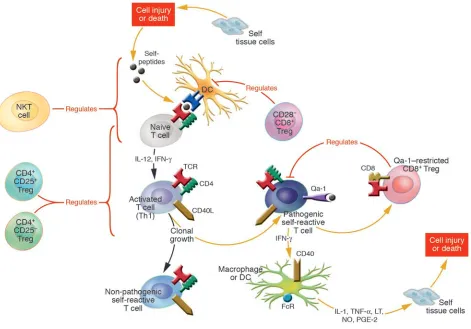
Tregs control the peripheral induction and clonal outgrowth of antigen-reactive T cells. This illustration shows various pathways of immunoregulation mediated by suppressor subsets of NKT, CD4+, and CD8+ T cell subsets. Each of the regulatory T cell subsets expresses
distinct receptors, employs different effector mechanisms, and functions predominately at different stages during the course of the peripheral immune response. The NKT and CD4+CD25+ regulatory cells are “natural suppressor cells”; they are present prior to antigen activation and
primarily function during the early “innate” and/or primary adaptive immune responses. In contrast, the CD8+ regulatory cells are induced to
In this regard, it is of great interest that a recently cloned tran-scription factor, termed Foxp3, a member of the forkhead family of DNA binding transcription factors, is not expressed in naive CD4+CD25– cells but is highly expressed in the naturally occur-ring CD4+CD25+ regulatory cells. Importantly, mutational defects in the Foxp3 gene result in the fatal autoimmune and inflamma-tory disorder of the scurfy mouse and in the clinical and molecu-lar features of the immunodysregulation, polyendocrinopathy, enteropathy, X-linked syndrome (IPEX syndrome) in humans. Both scurfy mice and IPEX patients have defects in T cell activation and reduced numbers and reduced suppressor functions mediated by the CD4+CD25+ T cells (91–93). In Foxp3–overexpressing mice, both CD4+CD25– and CD4–CD8+ T cells show suppressive activity, which suggests that expression of Foxp3 is linked to suppressor functions (94). Taken together, these data strongly support the idea that Foxp3 may uniquely define the subset of naturally occur-ring CD4+ suppressor T cells. However, the recent findings that Foxp3 can be expressed in CD4+CD25– cells following activation and are also expressed in activated CD8+ T cells suggest that Foxp3 is linked to functional suppression but not necessarily as a specific lineage marker (95–98).
Finally, the identity of the receptors that enable the CD4+CD25+ suppressor T cells to preferentially suppress self-reactive T cells and yet preserve normal immune functions remains a major unresolved issue. This apparent cognitive capacity to distinguish self from non-self is on the surface difficult to reconcile with a number of studies that have provided convincing evidence that the suppression medi-ated by CD4+CD25+ Tregs is not antigen specific (87). Thus, in ele-gant experiments, the Shevach laboratory showed that when T cells from TCR transgenic mice are activated with their peptide-MHC ligand and expanded in vitro in IL-2, the activated suppressors are subsequently capable of suppressing the responses of T cells from mice that express a different transgenic TCR (87, 99). Moreover, no MHC restriction is observed in the interaction of the activat-ed suppressors and the responding targets (100). Thus, the TCRs employed by these regulatory CD4+ T cells are likely to be quite diverse, and it is unknown whether their capacity to distinguish self from non-self is dependent of the TCR αβ they express. Insight into the mechanism by which these antigen-nonspecific T cells can still distinguish self from non-self may come from recent studies showing that the CD4+ T regulatory cells when nonspecifically activated by LPS express toll-like receptors (101). Thus, these natu-rally occurring Tregs may represent the regulatory component of the innate immune system responsive to “danger-like signals.” Perhaps self-reactive T cells preferentially express ligands that are recognized by toll-like receptors expressed by the regulatory cells. Clearly, the elucidation of the precise target structures recognized by CD4+CD25+ Tregs may help define the receptors employed by the regulatory cells to distinguish self from non-self. In contrast, as noted above, the Qa-1–restricted CD8+ suppressor T cells are not naturally occurring cells but instead are specifically induced during the primary adaptive immune response and are triggered to differ-entiate into effector-suppressor cells, which distinguish among the clones of autoreactive cells responding to a single peptide. Unlike the naturally occurring CD4+CD25+ suppressor T cells, Qa-1– restricted, CD8+ suppressor T cells employ their TCR αβ to recog-nize and distinguish targets of suppression.
NKT cells. NKT cells are a unique population of cells that express receptors of the NK lineage as well as a TCR αβ. Murine NKT cells express an invariant TCR α chain encoded by the Vα
14-Jα281 gene segment, paired preferentially to various Vβ chains (102). Human NKT cells express the Va24–JaQ invariant chain. The NKT cells recognize and kill tumor cells expressing lipid anti-gens structurally related to the glycolipid α-galactosylceramide (α-GalCer). These lipids are presented to NKT cells by the MHC class Ib molecule CD1d (103). Analogous CD1d-restricted NKT cells, which express the invariant Vα24-JαQ TCR, are also present in humans (104, 105). The CD1d-restricted NKT cells are mainly of CD4+ or CD4–CD8– phenotype (106), and although originally functionally defined, both in vivo and vitro, by their capacity to lyse a variety of tumor cells (107), they were later found also to be involved in the regulation of autoimmune diseases (108, 109). These in vivo roles in immune responses are thought to be linked to the fact that following TCR antigen triggering in vivo, NKT cells were observed to develop augmented killer cell activity and secrete large amounts of cytokines, including IL-4 and IFN-γ, as well as TGF-β and IL-10 (103, 110–112), known to be involved in the activation of cell types important in mediating both innate immunity and Th2-type adaptive immunity.
As a consequence, NKT cells have been shown to influence the course of autoimmune disease in a variety of animal models. Prom-inent among the diseases affected by NKT cells are those primarily induced by Th1 cells, including the NOD diabetes model and the EAE models of multiple sclerosis (109, 113, 114). In these diseases, the evidence strongly suggests that the Th2-favoring cytokines, IL-4 and IL-10, secreted by NKT cells play an important role (111, 115, 116). Adoptive transfer of cell populations enriched for NKT cells prevents T1D in NOD recipients (115, 117, 118). Moreover, depletion of NKT cells early in the evolution of diabetes in the NOD mice accelerates the onset of diabetes (119). For example, it was found that lack of CD1-restricted NKT cells promotes the development of diabetes, whereas activation of Vα14+ NKT cells by
α-GalCer suppresses disease in the NOD model. Similarly, mod-els of colitis or multiple sclerosis (EAE) depletion of NKT cells accelerates the onset of disease, whereas in vivo activation of NKT cells by treatment with the glycolipid ligand induces significant improvement or prevents disease. These effects are abrogated in CD1d-deficient mice.
Thus, as a member of the family of immunocompetent cells participating in the innate immune response, NKT cells are posi-tioned to influence and interact with other Tregs during the early phases of the autoimmune response (122). Their interaction with CD4+ T cells is implicit in their capacity to secrete cytokines such as IL-4 and IL-10 that may shift the Th1-Th2 balance. In addition, there is evidence that NKT cells also interact with CD8+ T cells in immunoregulation (123). The mechanisms through which NKT cells may regulate tolerance induction are complex and involve interactions with CD4+ and CD8+ regulatory cells. The complexity of how NKT cells are involved in regulating immune responses is the topic of the series review by Godfrey and Kronenberg (15).
An integrated overview of immunoregulation by NKT, CD4+CD25+, and Qa-1–restricted CD8+ T cell subsets
The resurgence of interest in immunosuppression mediated by T cells during the last decade has come from several distinct lines of investigation that have led to the concept that the immune system has evolved a variety of regulatory mechanisms mediated by distinct T cell subsets to suppress the outgrowth of poten-tially pathogenic self-reactive T cells. The regulation mediated by these suppressor subsets is superimposed on intrinsic regulatory mechanisms induced by the initial encounter of the TCR with antigen (Figure 3). These mechanisms include the induction of: (a) cell death if the encounter is of very high affinity; (b) nonre-sponsiveness if the encounter occurs in the absence of appropriate
costimulatory signals; and (c) the antigen-triggered differentiation into various Th subsets that secrete distinct arrays of Th1, Th2, Tr1, or Tr3 regulatory cytokines.
[image:9.585.54.532.109.409.2]Given this intricate set of intrinsic regulatory mechanisms, one can legitimately ask why the immune system evolved an extrinsic set of regulatory mechanisms dependent on suppressor cells. The answer is simply that in the absence of T cell suppression, the intrinsic mechanisms are not sufficient to prevent autoimmunity or dampen immune responses to prevent collateral immune inju-ry. To begin to understand the important interplay between the intrinsic and extrinsic mechanisms, it is important to emphasize that each of the extrinsic T cell suppressor subsets express distinct receptors, employ different effector mechanisms, and function at different stages during the evolution of immune responses (see Table 1). Thus, the NKT cells and CD4+CD25+ regulatory cells exist from the very early stages of life as “natural suppressor cells” prior to antigen activation and primarily function during the “innate” and/or primary immune responses. The NKT cells are endowed with pauciclonal TCR αβs consisting of an invariant Vα chain that specifically permits recognition of glycolipid mol-ecules often expressed by various pathogens and presumably also expressed by tumor cells, activated blasts, and injured apoptotic cells that arise at the inception of immune responses. These NKT cells are thus poised to secrete IL-4 and IL-10, which are known to influence the balance of Th1 or Th2 cells that emerge during the primary immune response.
Table 1
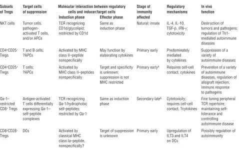
Properties of Treg subsets
Subsets Target cells Molecular interaction between regulatory Stage of Regulatory In vivo of Tregs of suppression cells and inducer/target cells immunity mechanisms function
Induction phase Effector phase affected
NKT cells Tumor cells, TCR recognizing Same as Natural; innate IL-4, IL-10, Destruction of
pathogen- CD1d/glycolipid; induction phase TGF-β, IFN-γ; tumors and pathogens;
activated T cells, restricted by CD1d cytotoxicity regulation of
Th1-and/or APCs mediated autoimmune
diseases CD4+CD25– T and B cells; Activated by MHC May function by Primary early Predominately Suppression of a
Tregs ?APCs class II–peptide elaborating cytokines mediated variety of
nonspecifically by cytokines autoimmune diseases
CD4+CD25+ T cells; Activated by Target and specificity Primary earlyA Requires cell-cell Prevention of a variety
Tregs ?APCs MHC class II–peptides is unknown; contact, cytokines of autoimmune
nonspecifically suppression is not diseases, regulation of
MHC restricted allograft rejection;
immune response to pathogens Qa-1– Antigen-activated TCR recognizing Same as induction Secondary lateA Cytotoxicity; Fine tuning peripheral
restricted T cells differentially Qa-1/hydrophobic phase requires cell-cell TCR repertoire;
CD8+ Tregs expressing Qa-1– self-peptides; contact, ?cytokines maintaining
self-self-peptide restricted by Qa-1 tolerance and
complexes controlling
autoimmune disease CD8+CD28– DCs Activated by Target of suppression Primary early Upregulation of Possibly regulation of
Tregs classical MHC is unknown ILT3 and ILT4 autoimmunity
class Ia–peptide, on DCs
nonspecifically?
Cells of the naturally occurring CD4+CD25+ suppressor subset, which are capable of suppressing the outgrowth of autoreactive cells, also exist in the peripheral lymphoid system. These cells, like the NKT cells, can function during the primary immune response and do not require specific induction. In vitro, the suppressor func-tion of these cells can be shown to dependent of cell-cell contact, but they can also express immunoregulatory cytokines including TGF-β, which may be involved in the their suppressor function in vivo. The precise specificity of these cells for their targets remains unknown, and it is not clear whether APCs and/or T cells are the targets of CD4+CD25+-mediated suppression. Moreover, although the CD4+CD25+ suppressor cells express conventional TCR αβ, the evidence suggests that these TCRs are not involved in the direct recognition of the targets of suppression.
In contrast to both the NKT cells and the naturally occurring CD4+CD25+ regulatory cells, the Qa-1–restricted CD8+ Tregs are not prevalent in naive animals prior to antigen encounter. As a consequence, adoptive transfer of CD8+ T cells from naive animals has no effect on the outcome of autoimmune responses, and deple-tion of CD8+ T cells prior the first induction of autoimmunity has no effect on the first episode of disease either. However, the CD8+ regulatory cells function like classical immunocompetent cells activated during adaptive immune responses. Thus, the they are induced by autologous CD4+ T cells activated during the primary immune response and differentiate into effector-suppressor cells, which function predominately during the secondary and memory phases of immunity. Thus, adoptive transfer of CD8+ T cells from self-antigen–primed mice will retard the outgrowth of the poten-tially pathogenic self-reactive T cells. In this regard, it is of inter-est that CD8+ Tregs are known to mediate resistance to autoim-munity following initial recovery from disease and to decrease the incidence and severity of relapse of the disease. In contrast to the CD4+CD25+ T cells, the CD8+ Tregs utilize their TCR αβs to direct-ly recognize target cells in an MHC-restricted fashion. Thus, the CD8+ Tregs are Qa-1 restricted and selectively downregulate cer-tain but not all activated T cells that preferentially express Qa-1– self-peptide(s) on their surface. They are poised to fine tune the immune response to suppress new or renewed outgrowth of
autoreactive cells during episodes of relapse from autoimmune disease. These suppressor cells thus represent more the adaptive suppressive response to immunity, whereas the CD4+ and NKT suppressor subsets mediate natural immunity to the outgrowth of potentially pathogenic cells.
In summary, in this introductory JCI series article, we empha-sized the idea that immunoregulation is carried out in an inte-grated manner by distinct suppressor T cells subsets that are superimposed on intrinsic homeostatic control mechanisms. Understanding the precise interplay between these intrinsic and suppressor mechanisms will undoubtedly be involved in the immunopathogenesis of a variety of diseases associated with abnormalities in immunoregulation. The evidence that distinct suppressor subsets are involved in many autoimmune states has been alluded to above and will be highlighted in greater detail in subsequent articles in this series (10, 13–15). In addition, the series will review the mounting evidence that suppressor cells are important in the immunopathogenesis of infectious and allergic diseases (11, 16) as well as allograft rejection (12). Finally, the idea that clinical induction of suppressor mechanisms may be impor-tant in the control of immunologic disease was conceived in the early days of the study of suppressor T cells. Induction of suppres-sor mechanisms by T cell vaccination to prevent autoimmunity, the study of which was pioneered by Irun Cohen in the early 1980s, was recently shown to involve suppressor T cells, as noted above. In this regard, the series will include a review of recent ideas concern-ing the immunobiology of T cell vaccination (17).
Acknowledgments
Research was supported by NIH grants AI39630 and AI39675 and National Multiple Sclerosis Society grant RG2938A (to H. Jiang), and by NIH grant U19 AI/46132 (to L. Chess).
Address correspondence to: Leonard Chess, Department of Medi-cine and Pathology, Rheumatology Division, Columbia University College of Physicians and Surgeons, 630 West 168th Street, PH8E, Suite 101, New York, New York 10032, USA. Phone: (212) 305-9986; Fax: (212) 305-4943; E-mail: [email protected].
1. Ehrlich, P. 1900. The Croonian lecture: on immu-nity with special reference to cell life. Proc. Royal Soc. London.66:424.
2. Jerne, N.K. 1955. The natural selection theory of anti-body formation. Proc. Natl. Acad. Sci. U. S. A.41:849. 3. Jerne, N.K. 1976. The immune system: a web of V
domains. In The Harvey Lectures 70. Academic Press. New York, New York, USA. 93–110.
4. Burnet, F.M. 1957. A modification of Jerne’s theory of antibody production using the concept of clonal selection. Australian Journal of Science.20:67–69. 5. Alt, F.W., Blackwell, T.K., DePinho, R.A., Reth,
M.G., and Yancopoulos, G.D. 1986. Regulation of genome rearrangement events during lymphocyte differentiation. Immunol. Rev.89:5–30.
6. Goldrath, A.W., and Bevan, M.J. 1999. Selecting and maintaining a diverse T-cell repertoire. Nature.
402:255–262.
7. Bouneaud, C., Kourilsky, P., and Bousso, P. 2000. Impact of negative selection on the T cell reper-toire reactive to a self-peptide: a large fraction of T cell clones escapes clonal deletion. Immunity.
13:829–840.
8. Kuchroo, V.K., et al. 2002. T cell response in experi-mental autoimmune encephalomyelitis (EAE): role of self and cross-reactive antigens in shaping, tuning, and regulating the autopathogenic T cell repertoire. Annu. Rev. Immunol.20:101–123.
9. Jiang, H., et al. 2003. Regulatory CD8+ T cells fine-tune the myelin basic protein-reactive T cell recep-tor V beta repertoire during experimental autoim-mune encephalomyelitis. Proc. Natl. Acad. Sci. U. S. A.100:8378–8383.
10. Fehérvari, Z., and Sakaguchi, S. 2004. CD4+ Tregs
and immune control. J. Clin. Invest.114:1209–1217. doi:10.1172/200423395.
11. O’Garra, A., Vieira, P.L., Vieira, P., and Goldfeld, A.E. 2004. IL-10 producing and naturally occur-ring Tregs: limiting collateral damage. J. Clin. Invest.
In press. doi:10.1172/JCI200423215.
12. Walsh, P.T., Taylor, D.K., and Turka, L.A. 2004. Tregs and transplantation tolerance. J. Clin. Invest.
In press. doi:10.1172/200423238.
13. Sarantopoulos, S., Lu, L., and Cantor, H. 2004. Qa-1 restriction of CD8+ suppressor T cells. J. Clin. Invest.
114:1218–1221. doi:10.1172/JCI200423152. 14. Kumar, V. 2004. Homeostatic control of
immu-nity by TCR peptide–specific Tregs. J. Clin. Invest.
114:1222–1226. doi:10.1172/JCI200423166. 15. Godfrey, D.I., and Kronenberg, M. 2004. Going
both ways: immune regulation via CD1d-depen-dent NKT cells. J. Clin. Invest. In press. doi:10.1172/ JCI200423594.
16. Robinson, D.S., Larché, M., and Durham, S.R. 2004. Tregs and allergic disease. J. Clin. Invest. In press. doi:10.1172/JCI200423595.
17. Cohen, I.R., Quintana, F.J., and Mimran, A. 2004. Tregs in T cell vaccination: exploring the regula-tion of regularegula-tion. J. Clin. Invest.114:1227–1232. doi:10.1172/JCI200423396.
18. Mueller, D.L., Jenkins, M.K., and Schwartz, R.H. 1989. Clonal expansion versus functional clonal inactiva-tion: a costimulatory signaling pathway determines the outcome of T cell antigen receptor occupancy [review]. Anænu. Rev. Immunol.7:445–480. 19. Janeway, C., Jr., and Bottomly, K. 1994. Signals
and signs for lymphocyte responses [review]. Cell.
76:275–285.
20. Lenschow, D.J., Walunas, T.L., and Bluestone, J.A. 1996. CD28/B7 system of T cell costimulation.
Annu. Rev. Immunol.14:233–258.
21. Savage, P.A., Boniface, J.J., and Davis, M.M. 1999. A kinetic basis for T cell receptor repertoire selec-tion during an immune response. Immunity.
10:485–492.
22. Davis, M.M., et al. 1998. Ligand recognition by alpha beta T cell receptors. Annu. Rev. Immunol.
16:523–544.
23. Lenardo, M., et al. 1999. Mature T lymphocyte apoptosis--immune regulation in a dynamic and unpredictable antigenic environment. Annu. Rev. Immunol.17:221–253.
induces contact-dependent B cell differentiation (help). J. Exp. Med.175:1091–1101.
25. Foy, T.M., Aruffo, A., Bajorath, J., Buhlmann, J.E., and Noelle, R.J. 1996. Immune regulation by CD40 and its ligand GP39 [review]. Annu. Rev. Immunol.
14:591–617.
26. Chess, L. 2000. Blockade of the CD40L/CD40 pathway. In Therapeutic Immunology. 2nd edition. K.F. Austen, S.J. Burakoff, F.S. Rosen, and T.B. Strom, editors. Blackwell Science, Inc. Cambridge, Massachusetts, USA. 441–456.
27. Caux, C., et al. 1994. Activation of human dendritic cells through CD40 cross-linking. J. Exp. Med.
180:1263–1272.
28. Klaus, S.J., et al. 1994. Costimulation through CD28 enhances T cell-dependent B cell activa-tion via CD40-CD40L interacactiva-tion. J. Immunol.
152:5643–5652.
29. Durie, F.H., Foy, T.M., Masters, S.R., Laman, J.D., and Noelle, R.J. 1994. The role of CD40 in the reg-ulation of humoral and cell-mediated immunity [review]. Immunol. Today.15:406–411.
30. Koulova, L., Clark, E.A., Shu, G., and Dupont, B. 1991. The CD28 ligand B7/BB1 provides costimulatory signal for alloactivation of CD4+ T cells. J. Exp. Med.173:759–762.
31. Coffman, R.L., and Mosmann, T.R. 1991. CD4+ T-cell subsets: regulation of differentiation and func-tion [review]. Res. Immunol.142:7–9.
32. Mosmann, T.R., Cherwinski, H., Bond, M.W., Gied-lin, M.A., and Coffman, R.L. 1986. Two types of murine helper T cell clone. I. Definition according to profiles of lymphokine activities and secreted proteins. J. Immunol.136:2348–2357.
33. Mosmann, T.R., and Coffman, R.L. 1989. TH1 and TH2 cells: different patterns of lymphokine secre-tion lead to different funcsecre-tional properties. Annu. Rev. Immunol.7:145–173.
34. Fitch, F.W., McKisic, M.D., Lancki, D.W., and Gajewski, T.F. 1993. Differential regulation of murine T lymphocyte subsets. Annu. Rev. Immunol.
11:29–48.
35. Seder, R.A., and Paul, W.E. 1994. Acquisition of lymphokine-producing phenotype by CD4+ T cells.
Annu. Rev. Immunol.12:635–673.
36. Groux, H., et al. 1997. A CD4+ T-cell subset inhibits antigen-specific T-cell responses and prevents coli-tis. Nature.389:737–742.
37. Levings, M.K., and Roncarolo, M.G. 2000. T-regu-latory 1 cells: a novel subset of CD4 T cells with immunoregulatory properties. J. Allergy Clin. Immunol.106:S109–S112.
38. Roncarolo, M.G., and Levings, M.K. 2000. The role of different subsets of T regulatory cells in controlling autoimmunity. Curr. Opin. Immunol.12:676–683. 39. Charlton, B., and Lafferty, K.J. 1995. The Th1/Th2
balance in autoimmunity. Curr. Opin. Immunol.
7:793–798.
40. Del Prete, G. 1998. The concept of 1 and type-2 helper T cells and their cytokines in humans. Int. Rev. Immunol.16:427–455.
41. O’Garra, A., and Vieira, P. 2004. Regulatory T cells and mechanisms of immune system control. Nat. Med.10:801–805.
42. Gershon, R.K., and Kondo, K. 1970. Cell interac-tions in the induction of tolerance: the role of thy-mic lymphocytes. Immunology.18:723–737. 43. Gershon, R.K., and Kondo, K. 1971. Infectious
immunological tolerance. Immunology.21:903–914. 44. Green, D.R., Flood, P.M., and Gershon, R.K. 1983.
Immunoregulatory T-cell pathways. Annu. Rev. Immunol.1:439–463.
45. Bloom, B.R., Salgame, P., and Diamond, B. 1992. Revisiting and revising suppressor T cells. Immunol. Today.13:131–136.
46. Cantor, H., and Boyse, E.A. 1975. Functional sub-classes of T lymphocytes bearing different Ly anti-gens. II. Cooperation between subclasses of Ly+
cells in the generation of killer activity. J. Exp. Med.
141:1390–1399.
47. Cantor, H., and Boyse, E.A. 1975. Functional subclasses of T-lymphocytes bearing different Ly antigens. I. The generation of functionally distinct T-cell subclasses is a differentiative process inde-pendent of antigen. J. Exp. Med.141:1376–1389. 48. Chess, L., and Schlossman, S.F. 1977. Human
lym-phocyte subpopulations. Adv. Immunol.25:213–241. 49. Reinherz, E.L., and Schlossman, S.F. 1980. The
differentiation and function of human T lympho-cytes. Cell.19:821–827.
50. Thomas, Y., et al. 1980. Functional analysis of human T cell subsets defined by monoclonal antibodies. I. Collaborative T-T interactions in the immunoregulation of B cell differentiation.
J. Immunol.125:2402–2408.
51. Stanton, T.H., et al. 1978. The Qa-1 antigenic sys-tem. Relation of Qa-1 phenotypes to lymphocyte sets, mitogen responses, and immune functions.
J. Exp. Med.148:963–973.
52. Eardley, D.D., et al. 1978. Immunoregulatory cir-cuits among T-cell sets. I. T-helper cells induce other T-cell sets to exert feedback inhibition. J. Exp. Med.147:1106–1115.
53. Shawar, S.M., Vyas, J.M., Rodgers, J.R., and Rich, R.R. 1994. Antigen presentation by major histo-compatibility complex class I-B molecules. Annu. Rev. Immunol.12:839–880.
54. Jiang, H., et al. 1995. Murine CD8+ T cells that spe-cifically delete autologous CD4+ T cells expressing V beta 8 TCR: a role of the Qa-1 molecule. Immu-nity.2:185–194.
55. Jiang, H., et al. 1998. T cell vaccination induces T cell receptor Vbeta-specific Qa-1- restricted regu-latory CD8(+) T cells. Proc. Natl. Acad. Sci. U. S. A.
95:4533–4537.
56. Hu, D., et al. 2004. Analysis of regulatory CD8 T cells in mice deficient in the Qa–1 class Ib mol-ecule, H2–T23. Nat. Immunol.5:516–523. 57. Moller, G. 1988. Do suppressor T cells exist? Scand.
J. Immunol.27:247–250.
58. Janeway, C.A., Jr. 1988. Do suppressor T cells exist? A reply. Scand. J. Immunol.27:621–623.
59. Jiang, H., Zhang, S.I., and Pernis, B. 1992. Role of CD8+ T cells in murine experimental allergic encephalomyelitis. Science.256:1213–1215. 60. Koh, D.-R., et al. 1992. Less mortality but more
relapses in experimental allergic encephalomyelitis in CD8–/– mice. Science.256:1210–1213. 61. Jiang, H., Braunstein, N.S., Yu, B., Winchester, R.,
and Chess, L. 2001. CD8+ T cells control the TH phenotype of MBP-reactive CD4+ T cells in EAE mice. Proc. Natl. Acad. Sci. U. S. A.98:6301–6306. 62. Panoutsakopoulou, V., et al. 2004. Suppression
of autoimmune disease after vaccination with autoreactive T cells that express Qa-1 peptide com-plexes. J. Clin. Invest.113:1218–1224. doi:10.1172/ JCI200420772.
63. Aldrich, C.J., et al. 1994. Identification of a Tap-dependent leader peptide recognized by allore-active T cells specific for a class Ib antigen. Cell.
79:649–658.
64. Jiang, S., et al. 1998. Induction of MHC-class I restricted human suppressor T cells by peptide priming in vitro. Hum. Immunol.59:690–699. 65. Chang, C.C., et al. 2002. Tolerization of dendritic
cells by T(S) cells: the crucial role of inhibitory receptors ILT3 and ILT4. Nat. Immunol.3:237–243. 66. Najafian, N., et al. 2003. Regulatory functions of
CD8+CD28- T cells in an autoimmune disease model. J. Clin. Invest.112:1037–1048. doi:10.1172/ JCI200317935.
67. Jiang, H., and Chess, L. 2000. The specific regula-tion of immune responses by CD8+ T cells restrict-ed by the MHC class IB molecule, QA-1. Annu. Rev. Immunol.18:185–216.
68. Li, J., Goldstein, I., Glickman-Nir, E., Jiang, H., and
Chess, L. 2001. Induction of TCR Vbeta-specific CD8+ CTLs by TCR Vbeta-derived peptides bound to HLA-E. J. Immunol.167:3800–3808.
69. Cantor, H., Shen, F.W., and Boyse, E.A. 1976. Sepa-ration of helper T cells from suppressor T cells expressing different Ly components. II. Activation by antigen: after immunization, antigen-specific suppressor and helper activities are mediated by dis-tinct T-cell subclasses. J. Exp. Med.143:1391–1340. 70. Sakaguchi, S., Fukuma, K., Kuribayashi, K., and
Masuda, T. 1985. Organ-specific autoimmune diseases induced in mice by elimination of T cell subset. I. Evidence for the active participation of T cells in natural self-tolerance; deficit of a T cell subset as a possible cause of autoimmune disease.
J. Exp. Med.161:72–87.
71. Sakaguchi, S., Sakaguchi, N., Asano, M., Itoh, M., and Toda, M. 1995. Immunologic self-tolerance maintained by activated T cells expressing IL-2 receptor alpha-chains (CD25). Breakdown of a single mechanism of self-tolerance causes various autoimmune diseases. J. Immunol.155:1151–1164. 72. Shevach, E.M. 2000. Regulatory T cells in
autoim-munity. Annu. Rev. Immunol.18:423–449. 73. Thomas, Y., et al. 1981. Functional analysis of
human T cell subsets defined by monoclonal anti-bodies. IV. Induction of suppressor cells within the OKT4+ population. J. Exp. Med.154:459–467. 74. Thomas, Y., et al. 1982. Functional analysis of
human T cell subsets defined by monoclonal anti-bodies. V. Suppressor cells within the activated OKT4+ population belong to a distinct subset.
J. Immunol.128:1386–1390.
75. Shevach, E.M. 2001. Certified professionals: CD4(+)CD25(+) suppressor T cells. J. Exp. Med.193: F41–F46.
76. Sakaguchi, S. 2000. Regulatory T cells: key controllers of immunologic self-tolerance. Cell.101:455–458. 77. Apostolou, I., Sarukhan, A., Klein, L., and von
Boehmer, H. 2002. Origin of regulatory T cells with known specificity for antigen. Nat. Immunol.
3:756–763.
78. Curotto de Lafaille, M.A., and Lafaille, J.J. 2002. CD4(+) regulatory T cells in autoimmunity and allergy. Curr. Opin. Immunol.14:771–778. 79. Lafaille, J.J., Nagashima, K., Katsuki, M., and
Tonegawa, S. 1994. High incidence of spontane-ous autoimmune encephalomyelitis in immuno-deficient anti-myelin basic protein T cell receptor transgenic mice. Cell.78:399–408.
80. Lafaille, J.J. 1998. The role of helper T cell subsets in autoimmune diseases. Cytokine Growth Factor Rev.
9:139–151.
81. Van de Keere, F., and Tonegawa, S. 1998. CD4(+) T cells prevent spontaneous experimental auto-immune encephalomyelitis in anti-myelin basic protein T cell receptor transgenic mice. J. Exp. Med.
188:1875–1882.
82. Furtado, G.C., et al. 2001. Regulatory T cells in spontaneous autoimmune encephalomyelitis.
Immunol. Rev.182:122–134.
83. Shevach, E.M. 2002. CD4+ CD25+ suppressor T cells: more questions than answers. Nat. Rev. Immunol.2:389–400.
84. Piccirillo, C.A., and Thornton, A.M. 2004. Corner-stone of peripheral tolerance: naturally occurring CD4+CD25+ regulatory T cells. Trends Immunol.
25:374–380.
85. Takahashi, T., et al. 2000. Immunologic self-toler-ance maintained by CD25(+)CD4(+) regulatory T cells constitutively expressing cytotoxic T lympho-cyte-associated antigen 4. J. Exp. Med.192:303–310. 86. Salomon, B., et al. 2000. B7/CD28 costimulation is
essential for the homeostasis of the CD4+CD25+ immunoregulatory T cells that control autoim-mune diabetes. Immunity.12:431–440.