Nephrolithiasis: site of the initial solid phase
David A. Bushinsky
J Clin Invest.
2003;111(5):602-605. https://doi.org/10.1172/JCI18016.
Most cases of nephrolithiasis are associated with the relatively common metabolic
abnormality of idiopathic hypercalciuria (1). These patients generally absorb an excess
amount of dietary calcium leading to increased urine calcium excretion and supersaturation
with respect to calcium oxalate and calcium phosphate; they subsequently form stones.
Other patients with nephrolithiasis, who have had an intestinal bypass procedure, absorb
oxalate in excess leading to increased urine oxalate excretion and supersaturation with
respect to calcium oxalate; they also subsequently form stones. In these and other causes of
nephrolithiasis, the site of the initial solid phase has long been the subject of debate. Over
65 years ago, A. Randall demonstrated that interstitial crystals located at, or adjacent to, the
papillary tip, Randall’s plaques, were common in stone formers (2). He found that these
crystals were composed not of calcium oxalate, the most common solid phase found in
patients with nephrolithiasis, but of calcium phosphate (3). He believed that the calcium
phosphate crystals formed in the papillary interstitium and then eroded into the urinary
space, serving as a heterogeneous nucleation surface for calcium oxalate. B. Finlayson
later argued that, due to rapid flow of the renal ultrafiltrate through the tubule, there was
insufficient time for formation of a lumen-obstructing solid phase (4), which also suggested
that an intratubular site of stone formation was unlikely. […]
Commentary
Find the latest version:
http://jci.me/18016/pdf
Part 1: clinical and pathophysiologic considera-tions. Obstet. Gynecol. Surv. 57:598–618. 3. Podjarny, E., Baylis, C., and Losonczy, G. 1999.
Ani-mal models of preeclampsia. Semin. Perinatol.
23:2–13.
4. Maynard, S.E., et al. 2003. Excess placental soluble fms-like tyrosine kinase 1 (sFlt1) may contribute to endothelial dysfunction, hypertension, and pro-teinuria in preeclampsia. J. Clin. Invest. 111:649–658. doi:10.1172/JCI200317189.
5. Eremina, V., et al. 2003. Glomerular-specific alter-ations of VEGF-A expression lead to distinct con-genital and acquired renal diseases. J. Clin. Invest.
111:707–716. doi:10.1172/JCI200317423. 6. King, B.F. 1987. Ultrastructural differentiation of
stromal and vascular components in early macaque placental villi. Am. J. Anat. 178:30–44. 7. Goldman-Wohl, D., and Yagel, S. 2002. Regulation of trophoblast invasion: from normal implantation to pre-eclampsia. Mol. Cell. Endocrinol. 187:233–238.
8. Zhou, Y., et al. 2002. Vascular endothelial growth factor ligands and receptors that regulate human cytotrophoblast survival are dysregulated in severe preeclampsia and hemolysis, elevated liver enzymes, and low platelets syndrome. Am. J. Pathol. 160:1405–1423.
9. Mattot, V., et al. 2002. Loss of the VEGF(164) and VEGF(188) isoforms impairs postnatal glomeru-lar angiogenesis and renal arteriogenesis in mice.
J. Am. Soc. Nephrol. 13:1548–1560.
See the related article beginning on page 607.
Nephrolithiasis: site of the initial
solid phase
David A. Bushinsky
University of Rochester School of Medicine and Dentistry and the Nephrology Unit, Strong Memorial Hospital, Rochester, New York, USA
J. Clin. Invest. 111:602–605 (2003). doi:10.1172/JCI200318016.
Most cases of nephrolithiasis are asso-ciated with the relatively common metabolic abnormality of idiopathic hypercalciuria (1). These patients gen-erally absorb an excess amount of dietary calcium leading to increased urine calcium excretion and supersatu-ration with respect to calcium oxalate and calcium phosphate; they subse-quently form stones. Other patients with nephrolithiasis, who have had an intestinal bypass procedure, absorb oxalate in excess leading to increased urine oxalate excretion and supersatu-ration with respect to calcium oxalate; they also subsequently form stones. In these and other causes of nephrolithi-asis, the site of the initial solid phase has long been the subject of debate. Over 65 years ago, A. Randall demon-strated that interstitial crystals located at, or adjacent to, the papillary tip, Randall’s plaques, were common in
stone formers (2). He found that these crystals were composed not of calcium oxalate, the most common solid phase found in patients with nephrolithiasis, but of calcium phosphate (3). He believed that the calcium phosphate crystals formed in the papillary inter-stitium and then eroded into the uri-nary space, serving as a heterogeneous nucleation surface for calcium oxalate. B. Finlayson later argued that, due to rapid flow of the renal ultrafiltrate through the tubule, there was insuffi-cient time for formation of a lumen-obstructing solid phase (4), which also suggested that an intratubular site of stone formation was unlikely. Howev-er, other investigators found that calci-um oxalate crystals adhered to cul-tured tubular cells (5), where they could either be endocytosed or remain on the cell surface, serving as a nidus for growth into larger, clinically signif-icant, calculi.
Site of the initial solid phase
Where is the site of initial crystalliza-tion — the interstitium, the tubular lumen, or perhaps the renal calyx, where supersaturated fluid awaits excretion into the ureter? Knowing the site of initial crystallization would improve understanding of the
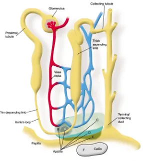
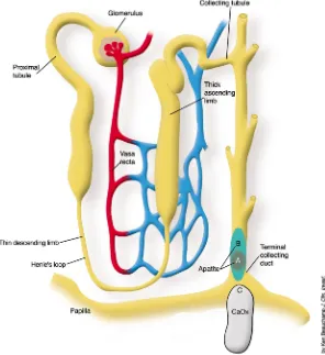
patho-genesis of stone formation and allow investigators to propose and test more focused hypotheses. This would help them to devise effective therapy aimed at preventing recurrent nephrolithia-sis, which afflicts approximately 50% of stone formers within five years of the initial stone (6). Yet until the ele-gant study by A.P. Evan et al. reported in this issue of the JCI(7), we did not have an answer to this rather elemen-tary question. These investigators per-formed kidney biopsies on stone-forming patients to determine the anatomical site and composition of the initial solid phase. They sampled areas adjacent to Randall’s plaques in patients undergoing percutaneous nephrolithotomy. In hypercalciuric calcium oxalate stone formers, they found initial calcium phosphate (apatite) crystallization in the base-ment membrane of the thin limbs of the loop of Henle (Figure 1) with sub-sequent extension to the vasa recta, then to the interstitial tissue sur-rounding the terminal collecting ducts, and finally, in the most severe cases, to the papillae. Erosion of this solid phase into the urinary space, which is supersaturated with respect to calcium oxalate, may have promot-ed heterogeneous nucleation and for-mation of kidney stones. In patients with hyperoxaluria resulting from intestinal bypass, the initial crystals were again a calcium phosphate com-plex, but these arose within the tubule lumens of terminal collecting ducts (Figure 2). Contact of these crystals with urine, supersaturated with respect to calcium oxalate, may have promoted heterogeneous nucleation and formation of kidney stones. Non-stone formers, subjected to nephrecto-my, had neither plaque nor crystals. Thus there are different sites of initial
Address correspondence to:David A. Bushinsky, University of Rochester School of Medicine and Dentistry, Nephrology Unit, Strong Memorial Hospital, 601 Elmwood Avenue, Box 675, Rochester, New York 14642, USA. Phone: (585) 275-3660;
Fax: (585) 442-9201; E-mail:
crystallization depending upon the metabolic abnormality leading to stone formation.
Potential mechanisms for stone formation
Why does the initial solid phase form in these distinct locations, and why are the initial crystals apparently only calcium phosphate? The basement membrane of the thin limb appears an unlikely site for initial crystallization in patients with idiopathic hypercalci-uria. It is not the site of either vectori-al cvectori-alcium or phosphorus transport (8) and, since even the transtubular permeabilities of these ions are very low (8), it is difficult to link supersat-uration within the thin limbs (9) to the surrounding interstitium. Howev-er, anatomically, the thin limbs are in very close proximity to the vasa recta
and the collecting ducts, and all are situated in a highly concentrated, hypertonic environment. One could propose a sequence of events which might lead to increased supersatura-tion and subsequent crystal forma-tion. Following ingestion and absorp-tion of dietary calcium, the renal-filtered load of calcium would increase, resulting in increased tubular calcium concentration (10). The medullary countercurrent mechanism would concentrate the calcium extracted from the thick ascending limb into the hypertonic papilla. The vasa recta, also with an increased calcium con-centration, would fail to readily remove calcium from the interstitium. The increased serum calcium would stimulate the calcium receptor and decrease reabsorption of water in the collecting duct (11), further
concen-trating the interstitium. Vectorial pro-ton transport into the collecting duct would alkalinize the interstitium. The pH of the vasa recta would also increase following gastric proton secretion, the so-called alkaline tide, resulting in less bicarbonate removal from the medullary interstitium. The increased pH would decrease the solu-bility of calcium phosphate complex-es. Perhaps an extracellular matrix protein, specific to the papillary inter-stitium, could provide a site promot-ing heterogeneous nucleation (12), which occurs with a lower degree of supersaturation than homogeneous nucleation. Future studies will be nec-essary to test these hypotheses.
Intraluminal crystal formation in the collecting duct appears a more likely site for initial crystallization in patients following intestinal bypass surgery. The collecting duct fluid can be hypertonic with elevated concen-trations of calcium leading to super-saturation. Yet the urine from the patients in Evan’s study was undersat-urated with respect to calcium phos-phate, indicating that, thermodynam-ically, a stone should not form. However, the lack of demonstrable supersaturation may be a function of the 24-hour urine collection; the max-imal supersaturation, and thus the propensity for stone formation, is never detected. While a 24-hour urine collection is an important predictor of the likelihood of forming stones, it is not the sole predictor. It seems proba-ble that supersaturation initiates crys-tal formation, but we still do not understand the relationship between the degree of urinary supersaturation and stone disease.
Future directions
Now that we know where the initial solid phase forms, what are the next questions? Investigators studying the kidney generally concentrate on the effects of transport on tubular fluid ion concentration; the current study will force us to look more carefully at the effects of basolateral membrane transport on interstitial ion concen-trations. We know little about super-saturation in this critical region of the kidney, yet this is where the majority of stones originate.
[image:3.576.83.369.49.372.2]Unneeded calcium and oxalate must be excreted in a minimal amount of urine to rid the body of these potential Figure 1
toxins while conserving extracellular fluid volume. Human urine and blood are supersaturated with respect to cal-cium oxalate (1) and calcal-cium hydro-gen phosphate (13), respectively, yet solid phases rarely form because we produce inhibitors to initial nucle-ation and subsequent aggregnucle-ation (12). In rats, stone formation occurs when the magnitude of the supersatu-ration overcomes this potent inhibi-tion (14). The current study should point us in the direction of investigat-ing the relationship between supersat-uration and inhibitor proteins not only in the urine but in the intersti-tium as well.
Crystals may stimulate production of proteins, such as osteopontin, which appear to regulate growth of the solid phase (15). In patients with idio-pathic hypercalciuria, there was evi-dence for crystal-induced cell injury in areas of dense crystal deposition, while in the bypass patients there was not
only cell injury but also cell death (7). Were levels of osteopontin increased in the medullary interstitium in either of these types of stone formers?
An animal model of stone formation
The genetic hypercalciuric stone– forming rat exhibits metabolic abnor-malities similar to patients with idio-pathic hypercalciuria in that these rats absorb excessive amounts of intestin-al cintestin-alcium, they fail to adequately reabsorb filtered calcium, and their bone resorption is uniquely sensitive to 1,25(OH)2D3. All of these
charac-teristics are apparently due to an increase in the number of receptors for vitamin D (16, 17). The genetic hypercalciuric stone-forming rats spontaneously form calcium phos-phate stones (18) similar to those found in this study unless their diet is augmented with an oxalate precursor (19). Both hypercalciuric rats and
humans appear to be predisposed to initially form calcium phosphate stones and not the commonly ob-served calcium oxalate stones. In both rats (20) and humans (21) the upper limit of metastability, that level of supersaturation at which a solid phase forms, increases with increasing calci-um oxalate, but not calcicalci-um phos-phate, supersaturation. Thus rats and humans appear protected against cal-cium oxalate stone formation unless a nucleation site, such as the more easi-ly formed calcium phosphate crystal, is present.
This study highlights the role of physician scientists working with basic scientists in medical research to joint-ly address important problems using sophisticated clinical and laboratory techniques and then applying these results to refine hypotheses for further testing. Agile movement between the bedside and the bench, as exemplified in this study, will provide insight into, and ultimately prevention of, disor-ders such as nephrolithiasis.
Acknowledgments
This work was supported in part by NIH Grants AR 46289, DK 57716, and DK 56788.
1. Monk, R.D., and Bushinsky, D.A. 2003. Kidney stones. In Williams textbook of endocrinology. P.R. Larsen, H.M. Kronenberg, S. Melmed, and K.S. Polonsky, editors. W.B.Saunders. Philadelphia, Pennsylvania, USA. 1411–1425.
2. Randall, A. 1937. The origin and growth of renal calculi. Ann. Surg.105:1009–1027.
3. Randall, A. 1940. Papillary pathology as a precur-sor of primary renal calculus. J. Urol.44:580–589. 4. Finlayson, B., and Reid, F. 1978. The expectation of free and fixed particles in urinary stone disease.
Invest. Urol.15:442–448.
5. Lieske, J.C., and Toback, F.G. 2000. Renal cell-uri-nary crystal interactions. Curr. Opin. Nephrol. Hypertens.9:349–355.
6. Asplin, J.R., Favus, M.J., and Coe, F.L. 2000. Nephrolithiasis. In The kidney. B.M. Brenner, edi-tor. W.B. Saunders Company. Philadelphia, Penn-sylvania, USA. 1774–1819.
7. Evan, A.P., et al. 2003. Randall’s plaque of patients with nephrolithiasis begins in basement membranes of thin loops of Henle. J. Clin. Invest.
111:607–616. doi:10.1172/JCI200317038. 8. Rocha, A.S., Magaldi, J.B., and Kokko, J.P. 1977.
Calcium and phosphate transport in isolated seg-ments of rabbit Henle’s loop. J. Clin. Invest.
59:975–983.
9. Asplin, J.R., Mandel, N.S., and Coe, F.L. 1996. Evi-dence for calcium phosphate supersaturation in the loop of Henle. Am. J. Physiol.270:F604–F613. 10. Bushinsky, D.A., and Monk, R.D. 1998. Calcium.
Lancet.352:306–311.
11. Hebert, S.C., Brown, E.M., and Harris, H.W. 1997. Role of the Ca2+-sensing receptor in divalent
[image:4.576.71.367.54.377.2]min-eral ion homeostasis. J. Exp. Biol.200:295–302. 12. Coe, F.L., and Parks, J.H. 1997. New insights into the pathophysiology and treatment of nephrolithiasis: new research venues. J. Bone.
Figure 2
Miner. Res.12:522–533.
13. Neuman, W.F., and Neuman, M.W. 1958. The chemical dynamics of bone mineral. University of Chicago Press. Chicago, Illinois, USA. 209 pp. 14. Bushinsky, D.A., Parker, W.R., and Asplin, J.R. 2000. Calcium phosphate supersaturation regu-lates stone formation in genetic hypercalciuric stone-forming rats. Kidney Int.57:550–560. 15. Xie, Y., et al. 2001. Expression, roles, receptors,
and regulation of osteopontin in the kidney. Kid-ney Int.60:1645–1657.
16. Bushinsky, D.A. 1999. Genetic hypercalciuric stone-forming rats. Curr. Opin. Nephrol. Hypertens.
8:479–488.
17. Li, X.-Q., Tembe, V., Horwitz, G.M., Bushinsky, D.A., and Favus, M.J. 1993. Increased intestinal vitamin D receptor in genetic hypercalciuric rats: a cause of intestinal calcium hyperabsorption.
J. Clin. Invest. 91:661–667.
18. Bushinsky, D.A., Grynpas, M.D., Nilsson, E.L., Nakagawa, Y., and Coe, F.L. 1995. Stone forma-tion in genetic hypercalciuric rats. Kidney Int.
48:1705–1713.
19. Bushinsky, D.A., et al. 2002. Calcium oxalate stone formation in genetic hypercalciuric stone-forming rats. Kidney Int.61:975–987.
20. Asplin, J.R., et al. 1997. Relationship between supersaturation and crystal inhibition in hyper-calciuric rats. Kidney Int.51:640–645.
21. Asplin, J.R., Parks, J.H., and Coe, F.L. 1997. Dependence of upper limit of metastablility on supersaturation in nephrolithiasis. Kidney Int.