Copyright © 1998, American Society for Microbiology. All Rights Reserved.
MINIREVIEW
Evolution of Streptococcus pneumoniae Serotypes and Antibiotic
Resistance in Spain: Update (1990 to 1996)
ASUNCIO´ N FENOLL,* ISABEL JADO, DOLORES VICIOSO, AMALIA PE´REZ,ANDJULIO CASAL
Laboratorio de Referencia de Neumococos, Servicio de Bacteriologı´a, Centro Nacional de Microbiologı´a, Instituto de Salud Carlos III, 28220 Majadahonda, Madrid, Spain
INTRODUCTION
The surveillance in Spain of Streptococcus pneumoniae iso-lated in hospitals from different regions of the country was initiated in our laboratory in 1979. In that year we found that 6% of the pneumococci isolated from blood, cerebrospinal fluid (CSF), or lower respiratory tract (LRT) specimens were not penicillin susceptible and that these strains were distrib-uted only among particular serogroups or serotypes (SGTs). We thus realized that although penicillin resistance was rare in most other countries, it was clearly significant in Spain (6). Rates of penicillin resistance increased continuously in the following years, reaching values up to 44.3% among invasive strains in 1989, and both highly penicillin-resistant and multi-drug-resistant strains reached high levels (15.3 and 7.9%, re-spectively) (11). In the present decade, resistance to penicillin has spread throughout the world and numerous countries are observing the same pattern that occurred in our country during the 1980s (1, 2, 16). The situation with antimicrobial resistance in Spain at the end of the 1980s caused concern (3, 22), and knowledge of its evolution could be of interest for those coun-tries where increasing resistance is being observed. This survey presents an update of the previous microbiological surveillance for pneumococcal infections. Here, we have included both invasive and noninvasive pneumococci isolated from 1990 to 1996 in Spanish hospitals. For comparative purposes, some tables and figures present data obtained in the period 1979 to 1989, including analysis of not only invasive strains (6, 11) but also noninvasive strains.
PNEUMOCOCCAL STRAINS AND LABORATORY DIAGNOSIS
From January 1990 to December 1996 a total of 9,243 pneu-mococcal isolates were received at the Pneupneu-mococcal Refer-ence Laboratory, Madrid, Spain, for typing purposes and an-tibiotic resistance surveillance. There was an increasing number of isolates every year, from 779 in 1990 to 1,633 in 1996. Pneumococci were isolated from patients and healthy carriers at 62 hospitals from 13 (of the 17) autonomous com-munities of Spain. Of the isolates received, 51% were from patients with systemic infections (pneumonia, sepsis, or men-ingitis) found mostly in the adult population. Among the pneu-mococci from other sources, approximately half (mostly iso-lated from sputum) belonged to adults and the other half were
from children with local (ear, sinus, and conjunctive) infections and from the nasopharynges of healthy carriers (Table 1). Among 8,740 isolates, 5,917 were from males and 2,823 were from females (ratio, 2.09). The ratio of males to females was 2.6 for adults and 1.3 for children.
All isolates were confirmed to be S. pneumoniae by colony morphology on blood agar, optoquine susceptibility, and so-dium deoxycholate solubility. Those strains that were negative for one or more of these tests (most of them unencapsulated) were studied by hybridization with two probes based on the autolysin and pneumolysin genes (12, 40). These two pneumo-coccal proteins have been demonstrated to be species specific (12, 15, 21, 32, 35, 45). Isolates resistant to optoquine and/or insoluble in deoxycholate but positive by hybridization were regarded as atypical pneumococcal strains and were included in the study. Serotyping was carried out by the Quellung reac-tion, with the use of 46 antisera provided by the Statens Seru-minstitut (Copenhagen, Denmark). Some of the pneumococci received in 1996 were serotyped by a dot blot assay using the same antisera (10). Susceptibilities to penicillin, tetracycline, chloramphenicol, erythromycin, cefotaxime, and vancomycin were determined by the agar dilution technique as previously described (11), according to criteria from the National Com-mittee for Clinical Laboratory Standards, guideline M-100-S6, for interpretation (28). In the present text, the term penicillin-resistant pneumococci (PRP) refers to both moderately and highly resistant strains, unless otherwise specified.
EVOLUTION OF SGTs
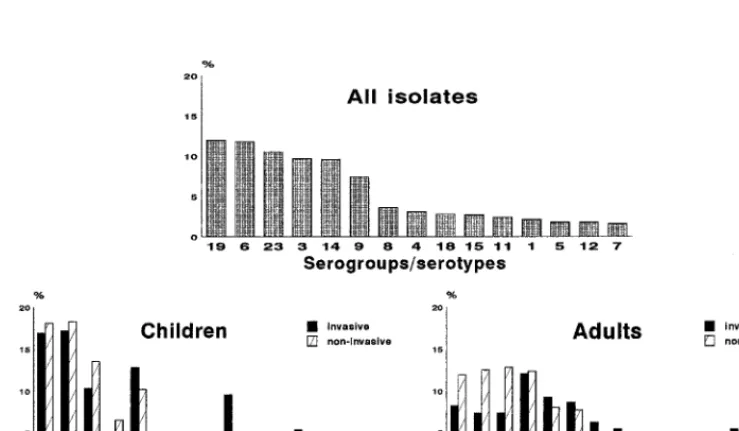
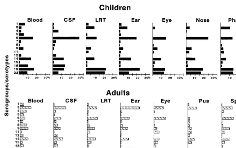
Thirty-eight different SGTs were found among the 9,243 pneumococci, but only 15 of them accounted for 83% of the strains (Fig. 1). The six SGTs found most frequently were SGTs 19 (12%), 6 (11.8%), 23 (10.5%), 3 (9.7%), 14 (9.6%), and 9 (7.4%), representing 61% of the pneumococci, while the remaining nine SGTs made up only 22% of isolates. The dis-tribution of pneumococci of these SGTs isolated from invasive and noninvasive diseases in children and adults is also shown in Fig. 1. Figure 2 shows details of the SGT distribution of iso-lates from different sources in children and adults. SGTs 1, 4, 5, 7, and 12 were isolated with a greater frequency from blood and CSF than from other sources, in both children and adults. In contrast, serogroup 18 was associated with blood and CSF only in children. SGTs 6, 14, 19, and 23 were found more frequently in children, regardless of source, and SGTs 3, 8, and 9 had an obvious predilection for adults. In adults, serotype 3 ranked first or second most common among invasive isolates and those from most other sources. However, in children with invasive disease serotype 3 isolates were rarely recovered, al-though these isolates were an important cause of otitis media
* Corresponding author. Mailing address: Laboratorio de Referen-cia de Neumococos, Servicio de Bacteriologı´a, Centro Nacional de Microbiologı´a, Instituto de Salud Carlos III, 28220 Majadahonda, Madrid, Spain. Phone: 34-915097975. Fax: 34-915097966. E-mail: [email protected].
3447
on May 15, 2020 by guest
http://jcm.asm.org/
and were the fourth most common serotype among isolates from the pharynges of carriers.
The predominant SGTs have remained the same over time, although the relative frequency of some of them has varied significantly. Figure 3 shows the changes in the prev-alence of invasive and noninvasive isolates of the 15 most-common SGTs since 1979. The prevalence of SGTs 9, 14,
[image:2.612.49.547.89.347.2]and 19 increased from 2 to 7%, 6 to 14%, and 5 to 7.5%, respectively. The prevalence of SGTs 1, 5, and 7 decreased from 10 to 2%, 7 to 1%, and 6 to 1%, respectively. The prevalence of serogroup 6 rose until 1987, when it reached 18.5%. It then slowly decreased but in the last 4 years has gradually risen again. The prevalence of serogroup 23 in-creased rapidly at the beginning of the 1980s from 5% to
[image:2.612.112.482.492.707.2]FIG. 1. Percentages of the 15 top-ranking pneumococcal SGTs isolated from all sources and ages (top) and from children and adult with invasive and noninvasive disease (bottom) in Spain from 1990 to 1996.
TABLE 1. Distribution of 9,243 pneumococcal strains according to source, age, and penicillin resistance in Spain from 1990 to 1996 No. (%) Penr
Strain group and source
Strains detected in:
Patients of all ages Children (0–14 yr) Adults (.14 yr) Patients of unknown age (total) Total No. (%) Penr Total No. (%) Penr Total No. (%) Penr
Invasive
Blood 3,319 1,143 (34.4) 424 216 (50.9) 2,160 681 (31.5) 735
CSF 384 166 (43.2) 80 46 233 89 (38.2) 71
LRTa 1,017 551 (54.2) 76 54 754 395 (52.4) 187
Total 4,720 1,860 (39.4) 580 316 (54.5) 3,147 1,165 (37.0) 993
Noninvasive
Sputum 1,447 863 (59.6) 44 26 1,145 669 (58.4) 258
Ear 691 426 (61.6) 512 325 (63.5) 107 54 (50.5) 72
Nose 492 332 (67.5) 386 265 (68.7) 64 40 42
Eye 668 396 (59.3) 368 244 (66.3) 200 95 (47.5) 100
Pharynx 447 238 (53.2) 343 175 (51.0) 33 23 71
Pus 147 85 (57.8) 33 26 91 46 23
Total 3,892 2,340 (60.1) 1,686 1,061 (62.9) 1,640 927 (56.5)
Otherb 631 327 129 74 300 134 202
Total 9,243 4,527 (49.0) 2,395 1,451 (60.6) 5,087 2,226 (43.7) 1,761
aLRT includes strains isolated from pleural fluid transtracheal puncture, bronchial protected brush, and pulmonary biopsy specimens.
bNo data (237 strains) and miscellaneous, (394 strains isolated from peritoneum, articular fluid, urine, vagina, Bartholin’s gland, and catheter specimens).
3448 MINIREVIEW J. CLIN. MICROBIOL.
on May 15, 2020 by guest
http://jcm.asm.org/
nearly 20% and then decreased, representing 10% of the total isolates in 1996.
EVOLUTION OF ANTIMICROBIAL RESISTANCE
Of the 9,243 strains studied, 4,527 (49%) showed decreased susceptibility to penicillin (MIC . 0.06mg/ml), 4,011 (43.4%) showed decreased susceptibility to tetracycline (MIC.2mg/ml), 2,852 (30.9%) showed decreased susceptibility to chlorampheni-col (MIC.4mg/ml), 2,077 (22.5%) showed decreased suscepti-bility to erythromycin (MIC. 0.25mg/ml), and 2,008 (21.7%) showed decreased susceptibility to cefotaxime (MIC. 0.5mg/ ml). All strains were susceptible to vancomycin (MIC# 0.5 mg/ml). Among pneumococci isolated from systemic infections, 39.4% were resistant to penicillin, 36% were resistant to tet-racycline, 25.7% were resistant to chloramphenicol, 16.7%
were resistant to erythromycin, and 18.6% were resistant to cefotaxime; for noninvasive pneumococci, the values were 59.4, 51.6, 36.5, 28.8, and 25.1%, respectively. Figure 4 shows how the percentages of resistant isolates have varied through the years. Among pneumococci causing systemic infections, there was an increasing trend towards penicillin resistance in the 1980s, reaching a maximum of 44.3% in 1989. From that year on, the rate decreased to 34.5% in 1992 and then rose again to around 42% in the final 3 years of the study. Tetra-cycline and chloramphenicol resistance followed the descend-ing pattern observed in previous years. In contrast, erythromy-cin resistance has been growing continuously, doubling from 10.2% (in 1990) to 23.8% (in 1996) in invasive pneumococci and from 20.7 to 40.4% in noninvasive pneumococci.
[image:3.612.104.495.70.316.2]Table 1 summarizes the percentage of PRP found in differ-ent specimens from children and adults. Blood pneumococci
[image:3.612.89.511.535.713.2]FIG. 2. Distribution of the 15 most-common pneumococcal SGTs from different specimen sources in children and adults in Spain from 1990 to 1996.
FIG. 3. Annual frequency of the 15 most-common SGTs in Spain from 1979 to 1996.
on May 15, 2020 by guest
http://jcm.asm.org/
presented the lowest rates of penicillin resistance (50.9% in children and 31.5% in adults), whereas nasal isolates showed the highest (68.7 and 62.5%, respectively). Rates of PRP were lower among adults than among children, regardless of the source of the specimen.
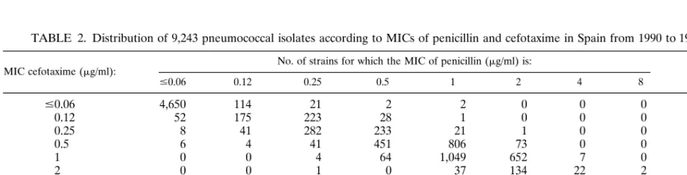
The MIC distribution for penicillin versus cefotaxime is shown in Table 2. Of the total isolates, 39% were moderately resistant and 10% were highly resistant to penicillin (of those, 31 and 8.4%, respectively, were invasive isolates). For cefo-taxime these percentages were 19.2 and 2.5%, respectively (of those, 16.6 and 2%, respectively, were invasive isolates). For most of the highly resistant pneumococci the MICs of the two b-lactams were 2 mg/ml. The MICs of cefotaxime correlated well with those of penicillin. For 6,764 isolates (73.2%) the MICs of the two antibiotics were identical, and for most other strains they differed by only 1 or 2 dilutions. For 264 (2.8%) strains, the MICs of cefotaxime were higher than those of penicillin. In contrast, the MICs of cefotaxime were lower in 2,215 (24%) strains. None of the penicillin-susceptible pneu-mococci were resistant to cefotaxime, but among the cefo-taxime-susceptible strains, 74 were resistant to penicillin. Ex-cept for one serogroup 19 strain (MIC of penicillin and cefotaxime58mg/ml) and a serotype 14 strain (MIC of pen-icillin50.25mg/ml; MIC of cefotaxime58mg/ml), all other
isolates for which the MICs were 8mg/ml were atypical pneu-mococci (noncapsulated, optoquine resistant, and/or bile neg-ative). Among isolates for which the MICs were 4mg/ml, 50% were atypical pneumococci and the rest belonged mostly to SGT 23. Seventy percent of the isolates for which the MICs were 2mg/ml belonged to SGTs 6, 14, and 23, while isolates of SGTs 9 and 19 were moderately resistant in the majority of cases.
[image:4.612.87.506.70.261.2]Table 3 summarizes the antibiotic resistance patterns found in this 7-year study, comparing them with those found in pre-vious periods. Among PRP, 72% were also resistant to other antibiotics, while among penicillin-susceptible pneumococci, only 21.5% were resistant to other drugs. Overall, 5,540 pneu-mococci were resistant to one or more drugs (60%), this being less than that found in previous years (74% in 1979 to 1984 and 68% in 1985 to 1989). Although the total percentage of pneu-mococci resistant to one or more drugs has decreased through the years, the number of strains with certain patterns of pen-icillin and/or erythromycin resistance has risen. Among them, the important and continuous increase in multidrug-resistant (PTCE) pneumococci (i.e., those resistant to penicillin, tetra-cycline, chloramphenicol, and erythromycin) is of particular concern. The average incidence of these isolates increased from 1.1% in 1979 to 1984 to 7.7% in 1985 to 1989 and 12.5%
FIG. 4. Development of antibiotic resistance in pneumococcal isolates in Spain from 1979 to 1996. From 1979 to 1983 only invasive isolates were studied. Cefotaxime susceptibility testing started in 1989.
TABLE 2. Distribution of 9,243 pneumococcal isolates according to MICs of penicillin and cefotaxime in Spain from 1990 to 1996a
MIC cefotaxime (mg/ml): No. of strains for which the MIC of penicillin (mg/ml) is: of strainsTotal no.
#0.06 0.12 0.25 0.5 1 2 4 8
#0.06 4,650 114 21 2 2 0 0 0 4,789
0.12 52 175 223 28 1 0 0 0 479
0.25 8 41 282 233 21 1 0 0 586
0.5 6 4 41 451 806 73 0 0 1,381
1 0 0 4 64 1,049 652 7 0 1,776
2 0 0 1 0 37 134 22 2 196
4 0 0 0 1 1 3 16 7 28
8 0 0 1 0 0 0 0 7 8
Total no. of strains 4,716 334 573 779 1,917 863 45 16 9,243
aSusceptibility categorizations for penicillin and cefotaxime: highly resistant, MIC$2mg/ml (both); moderately resistant, 0.12mg/ml#MIC#1mg/ml and MIC5
1mg/ml, respectively; and susceptible, MIC#0.06mg/ml and#MIC#0.5mg/ml, respectively.
3450 MINIREVIEW J. CLIN. MICROBIOL.
on May 15, 2020 by guest
http://jcm.asm.org/
[image:4.612.51.547.582.708.2]in 1990 to 1996. It should also be pointed out that within the period 1990 to 1996, the incidence of PTCE isolates increased notably (from 9.5% in 1990 to 16.6% in 1996).
RELATIONSHIP BETWEEN SGTs AND PENICILLIN RESISTANCE
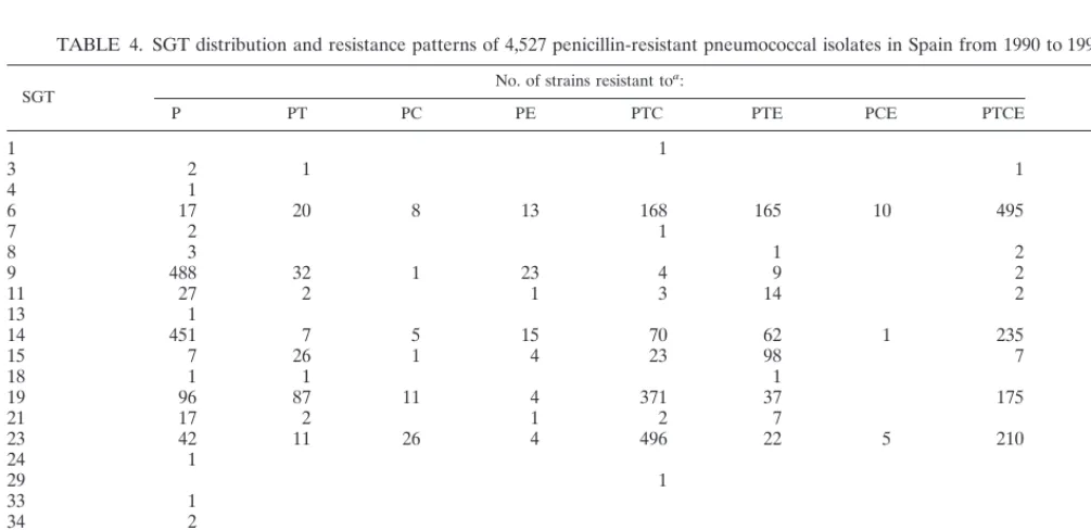
Table 4 shows the distribution of the PRP patterns by SGTs. Resistant pneumococci belonged to 20 different SGTs, but
86% of the isolates were confined to the 5 SGTs classically associated with penicillin resistance: SGTs 6 (19.7%), 9 (12.3%), 14 (18.7%), 19 (17.2%), and 23 (18%). Of the serogroup 6 resistant isolates, 55% demonstrated the PTCE pattern, 87% of serogroup 9 and 53% of serotype 14 were resistant only to penicillin, and 47% of serogroup 19 and 60% of serogroup 23 belonged to the PTC pattern (resistance to penicillin, tetracy-cline, and chloramphenicol).
The relative frequencies of SGTs 6, 9, 14, 19, and 23 within the PRP population throughout the years is shown in Fig. 5. At the beginning of the 1980s the majority of the PRP belonged to SGTs 6 and 23 (73% in 1985). The relative importance of SGTs 6 and 23 has decreased since then, even though in the last 2 years the percentage of PRP in SGT 6 has slightly increased. The other SGTs, which prior to 1985 were very infrequent among PRP, have subsequently increased in fre-quency, and at present the distributions of the five SGTs among the PRP population are quite similar.
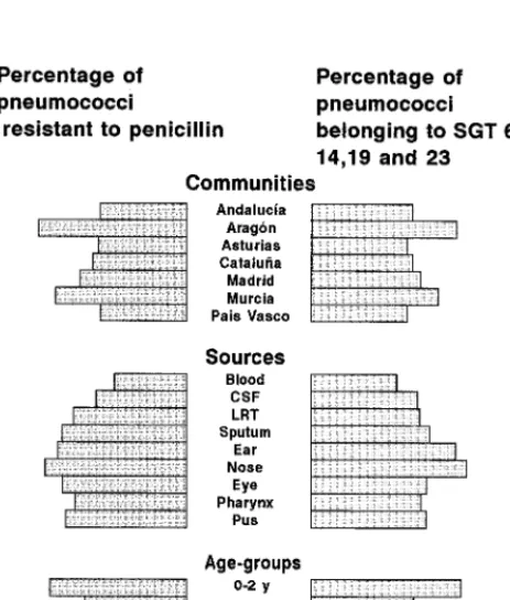
Pneumococci belonging to SGTs 6, 9, 14, 19, and 23 studied from 1990 to 1996 were almost all penicillin resistant. Penicillin resistance was found to be 95% for serotype 14 pneumococci, 84.5% for serogroup 23, 82% for serogroup 6, 80% for sero-group 9, and 70% for serosero-group 19. Figure 6 compares the percentages of PRP and of pneumococci belonging to the five SGTs recovered from different Spanish communities, sources, and age groups. Both distributions are quite similar, indicating that in Spain the differences in penicillin resistance rates are mainly due to differences in the prevalence of pneumococci belonging to SGTs 6, 9, 14, 19, and 23.
COMMENTS AND CONCLUSION
The epidemiological characteristics of the different pneumo-coccal SGTs are extremely complex. Although the leading SGTs causing illness are the same worldwide, for reasons that are unclear significant differences exist in their relative
fre-TABLE 3. Comparison of antibiotic resistance patterns of 9,243 pneumococcal isolates with those found in previous periods in Spain
Antibiotic(s)a
No. (%) of strains resistant during: 1979–1984b
(n51,381) 1985–1989 b
(n52,577) (n1990–199659,243)
P 63 (4.6) 207 (8.0) 1262 (13.7)
T 235 (17.0) 290 (11.3) 388 (4.2)
C 13 (0.9) 44 (1.7) 29 (0.3)
E 0 (—)c 7 (0.3) 53 (0.6)
PT 59 (4.3) 84 (3.3) 233 (2.5)
PC 9 (0.7) 27 (1.0) 55 (0.6)
PE 3 (0.2) 1 (—) 107 (1.2)
TC 342 (24.8) 264 (10.2) 337 (3.6)
TE 2 (0.1) 7 (0.3) 104 (1.1)
CE 0 (—) 0 (—) 4 (—)
PTC 252 (18.2) 570 (22.1) 1159 (12.5)
PTE 5 (0.4) 25 (1.0) 541 (5.9)
PCE 0 (—) 6 (0.2) 19 (0.2)
TCE 21 (1.5) 18 (0.7) 98 (1.1)
PTCE 15 (1.1) 199 (7.7) 1151 (12.5)
Total 1,019 (73.8) 1,749 (67.9) 5,540 (59.9)
aP, penicillin; T, tetracycline; C, chloramphenicol; and E, erythromycin. bData include invasive and noninvasive pneumococci.
[image:5.612.50.291.89.287.2]c—, 0% or an insignificant percentage.
TABLE 4. SGT distribution and resistance patterns of 4,527 penicillin-resistant pneumococcal isolates in Spain from 1990 to 1996
SGT No. of strains resistant to
a:
Total
P PT PC PE PTC PTE PCE PTCE
1 1 1
3 2 1 1 4
4 1 1
6 17 20 8 13 168 165 10 495 896
7 2 1 3
8 3 1 2 6
9 488 32 1 23 4 9 2 559
11 27 2 1 3 14 2 49
13 1 1
14 451 7 5 15 70 62 1 235 846
15 7 26 1 4 23 98 7 166
18 1 1 1 3
19 96 87 11 4 371 37 175 781
21 17 2 1 2 7 29
23 42 11 26 4 496 22 5 210 816
24 1 1
29 1 1
33 1 1
34 2 2
35 8 2 10
NTb 95 44 3 40 19 125 3 22 351
Total 1,262 233 55 107 1,159 541 19 1,151 4,527
aAbbreviations: P, penicillin; T, tetracycline; C, chloramphenicol; and E, erythromycin. bNT, nontypeable strains.
on May 15, 2020 by guest
http://jcm.asm.org/
[image:5.612.50.553.465.709.2]quencies as a function of time, geography, age of patient, and type of infection (14, 19, 34).
Regarding geographical variations, SGTs that are prevalent in some countries hardly cause illness in others. Serotypes 1 and 5 cause illness with much greater frequency in Latin Amer-ica than elsewhere but are especially uncommon in the United States and Canada (36). In Spain these two serotypes were important at the beginning of the 1980s but presently rank among the last places in frequency. Meanwhile, in some Eu-ropean countries, such as Denmark and Germany, serotype 1 isolates are the most frequent cause of infections (29, 33). The incidence of serotype 3 has decreased greatly in recent years in many countries (5, 18, 43); however, in Spain this type has maintained a high frequency throughout the last 18 years. An increasing proportion of serotype 14 isolates has been
ob-served in Spain and other countries (5, 18, 43) over the last 7 years. In general, geographical and temporal differences in the SGT distribution interfere with the development of one unique vaccine for worldwide use. In this context Sniadack et al. re-viewed pneumococcal SGT data from 16 countries on six con-tinents and estimated that the coverage for an optimal nano-valent vaccine for global use ranges from 44 to 90% of SGTs (38).
The differences found in SGT distribution by age coincide with previously published data (19). SGTs 6, 14, 18, 19, and 23 are most frequently associated with infection in children, while SGTs 3, 8, and 9 are more prevalent in adults. Analyzing the SGTs of 7,000 pneumococci isolated in 13 countries, Scott et al. observed as much variation in SGT distribution by age as one might expect between different species of respiratory bac-terial pathogens (36).
Although, in general, PRP have increased worldwide during recent years, the prevalence of resistance and the rates of emergence of resistance vary considerably from one country to another (1, 2, 16). Penicillin resistance increased sharply in Spain until 1989 (11), but it has since remained stable, despite annual variations, with a resistance rate among invasive pneu-mococci of about 42%. In many countries with low resistance rates during the previous decade, there has been a remarkable increase in the 1990s, similar to that which occurred in Spain in the 1980s. In Portugal penicillin resistance increased from 4.6% in 1989 to 17.9% in 1993 (31), in France it increased from 3.2% in 1987 to 20% in 1992 (4), and in the United States it increased from around 5% in the 1980s to 33.5% in 1996 (41). In contrast the incidence of PRP has remained stable at very low levels in other areas of Europe: Denmark (,1%), Ger-many (1.8%), Belgium (2 to 4%), Sweden (1.7%), Finland (1.7%), Great Britain (1.5 to 3.9%), and Italy (5.5%) (18, 20, 23, 29, 30, 33, 43). In the present situation, it is clear that in order to stop the increase and, if possible, reduce the fre-quency of PRP, an exhaustive surveillance of pneumococcal resistance is required, along with a strict control of antibiotic policy.
Association of penicillin resistance with only certain specific SGTs is a general finding. However, no clear explanation has been found for this association. Spanish penicillin-resistant isolates have been genetically characterized on numerous oc-casions, showing the existence of a number of distinct clones, the majority of which have spread globally (25, 27, 42). It has also been demonstrated that some of the resistant clones cir-culating in different countries (such as the multidrug-resistant serotype 19F) are serotype variants of the Spanish clone 23F, which originated by the horizontal transfer of capsular genes. In other cases new clones have emerged by the horizontal spread of altered pbp genes (e.g., the penicillin-resistant sero-type 9V clone) (7, 8). Although clonal spread, horizontal trans-fer, and antibiotic policy seem to have a definitive influence on PRP prevalence, there are many findings that are not easily explained by taking only these factors into account. Thus, it is difficult to explain how the Spanish clones 6B and 23F have spread to and become established in regions as remote as Iceland or South Korea (24, 39, 44) but have not become prevalent in England or Germany, in spite of millions of tour-ists coming through our country each year. In the 1980s, the Spanish clone 6B was detected in Iceland and Finland simul-taneously, and, surprisingly, the epidemic spread of clone 6B took place in Iceland but not in Finland (39).
[image:6.612.54.290.74.211.2]Molecular analyses of PRP isolates in different countries have demonstrated that just one or two clones are responsible for the majority of the resistant population. For instance, in France 50% of the PRP are the Spanish serotype 23F clone, in
FIG. 5. Relative frequencies of SGTs 6, 9, 14, 19, and 23 within the PRP population in Spain from 1979 to 1996.
FIG. 6. Comparison of PRP and SGTs by autonomous community, source, and age groups.
3452 MINIREVIEW J. CLIN. MICROBIOL.
on May 15, 2020 by guest
http://jcm.asm.org/
[image:6.612.60.292.433.705.2]Slovakia nearly all PRP are the serotype 14 clone, and in South Africa 71% of the PRP are serotype 19A and 6B clones (9, 13, 37). During the early 1980s in Spain, two major clones, 6B and 23F, were clearly predominant, but recently they have made way for other clones. On the other hand, and in spite of se-lective pressure exerted by the overuse of antibiotics in our country, certain penicillin-susceptible SGTs have not been dis-placed by these resistant clones. Currently, serotype 3 pneu-mococci are uniformly penicillin susceptible, although this has been one of the most prevalent serotypes in Spain for at least the past 18 years. This serotype not only has been isolated from adults but also is one of the main causes of otitis media in children. Curiously, serotype 3 pneumococci have been recov-ered with high frequency from infant carriers, and in spite of the extensive use of antibiotics in infants, for reasons that are not understood this serotype has neither acquired penicillin resistance nor decreased in prevalence over time.
Although highly resistant PRP (MICs, 8 to 32mg/ml) have been reported in some countries (17), in Spain the level of resistance remains relatively low and stable, with the penicillin MICs for the majority of the most-resistant isolates being 2 mg/ml. The finding in the United States of isolates with ex-tremely high resistance to cefotaxime but low resistance to penicillin and the proof that such resistance could be acquired by only one transformation step caused alarm among microbi-ologists due to the likely spread of this resistance pattern among pneumococcal populations (26). Fortunately this has not occurred, and currently this type of resistant isolate is very rare outside the United States. In our study, among the 9,243 strains analyzed, for only 3 was the MIC of cefotaxime signif-icantly greater than that of penicillin.
The results of the present study show that the alarming tendency toward increasing penicillin resistance in pneumo-cocci from Spain may have ended over the last few years. In contrast, erythromycin resistance has been growing continu-ously.
ACKNOWLEDGMENTS
We are grateful to B. G. Spratt for critical reading of the manuscript. We also express our appreciation to all Spanish microbiologists who sent strains to our laboratory.
REFERENCES
1. Appelbaum, P. C. 1992. Antimicrobial resistance in Streptococcus
pneu-moniae: an overview. Clin. Infect. Dis. 15:77–83.
2. Baquero, F. 1996. Epidemiology and management of penicillin-resistant pneumococci. Curr. Opin. Infect. Dis. 9:372–379.
3. Barnett, E. D., J. O. Klein, and D. W. Teele. 1992. Pneumococcal vaccine for Olympic athletes and visitors to Spain. N. Engl. J. Med. 326:1572. 4. Bedos, J. P., S. Chevret, C. Chastang, P. Geslin, B. Regnier, and French
Cooperative Pneumococcus Study Group.1996. Epidemiological features of and risk factors for infection by Streptococcus pneumoniae strains with di-minished susceptibility to penicillin: findings of a French survey. Clin. Infect. Dis. 22:63–72.
5. Butler, J. C., R. F. Breiman, H. B. Lipman, J. Hofmann, and R. R. Facklam. 1995. Serotype distribution of Streptococcus pneumoniae infections among preschool children in the United States, 1978–1994: implications for devel-opment of a conjugate vaccine. J. Infect. Dis. 171:885–889.
6. Casal, J. 1982. Antimicrobial susceptibility of Streptococcus pneumoniae: serotype distribution of penicillin-resistant strains in Spain. Antimicrob. Agents Chemother. 22:222–225.
7. Coffey, T. J., S. Berro´n, M. Daniels, M. E. Garcı´a-Leoni, E. Cercenado, E.
Bouza, A. Fenoll, and B. G. Spratt.1996. Multiple antibiotic-resistant
Strep-tococcus pneumoniae recovered from Spanish hospitals (1988–1994): novel
major clones of serotypes 14, 19F and 15F. Microbiology 142:2747–2757. 8. Coffey, T. J., C. G. Dowson, M. Daniels, and B. G. Spratt. 1995. Genetics and
molecular biology ofb-lactam-resistant pneumococci. Microb. Drug Resist.
1:29–33.
9. Doit, C., E. Denamur, B. Picard, P. Gaslin, J. Elion, and E. Bingen. 1996. Mechanisms of the spread of penicillin resistance in Streptococcus
pneu-moniae strains causing meningitis in children in France. J. Infect. Dis. 174:
520–528.
10. Fenoll, A., I. Jado, D. Vicioso, and J. Casal. 1997. Dot blot assay for the serotyping of pneumococci. J. Clin. Microbiol. 35:764–766.
11. Fenoll, A., C. Martı´n-Bourgon, R. Mun˜oz, D. Vicioso, and J. Casal. 1991. Serotype distribution and antimicrobial resistance of Streptococcus
pneu-moniae isolates causing systemic infections in Spain, 1979–1989. Rev. Infect.
Dis. 13:56–60.
12. Fenoll, A., J. V. Martinez-Suarez, R. Mun˜oz, J. Casal, and J. L. Garcı´a. 1990. Identification of atypical strains of Streptococcus pneumoniae by a specific DNA probe. Eur. J. Clin. Microbiol. Infect. Dis. 9:396–401.
13. Figueiredo, A. M., R. Austrian, P. Urbaskova, L. A. Teixeira, and A. Tomasz. 1995. Novel penicillin-resistant clones of Streptococcus pneumoniae in the Czech Republic and in Slovakia Microb. Drug Resist. 1:71–77.
14. Finland, M., and M. W. Barnes. 1977. Changes in occurrence of capsular serotypes of Streptococcus pneumoniae at Boston City Hospital during se-lected years between 1935 and 1974. J. Clin. Microbiol. 5:154–166. 15. Gillespie, S., C. Ullman, M. D. Smith, and V. Emery. 1994. Detection of
Streptococcus pneumoniae in sputum samples by PCR. J. Clin. Microbiol.
32:1308–1311.
16. Goldstein, F. W., J. F. Acar, and The Alexander Project Collaborative
Group.1996. Antimicrobial resistance among lower respiratory tract isolates of Streptococcus pneumoniae: results of a 1992-1993 Western Europe and USA Collaborative Surveillance Study. J. Antimicrob. Chemother. 38(Suppl. A):71–84.
17. Goldstein, F. W., J. Lin¨ares, J. M. Cristino, E. Malafiiej, M. Marcova, A.
Marton, J. Murphy, G. Schito, Y. Trupl, P. Urbaskova, R. Vanhoof, and the European Study Group, Hospital Saint Joseph, Paris.1996. Comparison of the intrinsic activity of 9 betalactams against 1131 strains of Streptococcus pneumoniae isolated in the 11 european countries between 1993 and 1995, abstr. T148. First European Congress of Antimicrobial Chemotherapy, Glas-gow, Scotland.
18. Hedlund, J., S. B. Svenson, M. Kalin, J. Henrichsen, B. Alsson-Liljequist, G.
Mo¨llerberg, and G. Ka´llenius.1995. Incidence, capsular types and antibiotic susceptibility of invasive Streptococcus pneumoniae in Sweden. Clin. Infect. Dis. 21:948–953.
19. Henrichsen, J. 1979. The pneumococcal typing system and pneumococcal surveillance. J. Infect. 1(Suppl. 2):31–37.
20. Johnson, A. P., D. C. E. Speller, R. C. George, M. Warner, G. Domingue, and
A. Efstratiou. 1996. Prevalence of antibiotic resistance and serotypes in pneumococci in England and Wales: results of observational surveys in 1990 and 1995. Br. Med. J. 312:1454–1456.
21. Kalin, M., K. Klanclerski, M. Granstrom, and R. Mo¨llby. 1987. Diagnosis of pneumococcal pneumonia by enzyme-linked immunosorbent assay of anti-bodies to pneumococcal hemolysin (pneumolysin). J. Clin. Microbiol. 25: 226–229.
22. Lancet. 1992. Pneumococcal vaccination for travel to Spain? Lancet 340:84– 85.
23. Marchese, A., E. A. Debbia, A. Arvigo, A. Pesce, and G. C. Schito. 1995. Susceptibility of Streptococcus pneumoniae strains isolated in Italy to peni-cillin and ten other antibiotics. J. Antimicrob. Chemother. 36:833–837. 24. McGee, L., K. P. Klugman, D. Friedland, and H. J. Lee. 1997. Spread of the
Spanish multi-resistant serotype 23F clone of Streptococcus pneumoniae to Seoul, Korea. Microb. Drug. Resist. 3:253–257.
25. Mun˜oz, R., T. Coffey, M. Daniels, C. G. Dowson, G. Laible, J. Casal, R.
Hakenbeck, M. Jacobs, J. M. Musser, B. G. Spratt, and A. Tomasz.1991. Intercontinental spread of a multiresistant clone of serotype 23F
Streptococ-cus pneumoniae. J. Infect. Dis. 164:302–306.
26. Mun˜oz, R., C. G. Dowson, M. Daniels, T. J. Coffey, C. Martin, R. Hakenbeck,
and B. G. Spratt.1992. Genetics of resistant to third-generation cephalo-sporins in clinical isolates of Streptococcus pneumoniae. Mol. Microbiol.
6:2461–2465.
27. Mun˜oz, R., J. M. Musser, M. Crain, D. E. Briles, A. Marton, A. J. Parkinson,
U. Sorensen, and A. Tomasz.1992. Geographic distribution of penicillin-resistant clones of 23F Streptococcus pneumoniae: characterization by peni-cillin-binding protein profiles, surface protein A typing, and multilocus en-zyme electrophoresis. Clin. Infect. Dis. 15:112–118.
28. National Committee for Clinical Laboratory Standards. 1995. Methods for dilution antimicrobial susceptibility tests for bacteria that grow aerobically. Approved standard M7-A3. National Committee for Clinical Laboratory Standards, Villanova, Pa.
29. Nielsen, S. V., and J. Henrichsen. 1996. Incidence of invasive pneumococcal disease and distribution of capsular types of pneumococci in Denmark, 1989–94. Epidemiol. Infect. 117:411–416.
30. Nissinen, A., M. Leinonen, P. Huovinen, E. Herva, M. L. Katila, S.
Konti-ainen, O. LiimatKonti-ainen, S. Oinonen, A. K. Takala, and P. H. Ma¨kela¨.1995. Antimicrobial resistance of Streptococcus pneumoniae in Finland, 1987–1990. Clin. Infect. Dis. 20:1275–1280.
31. Pato, M. V. V., C. B. Carvalho, A. Tomasz, and The Multicenter Study
Group.1995. Antibiotic susceptibility of Streptococcus pneumoniae isolates in Portugal. A multicenter study between 1989 and 1993. Microb. Drug. Resist.
1:59–69.
32. Pozzi, G., M. R. Oggioni, and A. Tomasz. 1989. DNA probe for identification of Streptococcus pneumoniae. J. Clin. Microbiol. 27:370–372.
on May 15, 2020 by guest
http://jcm.asm.org/
33. Reinert, R. R., A. Queck, A. Kaufhold, M. Kresken, and R. Lu¨tticken. 1995. Antimicrobial resistance and type distribution of Streptococcus pneumoniae isolates causing systemic infections in Germany, 1992–1994. Clin. Infect. Dis.
21:1398–1401.
34. Robbins, J. B., R. Austrian, C. J. Lee, S. C. Rastogi, G. Schiffman, J.
Hen-richsen, P. H. Ma¨kela¨, C. V. Broome, R. R. Facklam, R. H. Tiesjema, and J. C. Parke. 1983. Considerations for formulating the second-generation pneumococcal capsular polysaccharide vaccine with emphasis on cross-reac-tive types within groups. J. Infect. Dis. 148:1136–1159.
35. Rudolph, K. M., A. J. Parkinson, C. M. Black, and L. W. Mayer. 1993. Evaluation of polymerase chain reaction for diagnosis of pneumococcal pneumonia. J. Clin. Microbiol. 31:2661–2666.
36. Scott, J. A. G., A. J. Hall, R. Dagan, J. M. S. Dixon, S. J. Eykyn, A. Fenoll,
M. Hortal, L. P. Jette´, J. H. Jorgensen, F. Lamothe, C. Latorre, J. T. Macfarlane, D. M. Shlaes, L. E. Smart, and A. Taunay.1996. Serogroup-specific epidemiology of Streptococcus pneumoniae: associations with age, sex, and geography in 7,000 episodes of invasive disease. Clin. Infect. Dis.
22:973–981.
37. Smith, A. M., and K. P. Klugman. 1997. Three predominant clones identified within penicillin-resistant South African isolates of Streptococcus
pneu-moniae. Microb. Drug Resist. 3:385–389.
38. Sniadack, D. H., B. Schwartz, H. Lipman, J. Bogaerts, J. C. Butler, R.
Dagan, G. Echaniz-Aviles, N. Lloyd-Evans, A. Fenoll, N. I. Girgis, J. Hen-richsen, K. Klugman, D. Lehmann, A. K. Takala, J. Vandepitte, S. Cove, and R. F. Breiman.1995. Potential interventions for the prevention of childhood pneumonia: geographic and temporal differences in serotype and serogroup distribution of sterile site pneumococcal isolates from children—implications
for vaccine strategies. Pediatr. Infect. Dis. J. 14:503–510.
39. Soares, S., K. K. G. Kristinsson, J. M. Musser, and A. Tomasz. 1993. Evidence for the introduction of a multiresistant clone of serotype 6B
Strep-tococcus pneumoniae from Spain to Iceland in the late 1980s. J. Infect. Dis.
168:158–163.
40. Taira, S., E. Jalonen, J. C. Paton, M. Sarvas, and K. Runeberg-Nyman. 1989. Production of pneumolysin, an pneumococcal toxin, in Bacillus subtilis. Gene
77:211–218.
41. Thornsberry, C., P. Ogilvie, J. Kahn, Y. Mauriz, and the Laboratory
Inves-tigator Group.1997. Surveillance of antimicrobial resistance in Streptococcus
pneumoniae, Haemophilus influenza, and Moraxella catarrhalis in the United
States in 1996–1997 respiratory season. Diagn. Microbiol. Infect. Dis. 29: 249–257.
42. Tomasz, A. 1997. Antibiotic resistance in Streptococcus pneumoniae. Clin. Infect. Dis. 24(Suppl. 1):S85–S88.
43. Verhaegen, J., Y. Glupczynski, L. Verbist, M. Blogie, N. Verbiest, J.
Vande-ven, and E. Yourassowsky.1995. Capsular types and antibiotic susceptibility of pneumococci isolated from patients in Belgium with serious infections, 1980–1993. Clin. Infect. Dis. 20:1339–1345.
44. Versalovic, J., V. Kapur, E. O. Mason, U. Shah, T. Koeuth, J. R. Lupski, and
J. M. Musser. 1993. Penicillin-resistant Streptococcus pneumoniae strains recovered in Houston: identification and molecular characterization of mul-tiple clones. J. Infect. Dis. 167:850–856.
45. Virolainen, A., P. Salo, J. Jero, P. Karma, J. Eskola, and M. Leinonen. 1994. Comparison of PCR assay with bacterial culture for detecting Streptococcus
pneumoniae in middle ear fluid of children with acute otitis media. J. Clin.
Microbiol. 32:2667–2670.
3454 MINIREVIEW J. CLIN. MICROBIOL.