Low levels of tissue factor are compatible with
development and hemostasis in mice.
G C Parry, … , T Luther, N Mackman
J Clin Invest.
1998;
101(3)
:560-569.
https://doi.org/10.1172/JCI814
.
Tissue factor (TF) expression is associated with life-threatening thrombosis in a variety of
human diseases, including sepsis, cancer, and atherosclerosis. Recently, it was shown that
inactivation of the murine TF (mTF) gene results in embryonic lethality. To date, despite
extensive studies on the regulation of the TF promoter in vitro, no studies have examined
the cis-acting regulatory elements that control TF gene expression in vivo. Here we report
that a human TF (hTF) minigene containing the human TF promoter and human TF cDNA
directed a low level (approximately 1% relative to mouse TF) of both constitutive and
LPS-inducible human TF expression in transgenic mice. Importantly, the human TF minigene
rescued the embryonic lethality of murine TF null embryos, suggesting that human TF
substituted for murine TF during embryogenesis. Rescued mice (mTF-/-, hTF+), which
expressed low levels (approximately 1%) of TF activity, developed normally with no signs of
a bleeding diathesis, suggesting that low TF expression can maintain hemostasis
compatible with normal survival. These studies establish a novel mouse model system that
can be used to examine the regulation of the human TF gene in vivo and the impact of low
TF levels on the hemostatic balance in various thrombotic diseases.
Research Article
Find the latest version:
J. Clin. Invest.
© The American Society for Clinical Investigation, Inc. 0021-9738/98/02/0560/10 $2.00
Volume 101, Number 3, February 1998, 560–569 http://www.jci.org
Low Levels of Tissue Factor Are Compatible with Development and Hemostasis
in Mice
Graham C.N. Parry,* Jonathan H. Erlich,* Peter Carmeliet,‡ Thomas Luther,§ and Nigel Mackman*
*Departments of Immunology and Vascular Biology, The Scripps Research Institute, La Jolla, California 92037; ‡Center for Transgene
Technology and Gene Therapy, Interuniversity Institute for Biotechnology, Leuven, Belgium; and §Institute of Pathology, Technical
University Dresden, Dresden, Germany
Abstract
Tissue factor (TF) expression is associated with life-threat-ening thrombosis in a variety of human diseases, including sepsis, cancer, and atherosclerosis. Recently, it was shown that inactivation of the murine TF (mTF) gene results in em-bryonic lethality. To date, despite extensive studies on the reg-ulation of the TF promoter in vitro, no studies have
exam-ined the cis-acting regulatory elements that control TF gene
expression in vivo. Here we report that a human TF (hTF) minigene containing the human TF promoter and human TF
cDNA directed a low level (z 1% relative to mouse TF) of both
constitutive and LPS-inducible human TF expression in trans-genic mice. Importantly, the human TF minigene rescued the embryonic lethality of murine TF null embryos, suggest-ing that human TF substituted for murine TF dursuggest-ing
embryo-genesis. Rescued mice (mTF2/2, hTF1), which expressed
low levels (z 1%) of TF activity, developed normally with no
signs of a bleeding diathesis, suggesting that low TF expres-sion can maintain hemostasis compatible with normal sur-vival. These studies establish a novel mouse model system that can be used to examine the regulation of the human TF gene in vivo and the impact of low TF levels on the
hemo-static balance in various thrombotic diseases. (J. Clin.
In-vest. 1998. 101:560–569.) Key words: blood coagulation
fac-tors • gene expression • regulation • transgenic animals
Introduction
Tissue factor (TF)1 is the primary cellular initiator of blood
co-agulation (1, 2). Recent studies indicate that TF plays a role in
embryogenesis, metastasis, and tumor-associated angiogenesis (3–8). TF is expressed during the early stages of both human and murine embryogenesis and targeted disruption of the mu-rine TF (mTF) gene results in embryonic lethality between days E9.5 and 10.5 (8–11). TF null embryos exhibit abnormal yolk sac vasculature, suggesting that TF may play a role in blood vessel development (11). These studies may explain why there are no reports of humans lacking TF.
TF is constitutively expressed by perivascular and epithe-lial cells and plays a key role in hemostasis (12, 13). However, inducible expression of TF in a variety of pathological condi-tions, including gram-negative sepsis, atherosclerosis, and can-cer, is associated with life-threatening thrombosis (14–16). In sepsis, TF expression within the vasculature leads to dissemi-nated intravascular coagulation (17). TF is expressed by circu-lating monocytes and also by splenic microvascular endothelial cells in septic baboons (17, 18). Inhibition of TF activity in ani-mal models of sepsis prevents endotoxin-induced coagulation and lethality (19, 20). In atherosclerosis, plaque rupture and exposure of blood to TF expressed by macrophage-derived foam cells may be responsible for arterial thrombosis (21, 22). In addition, patients with unstable angina exhibit elevated lev-els of monocyte TF expression, which may contribute to a hypercoagulable state (16). Vascular smooth muscle cells also inducibly express TF after angioplasty and may initiate intra-vascular thrombosis (23). Furthermore, induction of TF activ-ity during postischemic reperfusion is associated with de-creased coronary flow (24). Indeed, inhibition of TF activity has been shown to reduce the frequency of occlusion in rabbit models of arterial thrombosis (25, 26).
Regulation of the human TF (hTF) gene has been charac-terized extensively in vitro (for a recent review see reference 27). LPS and cytokine induction in both monocytic cells and endothelial cells is mediated by a distal enhancer (2227 to 2172, relative to the start site of transcription) containing two AP-1 sites and a kB binding site (28–32). The kB site in the TF promoter selectively binds c-Rel-p65 heterodimers (29). The human, murine, rat, and porcine TF promoters are highly con-served (32–35), suggesting that the same or similar regulatory DNA elements control expression of the TF gene in the differ-ent mammalian species. However, to date no studies have ex-amined the regulatory regions within these promoters that confer cell type–specific and inducible TF expression in vivo. Studies on the regulation of TF gene expression in vivo may lead to the development of drugs that inhibit TF expression in diseases associated with thrombosis.
In this study, we characterized the in vivo expression of a human TF minigene, which contained the human TF promoter and cDNA. Our results indicated that regulatory elements in the minigene directed both tissue-specific and inducible ex-pression. Moreover, the human TF minigene rescued the em-bryonic lethality of murine TF null embryos. Adult rescued mice expressed low levels of human TF, suggesting that low TF Part of this work was presented at the 69th Scientific Session of the
American Heart Association in New Orleans, 10–13 November 1996 (1996. Circulation. 94:4059a) and the XVIth Congress of the Interna-tional Society on Thrombosis and Haemostasis in Florence, Italy, 6–12 June 1997 (1997. Thromb. Haemost. 2427a).
Address correspondence to Nigel Mackman, The Scripps Re-search Institute IMM-17, 10550 North Torrey Pines Rd., La Jolla, CA 92037. Phone: 619-784-8594; FAX: 619-784-8480; E-mail: nmackman @scripps.edu
Received for publication 5 June 1997 and accepted in revised form 24 November 1997.
levels can maintain hemostasis compatible with normal sur-vival.
Methods
Generation of transgenic and rescued mice.The human TF minigene was constructed in pBluescript (Stratagene Cloning Systems, La Jolla, CA) from DNA fragments spanning the human TF gene (34). All intermediates in the cloning were sequenced. The human TF minigene was excised on a 6.1-kbp KpnI-BstXI DNA fragment. The purified DNA was injected into the pronucleus of fertilized mouse embryos (strain CB6[C57BL/6 3 BALB/c]) by the Scripps Trans-genic Core facility. TransTrans-genic animals were identified by hybridizing Southern blots of EcoRI-digested tail DNA with a 641-bp human TF cDNA probe (36). Heterozygous F1 and F2 transgenic progeny were
obtained by mating the founder animals with C57BL/6J mice. To gen-erate rescued mice, we made a transgenic mouse homozygous for the hTF minigene (#47/47-5) (mTF1/1, hTF1/1) (C57BL/6[75%] 3 BALB/c[25%]) and crossed this mouse with a mouse heterozygous for the murine TF gene (mTF1/2, hTF2/2) (129/Sv[50%] 3 C57BL/ 6[50%]) (11) to create offspring heterozygous for both murine TF and the hTF minigene (mTF1/2, hTF1/2). Animals were studied be-tween 6 and 12 wk of age.
Isolation and culture of peritoneal exudate cells (PECs). Thiogly-collate-elicited PECs were harvested from litter-matched transgenic and nontransgenic mice. PECs were obtained by flushing the perito-neal cavity 3 d after intraperitoperito-neal injection with 3 ml of a 3% thioglycollate solution (Difco Laboratories Inc., Detroit, MI). After washing with PBS, PECs were resuspended to a final cell density of 106/ml in RPMI supplemented with 7% FBS (Gemini Bioproducts
Inc., Calabasas, CA), 2 mM L-glutamine, 50 mM 2-mercaptoethanol, 10 mM Hepes-buffered saline, pH 7.3, and penicillin-streptomycin. PECs were cultured for 2 or 5 h at 378C in the presence or absence of LPS (1 mg/ml; Escherichia coli serotype O111:B4; Calbiochem, La Jolla, CA). The protease inhibitor Na-tosylphenylalanyl chloromethyl ketone (TPCK) (Calbiochem) was used to inhibit the activation of NF-kB/Rel proteins.
Northern blotting and semiquantitative RT-PCR. RNA was iso-lated from mouse tissue or PECs using Trizol reagent (Life Technolo-gies Inc., Gaithersburg, MD) and quantitated by measuring absorp-tion at 260 nm. For analysis of mRNA expression in whole blood, 250 ml of whole blood was mixed with 12.5 ml of a lysis buffer (0.14 M NH4Cl, 0.017 M Tris, pH 7.2) to lyse the red blood cells. Next, cells
were washed with PBS and RNA isolated using Trizol reagent. The level of various mRNAs was determined by either Northern blotting or semiquantitative RT-PCR using primers specific for human TF (hTF), murine TF (mTF), TNF-a (Clontech), and glyceraldehyde
3-phosphate dehydrogenase (G3PDH) (Clontech). For hTF, two pairs of primers spanning intron 1 that yielded either a 628-bp prod-uct (59 primer 182–207 bp, 39 primer 781–810 bp) or a 363-bp product (59 primer 182–207 bp, 39 primer 524–545 bp) (numbering from refer-ence 37) were used. For mTF, we used a pair of primers that yielded a 242-bp product (59 primer 448–466 bp, 39 primer 671–690 bp) (num-bering from reference 38). Reverse transcription was performed us-ing a Superscript preamplification system (Life Technologies Inc.) with 2 mg of total RNA per cDNA synthesis reaction. The PCR am-plification reaction contained 10 mM Tris-HCl, pH 8.3, 1.5 mM MgCl2,
[image:3.612.59.399.55.229.2]50 mM KCl, 0.4 mM of each primer, 0.2 mM of each dNTP, 2 ml cDNA template, and 2.5 U of Taq polymerase (Boehringer Mannheim Bio-chemicals, Indianapolis, IN). 2 ml of cDNA product was subjected to increasing cycles of PCR to determine the linear range of the PCR re-action (not shown).
Figure 1. Schematic representation of the hTF minigene. Filled boxes indicate hTF cDNA sequences (the taller region repsents the coding region and the shorter re-gion represents the 59 and 39 noncoding re-gions). Genomic sequences are indicated by a single line and include intron 1. The bent arrow indicates the start site of tran-scription at 11. EcoRI sites in the mini-gene are indicated (E). The 641-bp human TF cDNA fragment (striped box) hybrid-izes with a 2.3-kbp EcoRI DNA fragment of the minigene. PCR primers (arrows) spanning intron 1 amplify a 628-bp DNA fragment derived from hTF mRNA.
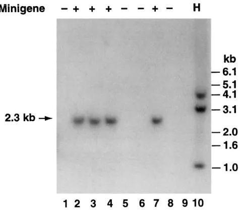
Figure 2. Southern blot analysis of DNA from the offspring of founder #63. DNA (10 mg) was digested with EcoRI and hybridized with a 32P-labeled hTF cDNA probe under conditions of high
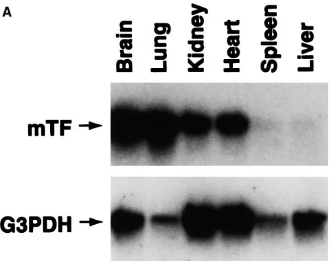
[image:3.612.318.556.444.652.2]Figure 3. Tissue-specific expression of the hTF mRNA. (A) Total RNA was extracted from brain, lung, kidney, heart, spleen, and liver of a nontransgenic mouse. The tissue distribution of mTF mRNA was determined by Northern blot analysis using an 818-bp mTF cDNA probe (35). The Northern blot was rehybridized with the housekeep-ing gene G3PDH. (B) Total RNA was extracted from tissues of a transgenic mouse (#47-37) and mRNA levels were determined by semiquantitative RT-PCR using mTF-specific primers (20 cycles), hTF-specific primers (30 cycles), or G3PDH-specific primers (16 cy-cles). Three independent PCR reactions were performed using differ-ent cDNA reactions. PCR products were hybridized with hTF, mTF, and G3PDH cDNA probes and band intensities were quantitated us-ing NIH Image. Data are shown as the mean of the three PCR reactions6standard error. Similar patterns of expression in these six tissues were observed in offspring from an independent founder line (#63-1). (C) Levels of mTF and hTF mRNAs in the kidney of a trans-genic mouse (#47-37) were determined by quantitative RT-PCR. De-termination of the levels of mTF mRNA (242 bp) used 10210 to 10216
mol of the competitor plasmid (370 bp) (lanes 2–8). Determination of the levels of hTF mRNA (363 bp) used 10213 to 10219 mol of the
com-petitor plasmid (330 bp) (lanes 2–8). Lane 1 contains molecular weight markers (M). Controls are mRNA alone (lane 9), competitor plasmid alone (lane 10), and buffer (lane 11).
Quantitative PCR. A synthetic DNA competitor template con-taining oligomers specific for mTF and hTF was constructed by PCR amplification of the plasmid pCCF, which contains a 282-bp interven-ing sequence (22). The PCR product was cloned into pBluescript (Stratagene) and was used as a DNA competitor to quantify cDNA expression. For hTF, the mRNA and competitor products were 363 and 330 bp, respectively. For mTF, the mRNA and competitor prod-ucts were 242 and 370 bp, respectively. PCR prodprod-ucts were quantified by competitive PCR essentially as described (39). PCR products were separated on 2 or 2.5% agarose gels and visualized by ethidium bro-mide staining. Images were captured using an Eagle-Eye II still video system (Stratagene) and band intensities were measured using NIH Image software. Log10 (absorbance control/absorbance mRNA) (y
axis) was plotted against log10 (number of control molecules) (x axis).
When the competitor and mRNA were present at equal amounts, the log10 (control/mRNA) equaled zero. Quantitative PCR results were
expressed as the number of cDNA molecules per nanogram of RNA. Determination of human TF antigen and functional activity. TF functional activity was determined from brain extracts or PECs in a one-stage clotting assay using either mouse (Sigma Chemical Co., St. Louis, MO) or human plasma as described (40). Brain extracts were prepared by homogenizing brain tissue in 15 mM octyl-b-D
-glucopy-ranoside, centrifuging at 12,000 g for 1 min, and incubating the super-natants at 378C for 15 min. PECs were solubilized with 15 mM
octyl-b-D-glucopyranoside at 378C for 15 min. TF functional activity was calculated in arbitrary units by reference to a standard curve. Human TF clots both human and mouse plasma, whereas mouse TF clots only mouse plasma (41). hTF activity was inhibited with anti–human TF monoclonal antibodies (TF8-5G9, TF8-6B4, and TF9-9C3) (40). hTF antigen was measured using a monoclonal antibody capture sys-tem ELISA (IMUBIND tissue factor ELISA kit; American Diagnos-tica Inc., Greenwich, CT), which does not detect murine TF.
Electrophoretic mobility shift assay (EMSA). Nuclear extracts were prepared from unstimulated and LPS-stimulated (1 mg/ml for 2 h) PECs (5 3 106 cells) as described (29). Nuclear extracts were
incu-bated with a radiolabeled TF kB site probe (29) and protein–DNA complexes were separated from free DNA probe by electrophoresis through 6% nondenaturing, polyacrylamide gels. Antibody supershift experiments were performed using anti-p65, anti-p50, and anti–c-Rel antibodies (Santa Cruz Biotechnology, Santa Cruz, CA) to identify proteins present in the complex (29).
on formalin-fixed, paraffin-embedded, 4-mM-thick sections. The goat anti–human TF antibody cross-reacts with mouse TF on Western blots (not shown). Control sections were stained with nonimmune goat immunoglobulin. Tissue sections were incubated with the pri-mary antibody overnight at 48C. Goat antibody was detected with the Vectastain Elite kit (Vector Laboratories, Burlingame, CA) using a biotinylated anti–goat immunoglobulin and 3,39-diaminobenzidine, which produced a brown reaction product. Human and murine TF antigens were also localized in murine tissues using an affinity-puri-fied rabbit anti–human TF polyclonal antibody on 6-mM fresh-frozen sections.
Results
Generation of transgenic mice. Our previous studies indicated that a transgene containing 1.0 kbp of the mTF promoter
[image:5.612.66.482.60.285.2]cloned upstream of the lacZ reporter gene was not expressed in transgenic mice (Drake, T.A., G.C.N. Parry, and N. Mack-man, unpublished data). This experience led us to include in-tron 1 in a hTF minigene (Fig. 1), which we believed would fa-cilitate efficient expression by promoting RNA splicing and may include regulatory elements that would contribute to TF expression in vivo. Screening of 93 offspring from the injection of the hTF minigene into fertilized mouse embryos identified 12 founder mice. Analysis of the offspring from crosses be-tween founder mice and wild-type C57BL/6 mice revealed germ-line transmission of the hTF minigene (Fig. 2) in 11 out of the 12 founders. The radiolabeled hTF cDNA did not hybridize with the mTF gene under the high stringency conditions used in the Southern blots. Comparison of the band intensities of the hTF minigene with that of human genomic DNA indicated
[image:5.612.52.560.519.742.2]that 9 of the 11 founders contained a single copy of the mini-gene. No phenotypic abnormalities were associated with the presence of the hTF minigene.
Tissue-specific expression of the hTF minigene. Expression of the human TF minigene was analyzed primarily in two founder lines (#47 and #63), although similar expression pat-terns were observed in five additional founder lines (#23, #27, #31, #45, and #76). Initial studies analyzed expression of hTF mRNA in the brain of transgenic mice. RT-PCR was used be-cause no signal was detected by Northern blotting. A 628-bp PCR product was observed with hTF-specific primers using total RNA from a transgenic mouse but not from a nontrans-genic littermate (not shown). This PCR product was depen-dent on reverse transcriptase and hybridized with a radio-labeled hTF cDNA, indicating that hTF mRNA was expressed in vivo.
Tissue-specific expression of hTF mRNA was examined in
transgenic mice. For comparison, mTF mRNA expression was measured by both Northern blotting and semiquantitative RT-PCR, which revealed highest levels in the brain and lung, inter-mediate levels in the kidney and heart, and low levels in the spleen and liver (Fig. 3, A and B). The housekeeping gene G3PDH is expressed at different levels in these six tissues. Various levels of hTF mRNA were observed by semiquantita-tive RT-PCR in tissues from transgenic mouse #47-37 (brain . kidney . spleen . lung . heart . liver) (Fig. 3 B). Similar patterns of hTF mRNA expression were observed in another founder line 63 (not shown), suggesting that the expression pattern of the hTF minigene was not affected by the site of in-tegration. To more accurately quantitate the expression level of the hTF minigene, hTF mRNA in transgenic mice was mea-sured by quantitative RT-PCR (Fig. 3 C). The brain and the kidney were chosen because they contained relatively high lev-els of hTF mRNA compared with the other tissues. The level of hTF mRNA (2.5 3 106 molecules/ng RNA) in the brain of
line #47-37 was 1.0% of the level of mTF mRNA (2.461.0 3 108
molecules/ng RNA). The level of hTF mRNA (1.860.9 3 106
molecules/ng RNA, mean6SD) in the kidneys of two trans-genic mice (#47-3 and #47-37) was 0.8% of the level of mTF mRNA (2.361.1 3 108 molecules/ng RNA, mean6SD, n 5 5).
Similar low levels of hTF mRNA (1.760.5 3 106 molecules/ng
RNA) were observed in the kidneys of two offspring (#63-1 and #63-9) from an independent founder. Taken together, these data suggest that the hTF was expressed at low levels in a tissue-specific manner.
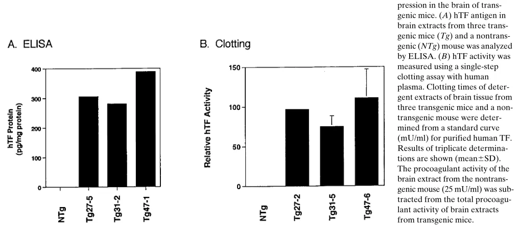
[image:6.612.59.247.56.354.2]Next, we determined the level of hTF antigen and func-tional activity in brain extracts from transgenic mice using an ELISA and clotting assay, respectively. hTF antigen was de-tected in brain extracts from transgenic mice but not from a nontransgenic mouse (Fig. 4 A). No statistically significant dif-ference in the procoagulant activity of brain extracts from transgenic and nontransgenic mice was observed using a clot-ting assay with mouse plasma (see Fig. 10). Therefore, we used a clotting assay with human plasma that detects human TF ac-tivity and not mouse TF (41). Brain extracts from transgenic mice contained hTF activity (Fig. 4 B), which was abolished by neutralizing anti–human TF monoclonal antibodies (not shown). LPS induction of the hTF minigene. To examine if expres-sion of the hTF minigene was inducible, we measured hTF mRNA levels by semiquantitative RT-PCR in unstimulated and LPS-stimulated PECs and whole blood from transgenic mice. LPS strongly induced hTF mRNA expression in both PECs and whole blood, presumably due to expression by peri-toneal macrophages and peripheral blood monocytes, respec-tively (Fig. 5). LPS also induced mTF mRNA expression in PECs from both nontransgenic and transgenic littermates and in whole blood from transgenic mice (Fig. 5), although the magnitude of induction of the endogenous mTF gene was sig-nificantly less than the induction of the hTF minigene. TNF-a mRNA expression was induced in PECs and whole blood treated with LPS. Expression of hTF antigen in PECs from transgenic mice was induced by LPS (Fig. 6 A). LPS stimula-tion also induced the expression of hTF activity in PECs from transgenic mice as measured in human plasma, which was abolished by anti–human TF monoclonal antibodies (Fig. 6 B). The role of NF-kB/Rel proteins in the LPS induction of the hTF minigene in PECs was assessed by EMSA and by the use of a specific NF-kB/Rel inhibitor. LPS stimulation of PECs in-duced the nuclear translocation of NF-kB/Rel proteins that Figure 5. LPS induction of the hTF mRNA expression.
bound to the TF kB site (Fig. 7 A). Antibody supershift exper-iments demonstrated that the TF complex was comprised of c-Rel and p65 (Fig. 7 B). Our previous studies indicated that TPCK, an inhibitor of NF-kB/Rel proteins, blocked LPS in-duction of TF expression in human monocytes and monocytic cells (42). Similarly, TPCK inhibited LPS induction of hTF an-tigen expression in PECs (Fig. 7 C). These studies indicate that LPS induces expression of the hTF minigene in murine PECs via an NF-kB/Rel–dependent mechanism.
[image:7.612.57.454.58.236.2]Rescue of murine TF null embryos with the human TF minigene. Recently, we demonstrated that targeted disruption of the mTF gene results in embryonic lethality between days E9.5 and 10.5 (11). Therefore, we examined if the hTF mini-gene could rescue the embryonic lethality in murine TF null embryos. Five independent crosses (mTF1/2, hTF1/2 3 mTF1/2, hTF1/2) were set up (Table I). If the hTF minigene rescued the embryonic lethality of mTF null embryos, 20% of weaned pups (3 wk) would be expected to survive due to the presence Figure 6. LPS induction of hTF expression in PECs from trans-genic mice. (A) hTF antigen in unstimulated and LPS-stimu-lated PECs from transgenic mice (#63-7 and #63-21) or nontrans-genic littermates was deter-mined by ELISA. Similar results were obtained with PECs from two independent founder lines (#27 and #47). (B) hTF activity with human plasma was deter-mined in unstimulated and LPS-stimulated (1 mg/ml for 5 h) PECs pooled from either trans-genic (#63-7 and #63-21) or non-transgenic littermates. hTF activity was abolished by incubation with monoclonal an-tibodies (Ab) (10 mg/ml) against human TF. Similar results were observed using transgenic mice #47-9 (not shown). The procoagulant activity of LPS-stimulated PECs from the nontransgenic mouse (4.3 mU/106 cells) was subtracted from the total procoagulant activity of unstimulated and
LPS-stimulated PECs from transgenic mice.
Figure 7. LPS induces expression of hTF in PECs via an NF-kB–dependent mechanism. (A) Nuclear extracts from PECs with or without LPS stimulation (1 mg/ml for 2 h) were incubated with a radiolabeled TF kB site. Protein–DNA complexes were analyzed by EMSA. Lanes 1 and 2 show PECs from a nontransgenic mouse and lanes 3 and 4 show PECs from a transgenic mouse. (B) Antibody supershift experiments were per-formed by incubating protein–DNA complexes with anti-p65, anti-p50, and anti–c-Rel antibodies (Santa Cruz Biotechnology). (C) hTF antigen levels were determined in unstimulated and LPS-stimulated PECs from transgenic mice (#47-7 and #47-8) in the presence or absence of TPCK (5
[image:7.612.58.550.424.673.2]of the human TF minigene. Screening of tail DNA from 117 weaned offspring identified 17 pups (14%) homozygous for the targeted mTF gene and containing the hTF minigene. A representative Southern blot is shown in Fig. 8. No mTF2/2, hTF2/2 mice have been observed at 3 wk. In a second breeding strategy, a rescued male heterozygous for the minigene (mTF2/2, hTF1/2) was crossed with a female heterozygous for murine TF and homozygous for the hTF minigene (mTF1/2, hTF1/1) (Table II). In this breeding, we expected 50% of the offspring to be rescued mice. So far, three litters have yielded 55% rescued offspring (12 of 22). These results demonstrate that the hTF minigene rescues murine TF null embryos, sug-gesting that human TF is expressed during embryonic develop-ment.
Analysis of hTF expression in rescued mice. The tissue-spe-cific expression of hTF in a rescued mouse (R47-23) was con-sistent with the pattern observed in transgenic mice (not
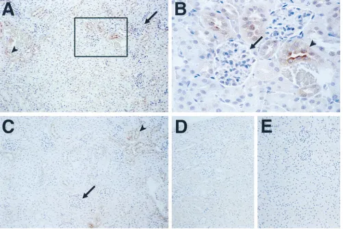
shown). Cell type–specific expression of the hTF minigene in rescued mice (R47-14 and R47-23) was analyzed by immuno-histochemistry. One of the most striking species-specific differ-ences in the pattern of TF expression between the human and the mouse occurs in the kidney where hTF is expressed in the glomeruli, whereas mTF is expressed in the tubules (8). In a rescued mouse (R47-14), the hTF minigene exhibited a mouse pattern of expression with hTF protein being strongly ex-pressed by tubular epithelial cells with little or no staining in glomerular cells (Fig. 9, A and B). A low level of nonspecific staining of renal tubules was observed with nonimmune goat immunoglobulin (Fig. 9, D and E). hTF protein was also ex-pressed in the epidermis of the tongue, adventitial cells sur-rounding blood vessels, bronchial epithelium, cardiac myo-cytes, and brain astrocytes in both formalin-fixed and frozen sections (data not shown) in a pattern similar to that of mTF and hTF protein (8, 43).
[image:8.612.57.555.76.174.2]Transgenic mice containing the hTF minigene expressed low levels of hTF mRNA (z 1%) relative to mTF mRNA. This low level expression of hTF may be due to some form of repression by the mTF gene. Therefore, the level of human TF expression was measured in a rescued mouse that did not con-tain a functional mTF gene. Brain extracts from a rescued mouse contained a low level of hTF antigen (263 pg/mg pro-tein), which is similar to the levels of hTF antigen in brain ex-tracts from transgenic mice. The total procoagulant activity of brain extracts from a rescued mouse (R47-23) was very low (z 0.7%) compared with a wild-type mouse in a clotting assay with mouse plasma (Fig. 10), indicating a low functional TF ac-tivity in rescued mice. The procoagulant acac-tivity of brain ex-tracts from transgenic and nontransgenic mice was not statisti-cally different using mouse plasma. These results indicate that transgenic and rescued mice express a similar low level of hTF mRNA and protein. Rescued mice developed normally, exhib-ited no excessive hemorrhage from tail transection for geno-typing or spontaneous hemorrhages, and were fertile. Rescued
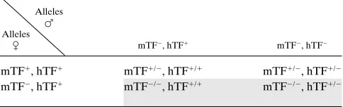
Table II. Offspring from mTF2/2, hTF1/23 mTF1/2, hTF1/1
Alleles F Alleles
C mTF2, hTF1 mTF2, hTF2
mTF1, hTF1 mTF1/2, hTF1/1 mTF1/2, hTF1/2 mTF2, hTF1 mTF2/2, hTF1/1 mTF2/2, hTF1/2
[image:8.612.57.280.438.619.2]Shaded boxes represent rescued mTF2/2 mice. Table I. Offspring from mTF1/2, hTF1/23 mTF1/2, hTF1/2
Alleles F Alleles
C mTF1, hTF1 mTF1, hTF2 mTF2, hTF1 mTF2, hTF2
mTF1, hTF1 mTF1/1, hTF1/1 mTF1/1, hTF1/2 mTF1/2, hTF1/1 mTF1/2, hTF1/2 mTF1, hTF2 mTF1/1, hTF2/1 mTF1/1, hTF2/2 mTF1/2, hTF1/2 mTF1/2, hTF2/2 mTF2, hTF1 mTF2/1, hTF1/1 mTF2/1, hTF1/2 mTF2/2, hTF1/1 mTF2/2, hTF1/2 mTF2, hTF2 mTF2/1, hTF2/1 mTF2/1, hTF2/2 mTF2/2, hTF2/1 mTF2/2, hTF2/2
Shaded boxes represent rescued mTF2/2 mice.
[image:8.612.315.557.647.723.2]mice exhibit no reduction in viability up to 7 mo of age. These data suggest that low levels of TF can maintain hemostasis that is compatible with normal survival.
Discussion
Our previous study failed to detect expression of b -galactosi-dase mRNA or protein from a transgene containing 1.0 kbp of the mTF promoter cloned upstream of the lacZ reporter (Drake, T.A., G.C.N. Parry, and N. Mackman, unpublished data). These results contrast with the expression of the hTF minigene shown here, indicating that additional DNA se-quences in the hTF promoter, intron 1, or the 39 flanking re-gion are required for TF gene expression in vivo. Many of the cis-acting regulatory elements that control TF gene expression in vitro are located in the proximal promoter region (2266 to 114) (27) and are highly conserved between the human and murine TF promoters (35), suggesting that the successful in vivo expression of the hTF minigene may be due to DNA ele-ments present in intron 1. Indeed, DNA eleele-ments that regulate cell type–specific expression in vivo have been reported in in-tronic sequences of genes such as PDGF-A and Tie 2 (44, 45).
Future studies will elucidate the relative contributions of the distal promoter region (22.1 to 21.0 kbp), intron 1, and the 39 flanking region to the regulation of TF expression in vivo.
The tissue-specific pattern of the hTF minigene was distinct from that of the endogenous mTF gene. Differences in the ex-pression patterns of hTF mRNA and mTF mRNA may be due, in part, to species-specific differences in TF expression and/or an absence of both positive and negative regulatory ele-ments in the minigene. Quantitation of the expression levels of the hTF minigene in two founder lines indicated that hTF mRNA in the kidney and brain of transgenic mice was # 1.0% of the level of mTF mRNA. Similarly, low levels of hTF anti-gen were detected in brain extracts of mice from three founder lines. This low level of expression of the hTF minigene in transgenic mice suggests that the minigene may lack a posi-tively acting enhancer or that the murine transcription factors inefficiently recognize regulatory elements in the human TF gene.
[image:9.612.58.559.60.395.2]the epidermis of the tongue, perivascular adventitial cells, car-diac myocytes, and brain astrocytes (data not shown). Of note was the pattern of expression in the kidney. Previous studies demonstrated that TF was expressed in glomerular cells in hu-man kidney and tubular cells in the murine kidney (8). In a res-cued mouse, hTF was strongly expressed in tubular cells with little or no staining in the glomerular cells. These results indi-cate that the species-specific pattern of TF expression in the kidney is not due to differences in the regulatory elements in the human and murine TF promoters, but rather is due to the expression of positively acting transcription factors in different cell types in the human and murine kidneys.
We have shown that LPS induction of the human TF gene in monocytic cells in vitro is mediated by a distal enhancer (2227 to 2172) and requires binding of c-Rel-p65 heterodimers to a kB site (28, 29). This enhancer region is present in the hTF minigene. Here, we demonstrate that LPS induced nuclear translocation of c-Rel-p65 heterodimers and hTF expression in PECs isolated from transgenic mice in a similar manner to hu-man monocytic cells (29). In addition, LPS induction of the hTF minigene was blocked by the protease inhibitor TPCK (42), suggesting that induction was mediated via an NF-kB/ Rel–dependent mechanism. Future studies will determine the precise role of the 2227 to 2172 enhancer in LPS induction of the hTF minigene in vivo.
TF expression during murine and human development sug-gested that TF may play a role in embryogenesis (8). Indeed, targeted disruption of the murine TF gene results in embry-onic lethality between days E9.5 and 10.5 (9–11). Our previous studies using a 129/Sv 3 C57BL/6 background indicated that only one mTF2/2 pup of 350 offspring (0.3%) survived to birth and then died of hemorrhage (11). More recently, it was re-ported that 2 mTF2/2 pups of 108 offspring (1.9%) survived to birth in the same 129/Sv 3 C57BL/6 background, whereas no
mTF2/2 embryos survived beyond day E10.5 in a 129/Sv back-ground, suggesting that genetic background can influence the survival of mTF2/2 pups (48). A single TF null pup was re-ported to live until 4 wk of age, although this mouse was deliv-ered by cesarean section (48). In our study, “rescued” mice were defined as mice that had been vaginally delivered and survive until weaning (3 wk). The genetic background of the offspring analyzed in this study is a mixture of C57BL/ 6[62.5%], 129/Sv[25%], and BALB/c[12.5%]. To date, screen-ing of 117 pups has not detected any mTF2/2 pups that lack the hTF minigene, suggesting that it is unlikely that our genetic background significantly contributes to the survival of mTF2/2, hTF1 pups observed in this study. Moreover, the rescue rate of 14% is consistent with the expected rate of 20%. In a second breeding strategy, we observed a rescue rate of 55% compared with the expected rate of 50%. Taken together, these data strongly suggest that expression of hTF during embryogenesis rescues TF2/2 embryos. At present, hTF expression levels in the visceral yolk sac have not been determined but we specu-late, based on the levels of hTF in adult mice, that only low lev-els of TF are required for rescue. The role of TF in embryo-genesis is controversial. Death of embryos has been attributed to fatal hemorrhage (10) or an abnormal yolk sac vasculature (11). We are currently attempting to distinguish between these two possibilities and to determine the role of TF in embryo-genesis by rescuing mTF2/2 embryos using modified versions of the hTF minigene.
Rescued mice (mTF2/2, hTF1) containing only low levels of human TF developed normally with no signs of a bleeding diathesis even after tail transection for genotyping. The proco-agulant activity of brain extracts of a rescued mouse was 0.7% of the level of a wild-type mouse. Low levels of total TF activ-ity of a tissue may not reflect the level of TF activactiv-ity at critical cell type–specific sites that are required for hemostasis. How-ever, if total TF activity does indeed reflect a low cell type– specific activity of TF at these sites, the viability of these mice suggests that, in the absence of additional challenge, low levels (, 1%) of TF can maintain hemostasis compatible with nor-mal growth and development. Rescued mice have survived normally up to 7 mo of age with no reduction in viability. This result is consistent with the lack of bleeding observed in exper-imental animals receiving large doses of inhibitory anti-TF an-tibodies (19, 20). A notable exception to the otherwise normal hemostasis observed in rescued mice was the uterine hemor-rhage during pregnancy and subsequent death of four rescued females. Although there are no reports of humans lacking TF, we speculate that the human population may contain individu-als either with low total TF or that express low levels of TF in the uterus during pregnancy that may be prone to excessive uterine bleeding.
Acknowledgments
We would like to acknowledge M. Smith and H. McClary for excel-lent technical assistance, Dr. K. Kono for advice on quantitative RT-PCR, Dr. W. Ruf and Dr. M. Ginsburg for stimulating discussions, Dr. L. Curtis and Dr. M. O’Connell for critical reading of the manu-script, and J. Robertson for preparing the manuscript.
[image:10.612.56.297.56.262.2]This research was supported by National Institutes of Health grants HL-48872 and HL-16411 (N. Mackman) and was performed during the tenure of an Established Investigatorship from the Ameri-can Heart Association (N. Mackman) and a Don and Lorraine Jaquot Figure 10. Procoagulant activity of a rescued mouse. Brain extracts
Travelling Fellowship from the Royal Australasian College of Physi-cians (J.H. Erlich).
References
1. Bach, R.R. 1988. Initiation of coagulation by tissue factor. Crit. Rev. Bio-chem. 23:339–368.
2. Edgington, T.S., N. Mackman, K. Brand, and W. Ruf. 1991. The struc-tural biology of expression and function of tissue factor. Thromb. Haemost. 66: 67–79.
3. Contrino, J., G. Hair, D.L. Kreutzer, and F.R. Rickles. 1996. In situ de-tection of tissue factor in vascular endothelial cells: correlation with the malig-nant phenotype of human breast disease. Nat. Med. 2:209–215.
4. Zhang, Y., Y. Deng, T. Luther, M. Müller, R. Ziegler, R. Waldherr, D.M. Stern, and P.P. Nawroth. 1994. Tissue factor controls the balance of angiogenic and antiangiogenic properties of tumor cells in mice. J. Clin. Invest. 94:1320– 1327.
5. Mueller, B.M., R.A. Reisfeld, T.S. Edgington, and W. Ruf. 1992. Expres-sion of tissue factor by melanoma cells promotes efficient hematogenous me-tastasis. Proc. Natl. Acad. Sci. USA. 89:1–6.
6. Bromberg, M.E., W.H. Konigsberg, J.F. Madison, A. Pawashe, and A. Garen. 1995. Tissue factor promotes melanoma metastasis by a pathway inde-pendent of blood coagulation. Proc. Natl. Acad. Sci. USA. 92:8205–8209.
7. Soifer, S.J., K.G. Peters, J. O’Keefe, and S.R. Coughlin. 1994. Disparate temporal expression of the prothrombin and thrombin receptor genes during mouse development. Am. J. Pathol. 144:60–69.
8. Luther, T., C. Flössel, N. Mackman, A. Bierhaus, M. Kasper, S. Albrecht, E.H. Sage, L. Iruela-Arispe, H. Grossmann, A. Ströhlein, et al. 1996. Tissue fac-tor expression during human and mouse development. Am. J. Pathol. 149:101–113. 9. Toomey, J.R., K.E. Kratzer, N.M. Lasky, J.J. Stanton, and G.J. Broze, Jr. 1996. Targeted disruption of the murine tissue factor gene results in embryonic lethality. Blood. 88:1583–1587.
10. Bugge, T.H., Q. Xiao, K.W. Kombrinck, M.J. Flick, K. Holmback, M.J.S. Danton, M.C. Colbert, D.P. Witte, K. Fujikawa, E.W. Davie, and J.L. Degen. 1996. Fatal embryonic bleeding events in mice lacking tissue factor, the cell-associated initiator of blood coagulation. Proc. Natl. Acad. Sci. USA. 93: 6258–6263.
11. Carmeliet, P., N. Mackman, L. Moons, T. Luther, P. Gressens, I. Van Vlaenderen, H. Demunck, M. Kasper, G. Breier, P. Evrard, et al. 1996. Role of tissue factor in embryonic blood vessel development. Nature. 383:73–75.
12. Fleck, R.A., L.V.M. Rao, S.I. Rapaport, and N. Varki. 1990. Localiza-tion of human tissue factor antigen by immunostaining with monospecific, poly-clonal anti-human tissue factor antibody. Thromb. Res. 57:765–781.
13. Drake, T.A., J.H. Morrissey, and T.S. Edgington. 1989. Selective cellu-lar expression of tissue factor in human tissues. Am. J. Pathol. 134:1087–1097.
14. Osterud, B., and T. Flæstad. 1983. Increased tissue thromboplastin ac-tivity in monocytes of patients with meningococcal infection: related to an unfa-vorable prognosis. Thromb. Haemost. 49:5–7.
15. Edwards, R.L., F.R. Rickles, and M. Cronlund. 1981. Abnormalities of blood coagulation in patients with cancer. J. Lab. Clin. Med. 98:917–928.
16. Leatham, E., P. Bath, J. Tooze, and A. Camm. 1995. Increased mono-cyte tissue factor expression in coronary disease. Br. Heart J. 73:10–13.
17. Drake, T.A., J. Cheng, A. Chang, and F.B. Taylor, Jr. 1993. Expression of tissue factor, thrombomodulin, and E-selectin in baboons with lethal Escher-ichia coli sepsis. Am. J. Pathol. 142:1–13.
18. Morrissey, J.H., and T.A. Drake. 1993. Procoagulant response of the en-dothelium and monocytes. In Pathophysiology of Shock, Sepsis and Organ Fail-ure. G. Schlag and H. Redl, editors. Springer-Verlag, Berlin. 564–574.
19. Levi, M., H. ten Cate, K.A. Bauer, T. van der Poll, T.S. Edgington, H.R. Büller, S.J.H. van Deventer, C.E. Hack, J. Wouter ten Cate, and R.D. Rosen-berg. 1994. Inhibition of endotoxin-induced activation of coagulation and fi-brinolysis by pentoxifylline or by a monoclonal anti-tissue factor antibody in chimpanzees. J. Clin. Invest. 93:114–120.
20. Taylor, F.B., Jr., A. Chang, W. Ruf, J.H. Morrissey, L. Hinshaw, R. Catlett, K. Blick, and T.S. Edgington. 1991. Lethal E. coli septic shock is pre-vented by blocking tissue factor with monoclonal antibody. Circ. Shock. 33: 127–134.
21. Wilcox, J.N., K.M. Smith, S.M. Schwartz, and D. Gordon. 1989. Local-ization of tissue factor in the normal vessel wall and in the atherosclerotic plaque. Proc. Natl. Acad. Sci. USA. 86:2839–2843.
22. Moreno, P.R., V.H. Bernardi, J. López-Cuéllar, A.M. Murcia, I.F. Pala-cios, H.K. Gold, R. Mehran, S.K. Sharma, Y. Nemerson, V. Fuster, and J.T. Fallon. 1996. Macrophages, smooth muscle cells, and tissue factor in unstable angina. Implications for cell-mediated thrombogenicity in acute coronary syn-dromes. Circulation. 94:3090–3097.
23. Marmur, J.D., M. Rossikhina, A. Guha, B. Fyfe, V. Friedrich, M. Mend-lowitz, Y. Nemerson, and M.B. Taubman. 1993. Tissue factor is rapidly induced in arterial smooth muscle after balloon injury. J. Clin. Invest. 91:2253–2259.
24. Golino, P., M. Ragni, P. Cirillo, V.E. Avvedimento, A. Feliciello, N. Es-posito, A. Scognamiglio, B. Trimarco, G. Iaccarino, M. Condorelli, et al. 1996. Effects of tissue factor induced by oxygen free radicals on coronary flow during
reperfusion. Nat. Med. 2:35–40.
25. Ragni, M., P. Cirillo, I. Pascucci, A. Scognamiglio, D. D’Andrea, N. Er-amo, M.D. Ezekowitz, A.B. Pawashe, M. Chiariello, and P. Golino. 1996. Mono-clonal antibody against tissue factor shortens tissue plasminogen activator lysis time and prevents reocclusion in a rabbit model of carotid artery thrombosis. Circulation. 93:1913–1918.
26. Jang, I.-K., H.K. Gold, R.C. Leinbach, J.T. Fallon, D. Collen, and J.N. Wilcox. 1992. Antithrombotic effect of a monoclonal antibody against tissue factor in a rabbit model of platelet-mediated arterial thrombosis. Arterioscler. Thromb. 12:948–954.
27. Mackman, N. 1995. Regulation of the tissue factor gene. FASEB (Fed. Am. Soc. Exp. Biol.) J. 9:883–889.
28. Mackman, N., K. Brand, and T.S. Edgington. 1991. Lipopolysaccharide-mediated transcriptional activation of the human tissue factor gene in THP-1 monocytic cells requires both activator protein 1 and nuclear factor kB binding sites. J. Exp. Med. 174:1517–1526.
29. Oeth, P.A., G.C.N. Parry, C. Kunsch, P. Nantermet, C.A. Rosen, and N. Mackman. 1994. Lipopolysaccharide induction of tissue factor gene expression in monocytic cells is mediated by binding of c-Rel/p65 heterodimers to a k B-like site. Mol. Cell. Biol. 14:3772–3781.
30. Parry, G.C., and N. Mackman. 1995. Transcriptional regulation of tissue factor expression in human endothelial cells. Arterioscler. Thromb. 15:612–621. 31. Bierhaus, A., Y. Zhang, Y. Deng, N. Mackman, P. Quehenberger, M. Haase, T. Luther, M. Müller, H. Böhrer, J. Greten, et al. 1995. Mechanism of the tumor necrosis factor a-mediated induction of endothelial tissue factor. J. Biol. Chem. 270:26419–26432.
32. Moll, T., M. Czyz, H. Holzmuller, R. Hofer-Warbinek, E. Wagner, H. Winkler, F.D. Bach, and E. Hofer. 1995. Regulation of the tissue factor pro-moter in endothelial cells. J. Biol. Chem. 270:3849–3857.
33. Taby, O., C.-L. Rosenfield, V. Bogdanov, Y. Nemerson, and M.B. Taubman. 1996. Cloning of the rat tissue factor cDNA and promoter: identifi-cation of a serum-response region. Thromb. Haemost. 76:697–702.
34. Mackman, N., J.H. Morrissey, B. Fowler, and T.S. Edgington. 1989. Com-plete sequence of the human tissue factor gene, a highly regulated cellular re-ceptor that initiates the coagulation protease cascade. Biochemistry. 28:1755–1762. 35. Mackman, N., S. Imes, W.H. Maske, B. Taylor, A.J. Lusis, and T.A. Drake. 1992. Structure of the murine tissue factor gene: chromosome location and conservation of regulatory elements in the promoter. Arterioscler. Thromb. 12:474–483.
36. Morrissey, J.H., H. Fakhrai, and T.S. Edgington. 1987. Molecular clon-ing of the cDNA for tissue factor, the cellular receptor for the initiation of the coagulation protease cascade. Cell. 50:129–135.
37. Morrissey, J.H., S.A. Gregory, N. Mackman, and T.S. Edgington. 1989. Tissue factor regulation and gene organization. Oxf. Surv. Eukaryotic Genes. 6: 67–84.
38. Stuhlmeier, K.M., V. Csizmadia, Q. Cheng, H. Winkler, and F.H. Bach. 1994. Selective inhibition of E-selectin, ICAM-1, and VCAM in endothelial cells. Eur. J. Immunol. 24:2186–2190.
39. Prud’Homme, G.J., D.H. Kono, and A.N. Theofilopoulos. 1995. Quanti-tative polymerase chain reaction analysis reveals marked overexpression of in-terleukin-1b, interleukin-10 and interferon-gamma mRNA in the lymph nodes of lupus-prone mice. Mol. Immunol. 32:495–503.
40. Morrissey, J.H., D.S. Fair, and T.S. Edgington. 1988. Monoclonal anti-body analysis of purified and cell-associated tissue factor. Thromb. Res. 52:247–261. 41. Fang, C.H., T.C. Lin, A. Guha, Y. Nemerson, and W.H. Konigsberg. 1996. Activation of factor X by factor VIIa complexed with human-mouse tis-sue factor chimeras requires human exon 3. Thromb. Haemost. 76:361–368.
42. Mackman, N. 1994. Protease inhibitors block lipopolysaccharide induc-tion of tissue factor gene expression in human monocytic cells by preventing ac-tivation of c-Rel/p65 heterodimers. J. Biol. Chem. 269:26363–26367.
43. Drake, T.A., J.H. Morrissey, and T.S. Edgington. 1989. Selective cellu-lar expression of tissue factor in human tissues. Implications for disorders of he-mostasis and thrombosis. Am. J. Pathol. 134:1087–1097.
44. Schlaeger, T.M., S. Bartunkova, J.A. Lawitts, G. Teichmann, W. Risau, U. Deutsch, and T.N. Sato. 1997. Uniform vascular-endothelial-cell-specific gene expression in both embryonic and adult transgenic mice. Proc. Natl. Acad. Sci. USA. 94:3058–3063.
45. Franklin, G.C., M. Donovan, G.I.R. Adam, L. Holmgren, S. Pfeifer-Ohlsson, and R. Ohlsson. 1991. Expression of the human PDGF-B gene is regu-lated by both positively and negatively acting cell type-specific regulatory elements located in the first intron. EMBO (Eur. Mol. Biol. Organ.) J. 10:1365– 1373.
46. Eddleston, M., J.C. de la Torre, M.B.A. Oldstone, D.J. Loskutoff, T.S. Edgington, and N. Mackman. 1993. Astrocytes are the primary source of tissue factor in the murine central nervous system. A role for astrocytes in cerebral hemostasis. J. Clin. Invest. 92:349–358.
47. Mackman, N., M.S. Sawdey, M.R. Keeton, and D.J. Loskutoff. 1993. Murine tissue factor gene expression in vivo: tissue and cell specificity and regu-lation by lipopolysaccharide. Am. J. Pathol. 143:76–84.