Kim L. Rice, Jonathan D. Licht
J Clin Invest.
2007;
117(4)
:865-868.
https://doi.org/10.1172/JCI31861
.
The deregulation of
homeobox
(
HOX
) genes in acute myeloid leukemia (AML) and the
potential for these master regulators to perturb normal hematopoiesis is well established. To
date, overexpression of
HOX
genes in AML has been attributed to specific chromosomal
aberrations and abnormalities involving mixed-lineage leukemia (MLL), an upstream
regulator of
HOX
genes. The finding reported in this issue of the
JCI
by Scholl et al. that
caudal-type homeobox transcription factor 2 (CDX2), which is capable of affecting
HOX
gene expression during embryogenesis, is overexpressed in 90% of patients with AML and
induces a transplantable AML in murine models provides an alternative mechanism for
HOX-induced leukemogenesis and yields important insights into the hierarchy of
HOX
gene
regulation in AML (see the related article beginning on page 1037).
Commentary
Find the latest version:
promotes tumor growth. Cancer Res. 65:2277–2286. 12. Covello, K.L., et al. 2006. HIF-2alpha regulates Oct-4:
effects of hypoxia on stem cell function, embry-onic development, and tumor growth. Genes Dev.
20:557–570.
13. Kondo, K., Kico, J., Nakamura, E., Lechpammer, M., and Kaelin, W.G.J. 2002. Inhibition of HIF is necessary for tumor suppression by the von Hip-pel-Lindau protein. Cancer Cell. 1:237–246. 14. Raval, R.R., et al. 2005. Contrasting properties of
hypoxia-inducible factor 1 (HIF-1) and HIF-2 in von Hippel-Lindau-associated renal cell carcinoma.
Mol. Cell. Biol. 25:5675–5686.
15. Park, S.-K., et al. 2003. Hypoxia-induced gene expres- sion occurs solely through the action of hypoxia-inducible factor 1a (HIF-1a): role of cytoplasmic trapping of HIF-2a. Mol. Cell. Biol. 23:4959–4971. 16. Hu, C.J., et al. 2006. Differential regulation of the
transcriptional activities of hypoxia-inducible fac-tor 1 alpha (HIF-1a) and HIF-2a in stem cells. Mol. Cell. Biol. 26:3514–3526.
17. Yu, A.Y., et al. 1999. Impaired physiological responses to chronic hypoxia in mice partially defi-cient for hypoxia-inducible factor 1a. J. Clin. Invest.
103:691–696.
18. Scortegagna, M., et al. 2005. HIF-2alpha regulates
murine hematopoietic development in an erythro-poietin-dependent manner. Blood. 105:3133–3140. 19. Warnecke, C., et al. 2004. Differentiating the func-tional role of hypoxia-inducible factor (HIF)-1alpha and HIF-2alpha (EPAS-1) by the use of RNA inter-ference: erythropoietin is a HIF-2alpha target gene in HepB and Kelly cells. FASEB J. 18:1462–1464. 20. Maxwell, P.H., et al. 1993. Identification of the
renal erythropoietin-producing cells using trans-genic mice. Kidney Int. 44:1149–1162.
21. Da Silva, J.L., et al. 1990. Tumour cells are the site of erythropoietin synthesis in human renal cancers associated with polycythemia. Blood. 75:577–582.
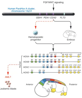
HOX
deregulation in acute myeloid leukemia
Kim L. Rice and Jonathan D. Licht
Division of Hematology/Oncology, Department of Medicine, Robert H. Lurie Comprehensive Cancer Center, Feinberg School of Medicine, Northwestern University, Chicago, Illinois, USA.
The deregulation of
homeobox
(
HOX
) genes in acute myeloid leukemia
(AML) and the potential for these master regulators to perturb normal
hematopoiesis is well established. To date, overexpression of
HOX
genes in
AML has been attributed to specific chromosomal aberrations and
abnor-malities involving mixed-lineage leukemia (MLL), an upstream regulator
of
HOX
genes. The finding reported in this issue of the
JCI
by Scholl et al.
that caudal-type homeobox transcription factor 2 (CDX2), which is capable
of affecting
HOX
gene expression during embryogenesis, is overexpressed
in 90% of patients with AML and induces a transplantable AML in murine
models provides an alternative mechanism for HOX-induced
leukemogen-esis and yields important insights into the hierarchy of
HOX
gene
regula-tion in AML (see the related article beginning on page 1037).
Nonstandard abbreviations used: AML, acute myeloid leukemia; ATRA, all-trans retinoic acid; CDX2, caudal-type homeobox transcription factor 2; ETV6, ets variant gene 6; FLT3, fms-like tyrosine kinase 3; HOX, homeobox;MEIS1, myeloid ecotropic viral integration site 1; MLL, mixed-lineage leukemia; PML, promyelocytic leukemia; RARa, retinoic acid receptor
a; RTK, receptor tyrosine kinase;RUNX1, runt-related transcription factor 1.
Conflict of interest: The authors have declared that no conflict of interest exists.
Citation for this article: J. Clin. Invest. 117:865–868 (2007). doi:10.1172/JCI31861.
Acute myeloid leukemia (AML) is a hetero-geneous disease in which hematopoietic progenitor cells acquire genetic lesions that lead to a block in differentiation, increased self-renewal, and unregulated proliferation. The emergence of leukemic blasts appears to require at least two major genetic “hits,” involving perturbations in growth factor signaling pathways and hematopoietic dif-ferentiation programs (1).
Among the receptor tyrosine kinases (RTKs), fms-like tyrosine kinase 3 (FLT3), which plays important roles in hema-topoietic progenitor cell survival and
proliferation, is overexpressed in a sig- nificant proportion of AMLs, and muta- tions resulting in the constitutive activa-tion of FLT3 occur in approximately 33% of patients (2). Mutations leading to the constitutive activation of a related RTK, c-KIT, and of signaling intermediates such as RAS, are also frequently described in AML (3). The dysregulation of associ-ated signaling pathways (e.g., Ras/MAPK, PI3K/AKT, and JAK/STAT) is thought to result in growth factor–independent pro- liferation and clonal expansion of hema-topoietic progenitors.
The second hit targets transcription fac-tors capable of disrupting hematopoietic cell differentiation. This may occur follow- ing the dysregulation of specific gene regu-lators as a result of gene amplification (e.g.,
v-myc myelocytomatosis viral oncogene homolog [MYC], mixed-lineage leukemia [MLL], genes at the chromosome 11q23 locus) (4); point mutations in transcriptional regula-tors (e.g., CCAAT/enhancer–binding protein
[C/EBP], runt-related transcription factor 1
[RUNX1]); and chromosomal
transloca-tions resulting in the fusion of promyelo-cytic leukemia (PML) and the retinoic acid receptor a (RARa) to yield the PML-RARa chimeric protein or fusion of RUNX1 and
runt-related transcription factor 1, translocated to 1 (RUNX1T1), which gives rise to the RUNX1-RUNX1T1 fusion protein (1). It is becoming increasingly clear that one set of genes commonly affected by these chimeri-cal and mutated transcriptional regulators are the homeobox (HOX) genes. Accordingly, overexpression of homeobox master tran-scription factors, which fulfill critical roles in embryonic development, organogenesis, and normal hematopoietic differentiation, is a common feature of AML (5).
HOX genes: from hematopoiesis to leukemia
to myeloid or erythroid lineages is accom-panied by global downregulation of HOX gene expression (7).
HOX genes may be dysregulated in AML by several different mechanisms. First, specific HOX genes can be disrupted via chromosomal translocation. Specifically,
HOXA9 and HOXD13 are dysregulated through the t(7;11) and t(2;11) transloca- tions, respectively (8), in both cases creat- ing fusion proteins between the HOX pro-tein and the nucleoporin 98 kDa (NUP98) nuclear protein. Overexpression of HOXA6,
HOXA7, HOXA9, and the HOX cofac-tor myeloid ecotropic viral integration site 1 (MEIS1) has also been correlated with chromosome 11q23 abnormalities involv-ing the MLL protein, which regulates the expression of HOX genes (9). A less fre-quently observed translocation in AML, t(8;16)(p11;p13), which results in the over-expression of the MYST3-CREBBP fusion protein, is also associated with overexpres-sion of HOXA9, HOXB9, HOXA10, and
MEIS1 (10). In addition, the expression of HOXC4 was shown to be upregulated in the NB4 PML-RARa cell line follow-ing all-trans
retinoic acid–induced (ATRA-induced) differentiation as well as in bone marrow from acute PML patients during ATRA treatment, strengthening the theory that dysregulated HOX expression is a sig-nature feature of AML (11).
CDX2 overexpression: a unifying theme in AML?
In keeping with the theme of dysregulated
HOX gene expression in AML, in this issue of the JCI Scholl et al. (12) demonstrate that caudal-type homeobox transcription fac-tor 2 (CDX2), a homeobox gene involved in anteroposterior axis definition and intesti-nal epithelial cell differentiation (13), but strikingly absent in hematopoietic progen-itors, is overexpressed in 90% of patients with AML. The leukemogenic potential of
CDX2 overexpression was first described by Chase et al. (14) following the identifi- cation of a novel chromosomal rearrange-ment, t(12;13)(p13;q12), in a patient with AML, which yielded the ets variant gene 6–CDX2 (ETV6-CDX2) fusion protein in addition to full-length CDX2. Significant-ly, transduction of murine hematopoi-etic progenitors with CDX2, but not the
ETV6-CDX2 fusion protein, resulted in a transplantable and fatal AML. Although ETV6-CDX2 induced myeloproliferation, it did not induce leukemia or accelerate CDX2-mediated leukemogenesis (15), suggesting that the primary oncogenic affect of this translocation is the ectopic expression of CDX2 in the hematopoietic compartment (Figure 1).
CDX2 belongs to the ParaHox A cluster, which consists of a three-gene complex including GS homeobox 1 (GSH1), pancreatic and duodenal homeobox 1 (PDX1),and CDX2 that exists on chromosome 13q12 (6) (Fig-ure 1). On the basis of sequence similarity,
CDX is more related to the posterior group of HOX genes, which includes paralo-gous groups 9–13, and during embryonic development is expressed in the posterior primitive streak to regulate HOX genes and in posterior signaling pathways to direct anteroposterior axis development and elongation. Given the role of CDX2 as a regulator of HOX genes in development, it is therefore not surprising that ectopic expression of CDX2 in hematopoietic pro-
genitors perturbs blood cell differentia-Figure 1
A model for CDX2-mediated
leukemogene-sis. CDX2 is expressed in the posterior
primi-tive streak during embryogenesis, where it directs anteroposterior axial development
and elongation by regulating HOX gene
expression. A study in this issue of the JCI
(12) reports that monoallelic expression of
CDX2 is observed in 90% of patients with
AML and may perturb hematopoiesis by
affecting HOX gene expression (red dashed
arrows indicate ectopic expression; black arrows indicate normal expression in
embry-onic development). GSH1, GS homeobox 1;
PDX1, pancreatic and duodenal
homeo-box 1. Figure modified with permission from
[image:3.585.67.351.79.418.2]tion programs strictly regulated by HOX genes. The finding by Scholl et al. (12) that overexpression of Cdx2 in primary murine hematopoietic progenitors resulted in transplantable AML in vivo, coinciding with a three-fold upregulation in Hoxb6 expression, is particularly intriguing given that overexpression of HOXB6 has been documented in approximately 40% of human AMLs that do not have chro-mosomal translocations (16). Identifying whether Cdx2 directly regulates Hoxb6 and providing evidence of the specific involve- ment of Hoxb6 in Cdx2-mediated leuke- mogenesis would strengthen the hypothe-sis that dysregulated HOX gene expression by CDX2 is a major pathway contributing to leukemogenesis.
The dysregulation of HOX genes by mem-bers of the caudal HOX family may prove to be a unifying theme in AML, particularly in light of a recent study by Bansal et al. (17) that revealed dysregulated CDX4 expression in 25% of AMLs. However, whereas CDX2 induced rapid AML in bone marrow trans-plant recipients, CDX4 induced a partially penetrant, long-latency AML, suggesting that these transcription factors regulate distinct sets of genes, including potentially different subsets of HOX genes. In addition to dysregulating HOX genes, CDX2 overex-pression may also affect signaling pathways.
Cdx1/2 mutant embryos demonstrate poste- rior body truncations that phenocopy loss-of-function Wnt3a and Fgfr1 mutants (18), suggesting an overlap among Cdx, Wnt, and Fgf pathways in axial patterning. This implies that some CDX2 target genes might include components of signaling pathways or that CDX2 itself may be a target of cell-signaling regulators, a point highlighted by Scholl et al. (12), given that related family members CDX1 and CDX4 are direct targets of the Wnt pathway commonly activated in AML. Indeed, Scholl et al. showed that knockdown of CDX2 slowed cellular pro-liferation specifically in CDX2-expressing AML cell lines. Taken together, these data suggest that gain-of-function mutations of CDX2 might augment cell signaling in addi-tion to disrupting differentiation programs. This may explain the rapid onset of AML in mice transplanted with CDX2-expressing hematopoietic progenitors.
Unraveling the mechanism
The molecular basis for the overexpres-sion of CDX2 remains elusive, and the absence of chromosomal rearrangements or mutations in the coding sequence or
proximal promoter of CDX2 suggest that other undefined regulatory regions driv-ing CDX2 expression may be a target for mutation. Indeed, aberrant expression of
CDX2 in the absence of genetic abnormali-ties on chromosome 13 was previously documented in a case of chronic myeloid leukemia blast crisis (14). Intriguingly, microarray analysis of 50 AML patient specimens revealed that the highest levels of HOX expression are associated with a subset of patients with intermediate cyto-genetics and elevated levels of FLT3 mRNA and FLT3 mutations. In the absence of mutations or amplifications, overexpres-sion of FLT3 was correlated with aberrant expression of multiple HOX genes, sug-gesting that deregulation of HOX genes may be due to aberrant growth factor sig-naling (19). Given the close proximity of the FLT3 gene to the CDX2 gene and the fact that both proteins are overexpressed in a high percentage of AMLs, it is tempt-ing to speculate that cis-acting activating mutations in promoter or enhancer ele-ments in the vicinity of these genes may contribute to the leukemic initiation and progression and may account for the link among FLT3, CDX2, and HOX gene over-expression in the absence of detectable cytogenetics (20). Detailed analyses of the regulatory sequences flanking CDX2 and FLT3 could provide insights into the role of these oncogenes in AML. It would also be important to correlate the level of
CDX2 overexpression in AML with HOX gene expression.
Identification of a HOX gene pathway disrupted in a substantial proportion of patients with AML (12) is a major clue to disease pathogenesis; however, more direct evidence of a link between CDX2 overex-pression and the dysregulation of specific HOX genes is needed. For example, demon-stration by chromatin precipitation that CDX2 occupies the promoters of genes such as HOXB8 and HOXB6 in CDX2-posi-tive AML cell specimens would reinforce the importance of CDX2 as an aberrant regulator of HOX genes. In addition, iden-tifying the mechanism by which CDX2 is overexpressed is crucial for unraveling the hierarchy of genetic pathways required for leukemia initiation and progression. While specific targeting of transcriptional regula-tors remains elusive, better understanding of the circuitry of the HOX genes in AML could present a new set of genetic targets, with potential therapeutic benefit for mul-tiple AML subtypes. Address correspondence to: Jonathan D. Licht, Division of Hematology/Oncology, Department of Medicine, Robert H. Lurie Comprehensive Cancer Center, Feinberg School of Medicine, Northwestern Uni-versity, 303 E. Superior Street, Lurie 5-123, Chicago, Illinois 60611, USA. Phone: (312) 503-0985; Fax: (312) 503-0189; E-mail: j-licht@northwestern.edu.
1. Licht, J.D., and Sternberg, D.W. 2005. The molecu-lar pathology of acute myeloid leukemia. Hematol-ogy Am. Soc. Hematol. Educ. Program. 2005:137–142.
2. Small, D. 2006. FLT3 mutations: biology and treat-ment. Hematology Am. Soc. Hematol. Educ. Program.
2006:178–184.
3. Goemans, B.F., et al. 2005. Mutations in KIT and RAS are frequent events in pediatric core-binding factor acute myeloid leukemia. Leukemia. 19:1536–1542. 4. Streubel, B., et al. 2000. Amplification of the MLL
gene on double minutes, a homogeneously stain-ing region, and ring chromosomes in five patients with acute myeloid leukemia or myelodysplastic syndrome. Genes Chromosomes Cancer. 27:380–386.
5. Ferrando, A.A., et al. 2003. Gene expression signa-tures in MLL-rearranged T-lineage and B-precursor acute leukemias: dominance of HOX dysregula-tion. Blood. 102:262–268.
6. Garcia-Fernandez, J. 2005. Hox, ParaHox, Proto-Hox: facts and guesses. Heredity. 94:145–152. 7. Pineault, N., Helgason, C.D., Lawrence, H.J., and
Humphries, R.K. 2002. Differential expression of Hox, Meis1, and Pbx1 genes in primitive cells throughout murine hematopoietic ontogeny. Exp. Hematol. 30:49–57.
8. Nakamura, T., et al. 1996. Fusion of the nucleopo-rin gene NUP98 to HOXA9 by the chromosome translocation t(7;11)(p15;p15) in human myeloid leukaemia. Nat. Genet. 12:154–158.
9. Schoch, C., et al. 2003. AML with 11q23/MLL abnormalities as defined by the WHO classifica- tion: incidence, partner chromosomes, FAB sub-type, age distribution, and prognostic impact in an unselected series of 1897 cytogenetically analyzed AML cases. Blood. 102:2395–2402.
10. Camos, M., et al. 2006. Gene expression profil-ing of acute myeloid leukemia with translocation t(8;16)(p11;p13) and MYST3-CREBBP rearrange- ment reveals a distinctive signature with a spe-cific pattern of HOX gene expression. Cancer Res.
66:6947–6954.
11. Kim, D.Y., et al. 2005. Upregulated hoxC4 induces CD14 expression during the differentiation of acute promyelocytic leukemia cells. Leuk. Lympho-ma. 46:1061–1066.
12. Scholl, C., et al. 2007. The homeobox gene CDX2 is aberrantly expressed in most cases of acute myeloid leukemia and promotes leukemogenesis. J. Clin. Invest. 117:1037–1048. doi:10.1172/JCI30182. 13. James, R., and Kazenwadel, J. 1991. Homeobox
gene expression in the intestinal epithelium of adult mice. J. Biol. Chem. 266:3246–3251. 14. Chase, A., et al. 1999. Fusion of ETV6 to the
caudal-related homeobox gene CDX2 in acute myeloid leukemia with the t(12;13)(p13;q12).
Blood. 93:1025–1031.
15. Rawat, V.P., et al. 2004. Ectopic expression of the homeobox gene Cdx2 is the transforming event in a mouse model of t(12;13)(p13;q12) acute myeloid leukemia. Proc. Natl. Acad. Sci. U. S. A. 101:817–822. 16. Giampaolo, A., et al. 2002. Expression pattern of
HOXB6 homeobox gene in myelomonocytic dif-ferentiation and acute myeloid leukemia. Leukemia.
16:1293–1301.
alone and in cooperation with Meis1a in a murine model. Proc. Natl. Acad. Sci. U. S. A. 103:16924–16929. 18. Partanen, J., Schwartz, L., and Rossant, J. 1998.
Opposite phenotypes of hypomorphic and Y766 phosphorylation site mutations reveal a function for Fgfr1 in anteroposterior patterning of mouse
embryos. Genes Dev. 12:2332–2344.
19. Roche, J., et al. 2004. Hox expression in AML identi-fies a distinct subset of patients with intermediate cytogenetics. Leukemia. 18:1059–1063.
20. Muller-Tidow, C., et al. 2004. High-throughput analysis of genome-wide receptor tyrosine kinase
expression in human cancers identifies potential novel drug targets. Clin. Cancer Res. 10:1241–1249.
21. Veraksa, A., Del Campo, M., and McGinnis, W. 2000. Developmental patterning genes and their conserved functions: from model organisms to humans. Mol. Genet. Metab. 60:85–100.
Hypoglycemia, functional brain failure,
and brain death
Philip E. Cryer
Division of Endocrinology, Metabolism and Lipid Research, Washington University School of Medicine, St. Louis, Missouri, USA.
Hypoglycemia commonly causes brain fuel deprivation, resulting in
func-tional brain failure, which can be corrected by raising plasma glucose
con-centrations. Rarely, profound hypoglycemia causes brain death that is not
the result of fuel deprivation per se. In this issue of the
JCI
, Suh and
col-leagues use cell culture and in vivo rodent studies of glucose deprivation
and marked hypoglycemia and provide evidence that hypoglycemic brain
neuronal death is in fact
increased
by neuronal NADPH oxidase activation
during glucose reperfusion (see the related article beginning on page 910).
This finding suggests that, at least in the setting of profound hypoglycemia,
therapeutic hyperglycemia should be avoided.
Nonstandard abbreviations used: T1DM, type 1 diabetes mellitus.
Conflict of interest: The author has served on advisory boards of Novo Nordisk Inc., Takeda Pharmaceuticals North America Inc., MannKind Corp., and Merck and Co. and as a consultant to TolerRx Inc., Amgen Inc., and Marcadia Biotech in recent years.
Citation for this article: J. Clin. Invest. 117:868–870 (2007). doi:10.1172/JCI31669.
Hypoglycemia, including iatrogenic hypo-glycemia in people with diabetes, causes brain fuel deprivation that initially trig- gers a series of physiological and behav-ioral defenses but if unchecked results in functional brain failure that is typically corrected after the plasma glucose concen-tration is raised. Rarely, profound, and at least in primates prolonged, hypoglycemia causes brain death.
Given the survival value of maintaining physiological plasma glucose concentra-tions, it is not surprising that mechanisms that normally very effectively prevent or rapidly correct symptomatic hypoglycemia have evolved (1). As a result, hypoglycemia is a distinctly uncommon clinical event except in people who use drugs that lower the plasma glucose concentration (2). Although there are other drugs, and sev-eral relatively uncommon conditions, that cause hypoglycemia (2), in the vast majority
of instances the offending drug is an insu-lin secretagogue or insulin used to treat diabetes mellitus (2, 3). As a result of the interplay of relative or absolute therapeu- tic insulin excess and compromised physi-ological and behavioral defenses against falling plasma glucose concentrations, hypoglycemia is the limiting factor in the glycemic management of diabetes (3). It causes recurrent morbidity in most people with type 1 diabetes mellitus (T1DM) and in many with advanced T2DM and is some-times fatal. Furthermore, hypoglycemia, as well as prior exercise and sleep, further compromise glycemic defenses by causing hypoglycemia-associated autonomic fail-ure and thus a vicious cycle of recurrent hypoglycemia. Finally, the barrier of hypo- glycemia precludes maintenance of eugly-cemia over a lifetime of diabetes and thus full realization of the long-term vascular benefits of glycemic control.
Functional brain failure
Recent interest in alternative brain fuels (including lactate derived from glucose largely within the brain; refs. 4–6) not- withstanding, glucose is an obligate meta-bolic fuel for the brain under physiological conditions (7). Because the brain cannot synthesize glucose or store substantial amounts as glycogen in astrocytes, the brain requires a virtually continuous supply
of glucose from the circulation. Facilitated diffusion of glucose from the blood into the brain is a direct function of the arterial plasma glucose concentration. The rate of blood-to-brain glucose transport exceeds the rate of brain glucose metabolism at normal (or elevated) plasma glucose levels, but it falls and becomes limiting to brain glucose metabolism when arterial glucose concentrations fall to low levels (8). Thus, hypoglycemia causes brain fuel deprivation and, as a result, functional brain failure.