Citation:
Wittkopf, PG and Lloyd, DM and Johnson, MI (2018) The effect of visual feedback of body parts on pain perception: A systematic review of clinical and experimental studies. European Journal of Pain, 22 (4). pp. 647-662. ISSN 1090-3801 DOI: https://doi.org/10.1002/ejp.1162
Link to Leeds Beckett Repository record: http://eprints.leedsbeckett.ac.uk/4571/
Document Version: Article
The aim of the Leeds Beckett Repository is to provide open access to our research, as required by funder policies and permitted by publishers and copyright law.
The Leeds Beckett repository holds a wide range of publications, each of which has been checked for copyright and the relevant embargo period has been applied by the Research Services team.
We operate on a standard take-down policy. If you are the author or publisher of an output and you would like it removed from the repository, please contact us and we will investigate on a case-by-case basis.
Title: The effect of visual feedback of body parts on pain perception: A systematic review of clinical and experimental studies
Running head: The effect of visual feedback of body parts on pain
Authors:
P. G. Wittkopf [1] D. M. Lloyd [2] M. I. Johnson [1]
Affiliations:
1. Centre for Pain Research, School of Clinical and Applied Sciences, Leeds Beckett University City Campus, Leeds LS1 3HE, UK.
2. School of Psychology, University of Leeds, Lifton Place, Leeds LS2 9JT, UK.
Address for correspondence: Priscilla G. Wittkopf
Centre for Pain Research, School of Clinical and Applied Sciences, Leeds Beckett University City Campus, Leeds LS1 3HE, United Kingdom
Telephone: +44 113 2025114.
e-mail: [email protected]
Category: Review
Funding: Priscilla Wittkopf is funded by conselho nacional de desenvolvimento científico e tecnológico Brazilian Government.
Competing interests: None
ABSTRACT
Background and Objective: The aim of this systematic review was to evaluate the effect of visual feedback techniques on pain perception by analysing the effect of normal-sized, magnified or minified visual feedback of body parts on clinical and experimentally-induced pain. Databases and Data Treatment: Databases searched: Medline, Embase, PsychInfo, PEDro, CINAHL, CENTRAL and OpenSIGLE. Studies investigating pain patients and pain-free participants exposed to experimentally-induced pain were analysed separately. Risk of bias was assessed and data meta-analysed. Results: 34 studies were included. A meta-analysis of clinical data favoured mirror visual feedback (6 trials; mean difference=-13.06mm; 95%CI -23.97, -2.16). Subgroup analysis favoured mirror visual feedback when used as a course of treatment (3 trials; mean difference=-12.76mm; 95% CI -24.11, -1.40), and for complex regional pain syndrome (3 trials; standard mean difference=-1.44; 95%CI -1.88, -0.99). There is insufficient evidence to determine differences between normal-sized view and a size-distorted view of the limb. Mirror visual feedback was not superior to object view or direct view of the hand on reducing experimental pain in pain-free participants. There were inconsistencies in study findings comparing normal-sized reflection of a body part and a reflection of an object, or a magnified or minified reflection. Conclusions: There is tentative evidence that mirror visual feedback can alleviate pain when delivered as a course of treatment, and for patients with complex regional pain syndrome. It was not possible to determine whether normal-sized, magnified or minified visual feedback of body parts affects pain perception because of contradictory findings in primary studies.
INTRODUCTION
Visual feedback (VF) of body parts has been used as a therapeutic technique to reduce pain and improve function (Thieme et al. 2012; Thieme et al. 2016). Visual feedback has been used in the rehabilitation of conditions where body parts feel large and swollen (e.g., complex regional pain syndrome (CRPS)) or small and withered (e.g., osteoarthritic hands)(Boesch et al. 2016). It is hypothesised that VF techniques facilitate re-organisation of neural circuits to their pre-pain state (Apkarian et al. 2011; Flor et al. 2006; Moseley et al. 2012; Ramachandran and Altschuler 2009; Wand et al. 2011). For example, Foell et al. (2014) investigated the use of VF for phantom limb pain (PLP) and found that the reduction in severity of PLP correlated with a reduction of dysfunctional reorganisation in the somatosensory cortex.
Visual feedback techniques include the use of mirrors, virtual reality, and real-time video capture. Generally, a normal-sized VF of a limb is used, although clinicians have attempted to improve efficacy by minifying the appearance of painfully swollen limbs and magnifying painfully withered limbs (Wittkopf and Johnson 2016). Moseley et al. (2008) studied individuals with chronic painful arms and found that magnifying the appearance of their affected arm increased movement-induced pain, and minifying the appearance of the arm reduced movement-induced pain. Experiments investigating pain-free individuals exposed to painful stimuli have been used to explore the factors influencing response to VF. Mancini et al. (2011) found that a magnified reflection of the hand reduced contact heat pain whereas a minified reflection increased pain. It is possible that mechanisms involved in visually-induced analgesia differ between patients with pain and
experimentally-induced pain in pain-free participants. Cortical reorganisation has been related to pain reduction in patients with persistent pain (Diers et al. 2010; Foell et al. 2014). Reduction in activation of specific areas related to pain processing has been identified in healthy participants (Longo et al. 2012; Torta et al. 2015).
experimentally-induced pain in pain-free participants. We compared normal-sized visual feedback of body parts, using mirrors and other visual feedback techniques, against controls. We also compared normal-sized visual feedback of body parts against magnified and minified views of the body part.
METHODS
Search methods for identification of studies
This systematic review process was guided by the Cochrane Collaboration of Systematic Reviews (Higgins and Green 2011) and the Preferred Reporting Items for Systematic Review and Meta-analysis (PRISMA) statement (Moher et al. 2015). The following databases were searched from inception between 1 and 8 March 2017: Medline, Embase, PsychInfo, PEDro, CINAHL, CENTRAL, and OpenSIGLE. For the search strategy, a combination of controlled vocabulary (i.e., medical subject headings) and free-text terms were used to identify manuscripts (supporting material Appendix S1-Medline search strategy). Hand searches of the reference lists of included studies and previously published systematic reviews were conducted.
Criteria for considering studies for this review
Studies investigating participants with clinical pain or pain-free healthy subjects were included. Studies evaluating VF of any body part using mirrors, lenses, binoculars, virtual reality or video manipulations were included provided they had a control condition. Studies that did not evaluate the view of a body part on a first person perspective and/or a representation of a body part (i.e., prostheses, rubber hand, mannequins) were excluded. Studies that investigated virtual reality as a distraction and not as VF of a body part were also excluded. Studies were eligible if they were randomised controlled trials or quasi-randomised trials. Cross-over (within-subject) and parallel-group (between-subject) designs were included. Reviews, thesis, abstracts, and case studies were excluded.
Study selection
Two reviewers (PGW and MIJ) screened titles and abstracts obtained from database searches to identify potentially relevant studies, and then the full text. A third reviewer (DML) acted as arbiter. Information extracted from included studies was: study design, type of participants, patient population, sample size, type and nature of control, type and duration of visual feedback
Risk of bias assessment
Two reviewers independently assessed risk of bias in the studies (PGW and MIJ). It consisted of assessment of selection bias, attrition bias, blinding, and sample size. Additionally, for studies with a repeated measures design measures taken to control for cross-over effects were analysed
(supporting material Table S1). For randomised controlled trials the way in which investigators dealt with drop-outs were assessed by checking the presence of intention to treat analysis. For clinical studies the Cochrane Collaboration’s assessment tool was used (Higgins and Green 2011). For experimental studies the Cochrane Collaboration’s assessment tool was used but adapted to account for differences in the design of the experimental studies according to their purpose. If consensus could not be reached a third reviewer (DML) acted as arbiter.
Data synthesis and analysis
The studies investigating a clinical pain condition and the studies investigating pain-free subjects were analysed separately using an identical protocol. Studies were analysed according to the use of mirrors or other VF techniques (e.g., real-time video, virtual reality, and lenses). Some studies did not use a normal-sized VF of a body part as the experimental condition but as control condition. For consistency in reporting and analysis, in these cases the control condition was classified as
experimental condition. Therefore, our condition of interest was normal-sized VF of a body part.
We planned to conduct a meta-analysis if there were more than two studies using similar techniques and outcome measures (e.g., pain intensity). The number of participants and pain outcome measure mean and standard deviation post intervention was pooled and analysed using Revman 5.1
software. If it was not possible to conduct a meta-analysis, studies were individually analysed and effects sizes calculated for comparisons within each study. When further details about studies were needed the corresponding author of each study was contacted. When trials provided pain outcome results in median and range the data was transformed into mean and standard deviation following the formula proposed by Hozo et al. (2005). The mean difference (MD) and 95% confidence intervals (CI) was calculated using a random effects model in studies with parallel groups and pain intensity measured using a visual analogue scale (VAS). For pain intensity a minimally important difference of 10 mm in a 100 mm VAS was considered clinically relevant (Busse et al. 2015). Studies with multiple comparison groups were included combining the control groups creating a single pair-wise
of the SMD was calculated imputing a correlation coefficient and to allow comparisons between parallel groups and cross-over studies a correlation coefficient was imputed for both. Correlation coefficients were calculated from raw data when available, and when data were not available the correlation coefficient from a study with similar design and comparisons was used. A sensitivity analysis was conducted when a correlation coefficient was imputed, as instructed in the Cochrane Handbook for Systematic Reviews of Interventions (Higgins and Green 2011). When analyses resulted in a significant effect (p ≤ 0.05) the SMD was interpreted according to Cohen’s d effect size in which 0.2 = small effect, 0.5 = medium effect, and 0.8 = large effect (Cohen 1998; Higgins and Green 2011). No subgroup analysis was predetermined. We only used the data analysed in the trial for analysis in cases of missing data due to withdrawals or drop-outs. Heterogeneity between comparable trials was assessed using a standard Chi² test and I2 statistics. When Chi² resulted in a p value < 0.1, statistically significant heterogeneity was considered present. When I2 > 60% substantial heterogeneity was considered present (Higgins and Green 2011). We planned to analyse the
potential for publication bias by examining funnel plots in the case of sufficient pooled data.
RESULTS
The search found 5442 records, of which 712 were duplicates. Of the 4730 records screened by title and abstracts, 106 were potentially relevant and full reports obtained and screened. Of these, 72 reports were excluded with reasons. Thus, there were 34 reports of studies that met our eligibility criteria and were included for review (supporting material Figure S1). Twenty-three studies were categorised as including a sample of individuals with clinical pain (607 participants). Three of these clinical studies included a sample of pain-free healthy participants that were not exposed to
experimentally-induced pain (Daenen et al. 2012a; Daenen et al. 2012b; McCabe et al. 2007). These studies were included only in the analysis of clinical pain studies. Two clinical studies included a sample of pain-free healthy participants that were exposed to experimentally-induced pain (De Kooning et al. 2017; Diers et al. 2013), and were included in the analysis of clinical pain and the analysis of experimentally-induced pain in pain-free participants. Thus, 13 studies were categorised as including a sample of pain-free individuals exposed to experimentally-induced pain (310
participants).
Studies investigating participants with a clinical pain condition Characteristics of included studies
repeated-measures design experiments with a primary aim to evaluate the clinical efficacy of mirror VF. Three studies with a within-subjects repeatedmeasures design used mirror VF to investigate whether visually-mediated incongruence between sensory feedback and motor output evoked pain. Six clinical studies investigated the effects of other VF techniques on pain. Study sample sizes were between 6 to 80 participants with group (trial arm) sizes between 6 to 41 participants. The
characteristics of participants of each individual study (clinical condition, age, sex) are presented in Table 1.
[Insert Table 1 here]
Risk of bias
All studies had a high or unclear risk of bias associated with blinding of participants (supporting material Table S2). The outcome assessor was not blinded in 6 (23%) studies, and it was unclear whether the assessor was blinded in 10 (43.4%) studies. A sample size calculation was not reported in 9 studies (39%). Five (41.6%) of the studies with a repeated-measures design adequately
controlled for crossover effects.
Analysis of mirror VF
Seventeen studies used mirror VF. Fourteen studies investigated the effects of mirror VF using a normal-sized reflection of a body part on clinical pain. Three studies investigated the presence of sensations (i.e., pain, tightness, tiredness, weight changes) whilst participants performed congruent and incongruent arm movements while looking at a normal-sized reflection of the arm.
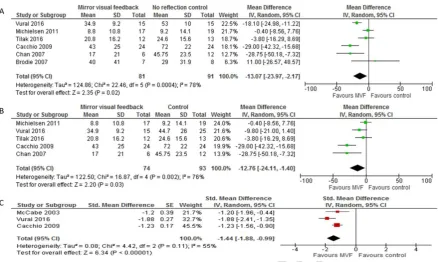
Data for pain intensity could be pooled from 10 trials. Three studies did not measure pain intensity (Daenen et al. 2012a; Daenen et al. 2012b; Dohle et al. 2009), and the other four studies did not report all relevant information needed for analysis (Bayon-Calatayud et al. 2016; Cacchio et al. 2009a; Hunter et al. 2003; McCabe et al. 2007). Data from 6 trials comparing mirror VF using a normal-sized reflection of a body part against a no-reflection control could be analysed and resulted in a significant effect in favour of mirror VF using a random-effects model (Z=2.35, p=0.02; Figure 1(a)). The MD was -13.07 (95%CI=-23.97, - 2.17) mm on a 100mm VAS, which is considered clinically relevant. However, there was substantial heterogeneity (I2=78%).
A subgroup analysis was conducted comparing mirror VF using a normal-sized reflection of a body part with covered mirror control. Data for pain intensity were pooled from 3 trials and resulted in no significant overall effect (Z=1.78, p=0.08) (supporting material Figure S2(a)).
Subgroup analyses were conducted in studies in which mirror VF was delivered in one session and as a prolonged treatment. When mirror VF was administered in one session there was no significant overall effect (Z=1.03, p=0.30, supporting material Figure S2(b)). The analysis including 5 trials in which mirror VF using a normal-sized reflection of a limb was delivered in multiple sessions resulted in a significant overall effect (Z=2.20, p=0.03), but with substantial heterogeneity (I2 =76%). The MD was -12.76 (95%CI=-24.11, -1.40) mm on a 100mm VAS using a random-effects model (Figure 1(b)).
Data from studies investigating patients with PLP and CRPS could be pooled and analysed separately. Data for pain intensity were pooled from 3 trials investigating PLP patients and resulted in no
significant overall effect (Z=1.00, p=0.32, supporting material Figure S2(c)). Data for pain intensity were pooled from 3 trials investigating CRPS patients and resulted in a significant large effect in favour of mirror VF using a normal-sized reflection (Z=6.34, p<0.001 SMD=-1.44; 95%CI=-1.88, -0.99) The I2 statistic (55%) suggested moderate heterogeneity using a random-effects model (Figure 1(c)).
A funnel plot was created to analyse publication bias (supporting material Figure S3) but there is an insufficient number of trials to allow a meaningful conclusion. Publication bias cannot be discounted.
Two studies could not be included in the meta-analysis. A study using 15 amputees with PLP showed no significant pain reduction comparing mirror VF using a normal-sized reflection with no treatment (SMD=-0.08; 95%CI=-1.10, 0.93, (Anghelescu et al. 2016). The study including 6 amputees with PLP showed that the combination of mirror VF using a normal-sized reflection with synchronised stroking of the stump and the hand in front of the mirror significantly reduced pain compared with only mirror VF using a normal-sized reflection (SMD=-1.58, 95%CI=-2.50, -0.66, (Schmalzl et al. 2013).
Follow up data from two studies were pooled. There was a significant large effect in favour of mirror VF using a normal-sized reflection in a follow-up of 6 months in the study conducted by Cacchio et al. (2009a) (SMD=-1.46, 95%CI=-1.83, -1.09). The other study showed no significant effect of mirror VF using a normal-sized reflection in a follow-up of 6 months (SMD=-0.34, 95%CI=-0.75, 0.07,
Data could not be extracted from 4 study reports. Cacchio et al. (2009b) reported that a course of 4 weeks of mirror VF using a normal-sized reflection reduced pain intensity in patients with CRPS when compared with covered mirror and mental imagery. Bayon-Calatayud et al. (2016) investigated patients with closed distal radial fracture and found no difference in pain intensity comparing mirror VF using a normal-sized reflection with direct view of the arm. Hunter et al. (2003) investigated whether mirror VF associated with tactile stimulation was more effective than mirror VF on its own in 13 amputees. It was found that 2 participants reported pain during mirror VF, whilst no
participants reported pain during mirror VF combined with tactile stimuli. McCabe et al. (2007) investigated the effect of sensory-motor mismatch in patients with fibromyalgia by asking patients to perform congruent and incongruent movements while observing the reflection of a limb or observing a white board. When observing a mirror reflection of the limb, 6 participants reported pain during congruent movements and 9 participants reported pain during incongruent movements. When observing a whiteboard 9 participants reported pain during congruent movement and 11 participants reported pain during incongruent movements.
Analyses of other VF techniques
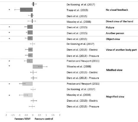
Six of the twenty-three clinical studies evaluated other VF techniques. Differences in study designs, VF techniques and controls prevented meta-analysis. Studies were individually analysed and effects sizes calculated for comparisons within each study (Figure 2).
[Insert Figure 2 here]
Studies investigating pain-free healthy subjects exposed to experimentally-induced pain Characteristics of included studies
Thirteen studies (310 participants) were included for review (Table 2). Seven studies used mirror VF, three studies used virtual reality, two studies used real-time video, and one study used lenses. Study sample sizes varied from 10 to 44 participants and group sizes from nine to 34 participants. The mean age of participants of each individual study varied from 21.6 to 54.69 years (Table 2).
Subjective characteristics of pain free subjects were recorded in two studies using the Neck Disability Index, Pain Catastrophizing Scale (De Kooning et al. 2017), and Centre for Epidemiological Studies Depression Scale (Diers et al. 2013). There were no analyses investigating the effect of these subjective characteristics on experimentally-induced pain outcomes.
[Insert Table 2 here]
Risk of bias
All studies presented high risk of bias (supporting material Table S4). The outcome assessor was not blinded in two (15.3%) studies, and it was unclear whether the outcome assessor was blinded in 10 (80%) studies. A sample size calculation was not provided in any study report. Random sequence generation of conditions was reported in 11 studies (84.6%). Eight (61.5%) of the studies with a repeated-measures design adequately controlled for crossover effects.
Analysis of mirror VF
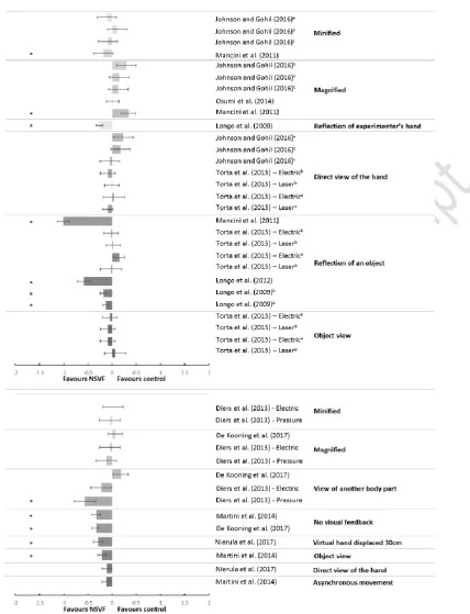
Seven studies compared mirror VF using a normal-sized reflection of a body part with a control. There was sufficient information to analyse the effect of mirror VF on pain in six studies. Due to difference in study designs, VF techniques and controls a meta-analysis could not be conducted. Studies were individually analysed and effects sizes calculated for comparisons within each study (Figure 3).
[Insert Figure 3 here]
Mirror VF vs. control
compared with an object view in 4 comparisons (supporting material Table S5, (Torta et al. 2015). There was no significant effect of mirror VF compared with direct view of the body part (Johnson and Gohil 2016; Torta et al. 2015). There was a significant moderate effect size in favour of mirror VF compared with the reflection of the hand of the experimenter (SMD=-0.26; 95%CI=-0.40, -0.12 (Longo et al. 2009).
There were eight comparisons of mirror VF using a normal-sized reflection of the hand against the reflection of an object in four studies. There was a significant small effect in favour of mirror VF in two comparisons in the study by Longo et al. (2009) (Experiment 1: SMD=-0.13, 95%CI=-0.25, -0.01. Experiment 2: SMD=-0.18, 95%CI=-0.32, -0.04). There was a significant moderate (SMD=-0.59; 95%CI=-0.84, -0.34) and large effect (SMD=1.01; 95%CI=0.77, 1.25) in favour of mirror VF in two studies (Longo et al. 2012; Mancini et al. 2011). The other 4 comparisons resulted in a non-significant effect (Torta et al. 2015). Data could not be extracted from the study conducted by Mancini et al. (2013) and they reported a significant reduction in pain intensity using a normal-sized mirror reflection of the limb compared with a reflection of an object.
Three studies used mirrors to magnify and minify the size of the body part. In the study conducted by Mancini et al. (2011) there was a significant moderate effect in favour of a magnified reflection compared with a normal-sized reflection (SMD=-0.34, 95%CI=-0.63, -0.05), and a small effect in favour of mirror VF using a normal-sized reflection compared with a minified reflection (SMD=0.18, 95%CI=0.01,0.37). There were no significant differences in the other 7 comparisons (Johnson and Gohil 2016; Osumi et al. 2014).
Analysis of other VF techniques
Six of the 13 included experimental studies evaluated other VF techniques. Due to differences in study designs, VF techniques, and controls a meta-analysis could not be conducted. Studies were individually analysed and effects sizes calculated for comparisons within each study (Figure 3).
Kooning et al. 2017). Observing a magnified or minified body part did not affect pain perception. Data could not be extracted from two studies (Romano et al. 2016; Romano and Maravita 2014). Results for individual comparisons are provided in Figure 3 and Table S5.
DISCUSSION
This systematic review included 23 clinical studies and 13 experimental studies. Our meta-analysis of data from 8 clinical studies provides tentative evidence of pain reduction when mirror VF is
delivered as a course of treatment, and with patients with CRPS. There was also an effect on pain reduction in favour of mirror VF using a normal-sized reflection of a body part when compared with a no reflection control. Studies that used real-time video of the back of patients with back pain found that observing a real-time video of the back alleviated back pain at rest and during movement but did not affect pressure-evoked pain. This systematic review was unable to determine whether normal-sized, magnified and minified VF of body parts affects pain perception because of
contradictory findings in primary studies.
Mirror VF using a normal-sized reflection was not superior to object view or direct view of the hand on reducing experimental pain. There was no consistency in the findings from 17 comparisons from 6 studies to determine whether there were differences experimentally-induced pain between mirror VF using a normal-sized reflection of a body part and a reflection of an object, or a magnified, or minified reflection of the body part. Inconsistent results were also obtained with the analysis of virtual reality studies.
Our meta-analysis of clinical data found a MD of -13.07mm on a 100mm VAS in favour of mirror VF using a normal-sized reflection of a body part when compared with a no reflection control. Mirror VF delivered as a course of treatment resulted in a MD of -12.76mm on a 100mm VAS in favour of mirror VF using a normal-sized reflection. Mirror VF using a normal-sized reflection showed a
meta-analysis of two RCTs, and found that a 4 week course of mirror VF using a normal-sized reflection of body parts reduced pain, but a meta-analysis of 3 studies analysing one session of mirror VF did not. Substantial statistical heterogeneity was present in all of these meta-analyses. Our meta-analysis extends these findings by including three additional clinical studies.
We used broad inclusion criteria to improve statistical power but at the expense of substantial statistical heterogeneity, with studies having a high risk of bias affecting the credibility of effect sizes. Small sample sizes and underpowered primary studies were the norm, with sample size calculations provided in only 40% of clinical studies and none of the experimental studies. We used a random-effects model, which assumes effect sizes are a random sample drawn from a population of effect sizes, and variation is due to population variance plus sampling error (Borenstein et al. 2009; Higgins and Green 2011). We estimated mean and standard deviation from median, range and sample size for the RCT conducted by Vural et al. (2016) because this approach has been extensively used in previous meta-analyses and unlikely to introduce inaccuracies into statistical estimates (Bland 2015; Hozo et al. 2005; Koenig and Thayer 2016). We chose to include a variety of painful conditions and body parts as determined by the investigators of primary studies, with no reason to suspect that any of the conditions would not respond to VF.
The use of broad inclusion criteria in systematic reviews has been challenged as it can lead to misleading conclusions in favour of the intervention (Carroll et al. 2000). However, this is not always the case. Bennett et al. (2011) demonstrated that potential sources of bias occur in both directions especially for treatments where the optimal technique and dosage are not known, as is the case for VF techniques. It is likely that sub-optimal VF techniques contributed to negative outcome studies. Frequency and time of exposure seems to be an important aspect of VF, and it has been
recommended that mirror VF should be performed little and often. A single half hour session once a day or once a week is not encouraged (McCabe 2011). Studies included in our systematic review used a variety of VF protocols ranging from a session of 1 minute to 1 hour 5 days a week during 6 weeks. Visually distorting the size of painful body parts is another component of optimal technique that has aroused interest, despite few available studies on which to judge efficacy (Wittkopf and Johnson 2016). Likewise, embodiment of the viewed body part, which describes the subjective experience of having a sense of one’s own body, including a sense of ownership of body parts (de Vignemont 2011; Longo et al. 2008), is considered an important determinant of outcome but rarely assessed in trials (Foell et al. 2014; McCabe 2011; Wittkopf et al. 2017).
In conclusion, it was not possible to determine whether normal-sized, magnified and minified VF of body parts affects pain perception in patients or pain-free participants because of contradictory findings in primary studies. The most likely explanation for the contradictory nature of the findings is variability in study methodology and a high risk of bias. Rather than continuing to undertake meta-analyses of many underpowered small scale studies of VF techniques it would be more appropriate to undertake one robust multi-centred RCT to determine clinical efficacy against a standard
treatment or a pragmatic trial to determine effectiveness versus usual care. Such a trial should include sample sizes of >200 per treatment arm, as recommended by the Cochrane collaboration, to generate findings with sufficient confidence (and low risk of bias) to generalise to clinical practice. The likelihood of a multi-centred RCT being realised is low because funding councils do not consider these types of interventions high priority. Thus, meaningful synthesis of the findings of studies that evaluate VF techniques will continue to be descriptive.
Author Contributions
All authors contributed to conception, design, analysis and interpretation of data. The first author drafted the manuscript. All authors critically revised the article for important intellectual content. All authors gave final approval for publication.
REFERENCES
Anghelescu DL, Kelly CN, Steen BD, Wu J, Wu H, DeFeo BM, Scobey K, Burgoyne L. Mirror therapy for phantom limb pain at a pediatric oncology institution. Rehabil Oncol 2016;34: 104-110. Apkarian AV, Hashmi JA, Baliki MN. Pain and the brain: specificity and plasticity of the brain in clinical
chronic pain. Pain 2011;152: S49-64.
Bayon-Calatayud M, Benavente-Valdepenas AM, Del Prado Vazquez-Munoz M. Mirror therapy for distal radial fractures: A pilot randomized controlled study. J Rehabil Med 2016;48: 829-832. Bennett MI, Hughes N, Johnson MI. Methodological quality in randomised controlled trials of
transcutaneous electric nerve stimulation for pain: low fidelity may explain negative findings. Pain 2011;152: 1226-1232.
Bland M. Estimating mean and standard deviation from the sample size, three quartiles, minimum, and maximum. Int J Stat Med Res 2015;4: 57.
Boesch E, Bellan V, Moseley GL, Stanton TR. The effect of bodily illusions on clinical pain: a systematic review and meta-analysis. Pain 2016;157: 516-529.
Borenstein M, Hedges LV, Higgins J, Rothstein HR. Introduction to Meta-Analysis Wiley Online Library. 2009.
Bowering KJ, O'Connell NE, Tabor A, Catley MJ, Leake HB, Moseley GL, Stanton TR. The effects of graded motor imagery and its components on chronic pain: a systematic review and meta-analysis. J Pain 2013;14: 3-13.
Brodie EE, Whyte A, Niven CA. Analgesia through the looking-glass? A randomized controlled trial investigating the effect of viewing a 'virtual' limb upon phantom limb pain, sensation and movement. Eur J Pain 2007;11: 428-436.
Reporting Pain in Clinical Trials and Systematic Reviews: Recommendations from an OMERACT 12 Workshop. J Rheumatol 2015;42: 1962-1970.
Cacchio A, Blasis E, Blasis V, Santilli V, Spacca G. Mirror therapy in complex regional pain syndrome type 1 of the upper limb in stroke patients. Neurorehabil Neural Repair 2009a;23: 792-799. Cacchio A, Blasis E, Necozione S, Orio F, Santilli V. Mirror therapy for chronic complex regional pain
syndrome type 1 and stroke. N Engl J Med 2009b;361: 634-636.
Carroll D, Moore R, McQuay H, Fairman F, Tramer M, Leijon G. Transcutaneous electrical nerve stimulation (TENS) for chronic pain. The Cochrane Library 2000.
Chan BL, Witt R, Charrow AP, Magee A, Howard R, Pasquina PF, Heilman KM, Tsao JW. Mirror therapy for phantom limb pain. N Engl J Med 2007;357: 2206-2207.
Cohen J. Statistical Power Analysis for the Behavioral Sciences. Second Edition Lawrence Erlbaum Associates. 1998.
Daenen L, Nijs J, Roussel N, Wouters K, Cras P. Altered perception of distorted visual feedback occurs soon after whiplash injury: an experimental study of central nervous system processing. Pain Physician 2012a;15: 405-413.
Daenen L, Nijs J, Roussel N, Wouters K, Van Loo M, Cras P. Sensorimotor incongruence exacerbates symptoms in patients with chronic whiplash associated disorders: an experimental study. Rheumatology (Oxford) 2012b;51: 1492-1499.
De Kooning M, Daenen L, Verhelpen S, Don S, Voogt L, Roussel N, Ickmans K, Van Loo M, Cras P, Nijs J. Abnormal Pain Response to Visual Feedback During Cervical Movements in Chronic Whiplash: An Experimental Study. Pain Pract 2017;17: 156-165.
de Vignemont F. Embodiment, ownership and disownership. Conscious Cogn 2011;20: 82-93.
Diers M, Christmann C, Koeppe C, Ruf M, Flor H. Mirrored, imagined and executed movements differentially activate sensorimotor cortex in amputees with and without phantom limb pain. Pain 2010;149: 296-304.
Diers M, Kamping S, Kirsch P, Rance M, Bekrater-Bodmann R, Foell J, Trojan J, Fuchs X, Bach F, Maass H, Cakmak H, Flor H. Illusion-related brain activations: a new virtual reality mirror box system for use during functional magnetic resonance imaging. Brain Res 2015a;1594: 173-182. Diers M, Loffler A, Zieglgansberger W, Trojan J. Watching your pain site reduces pain intensity in
chronic back pain patients. Eur J Pain 2015b.
Diers M, Zieglgänsberger W, Trojan J, Drevensek AM, Erhardt-Raum G, Flor H. Site-specific visual feedback reduces pain perception. Pain 2013;154: 890-896.
Djavadkhani Y, Marshall NS, D'Rozario AL, Crawford MR, Yee BJ, Grunstein RR, Phillips CL. Ethics, consent and blinding: lessons from a placebo/sham controlled CPAP crossover trial. Thorax 2015;70: 265-269.
Dohle C, Püllen J, Nakaten A, Küst J, Rietz C, Karbe H. Mirror therapy promotes recovery from severe hemiparesis: a randomized controlled trial. Neurorehabil Neural Repair 2009;23: 209-217. Flor H, Denke C, Schaefer M, Grusser S. Effect of sensory discrimination training on cortical
reorganisation and phantom limb pain. Lancet 2001;357: 1763-1764.
Flor H, Nikolajsen L, Staehelin Jensen T. Phantom limb pain: a case of maladaptive CNS plasticity? Nat Rev Neurosci 2006;7: 873-881.
Foell J, Bekrater-Bodmann R, Diers M, Flor H. Mirror therapy for phantom limb pain: brain changes and the role of body representation. Eur J Pain 2014;18: 729-739.
Gracely R. Studies of pain in human subjects. In: Melzack andWall’s textbook of pain.Philadelphia: Elsevier Churchill Livingstone; 2006; 267-290.
Handwerker H and Kobal G. Psychophysiology of experimentally induced pain. Physiol Rev 1993;73: 639-671.
Higgins JPT and Green S.Cochrane Handbook for Systematic Reviews of Interventions Version 5.1. 0 [Updated March 2011]. 2011.
Hunter JP, Katz J, Davis KD. The effect of tactile and visual sensory inputs on phantom limb awareness. Brain 2003;126: 579-589.
Johnson MI and Gohil M. An investigation into enlarging and reducing the size of mirror reflections of the hand on experimentally-induced cold-pressor pain in healthy human participants. Scan J Pain 2016;10: 19-25.
Koenig J and Thayer JF. Sex differences in healthy human heart rate variability: A meta-analysis. Neurosci Biobehav Rev 2016;64: 288-310.
Lewis JS, Kersten P, McCabe CS, McPherson KM, Blake DR. Body perception disturbance: A contribution to pain in complex regional pain syndrome (CRPS). Pain 2007;133: 111-119. Longo MR, Betti V, Aglioti SM, Haggard P. Visually induced analgesia: seeing the body reduces pain. J
Neurosci 2009;29: 12125-12130.
Longo MR, Iannetti GD, Mancini F, Driver J, Haggard P. Linking pain and the body: neural correlates of visually induced analgesia. J Neurosci 2012;32: 2601-2607.
Longo MR, Schuur F, Kammers MP, Tsakiris M, Haggard P. What is embodiment? A psychometric approach. Cognition 2008;107: 978-998.
Mancini F, Longo MR, Canzoneri E, Vallar G, Haggard P. Changes in cortical oscillations linked to multisensory modulation of nociception. Eur J Neurosci 2013;37: 768-776.
Mancini F, Longo MR, Kammers MP, Haggard P. Visual distortion of body size modulates pain perception. Psychol Sci 2011;22: 325-330.
Martini M, Perez‐Marcos D, Sanchez‐Vives MV. Modulation of pain threshold by virtual body ownership. Eur J Pain 2014;18: 1040-1048.
McCabe C. Mirror visual feedback therapy. A practical approach. J Hand Ther 2011;24: 170-178; quiz 179.
McCabe CS, Cohen H, Blake DR. Somaesthetic disturbances in fibromyalgia are exaggerated by sensory - Motor conflict: Implications for chronicity of the disease? Rheumatology 2007;46: 1587-1592.
McCabe CS, Haigh RC, Blake DR. Mirror visual feedback for the treatment of complex regional pain syndrome (type 1). Curr Pain Headache Rep 2008;12: 103-107.
Michielsen ME, Selles RW, Geest JN, Eckhardt M, Yavuzer G, Stam HJ, Smits M, Ribbers GM, Bussmann JB. Motor recovery and cortical reorganization after mirror therapy in chronic stroke patients: a phase II randomized controlled trial. Neurorehabil Neural Repair 2011;25: 223-233.
Moher D, Hopewell S, Schulz KF, Montori V, Gotzsche PC, Devereaux PJ, Elbourne D, Egger M, Altman DG. CONSORT 2010 explanation and elaboration: updated guidelines for reporting parallel group randomised trials. Int J Surg 2012;10: 28-55.
Moher D, Shamseer L, Clarke M, Ghersi D, Liberati A, Petticrew M, Shekelle P, Stewart LA. Preferred reporting items for systematic review and meta-analysis protocols (PRISMA-P) 2015 statement. Systematic reviews 2015;4: 1.
Moseley GL. Distorted body image in complex regional pain syndrome. Neurology 2005;65: 773. Moseley GL, Gallace A, Spence C. Bodily illusions in health and disease: Physiological and clinical
perspectives and the concept of a cortical 'body matrix'. Neurosci Biobehav Rev 2012;36: 34-46.
Moseley GL, Parsons TJ, Spence C. Visual distortion of a limb modulates the pain and swelling evoked by movement. Curr Biol 2008;18: R1047-1048.
Nierula B, Martini M, Matamala-Gomez M, Slater M, Sanchez-Vives MV. Seeing an Embodied Virtual Hand is Analgesic Contingent on Colocation. J Pain 2017;18: 645-655.
Osumi M, Imai R, Ueta K, Nakano H, Nobusako S, Morioka S. Factors associated with the modulation of pain by visual distortion of body size. Front Hum Neurosci 2014;8: 137.
Preston C and Newport R. Analgesic effects of multisensory illusions in osteoarthritis. Rheumatology (Oxford) 2011;50: 2314-2315.
Ramachandran VS, Rogers-Ramachandran D, Cobb S. Touching the phantom limb. Nature 1995;377: 489-490.
Romano D, Llobera J, Blanke O. Size and Viewpoint of an Embodied Virtual Body Affect the Processing of Painful Stimuli. J Pain 2016;17: 350-358.
Romano D and Maravita A. The visual size of ones own hand modulates pain anticipation and perception. Neuropsychologia 2014;57: 93-100.
Schmalzl L, Ragno C, Ehrsson HH. An alternative to traditional mirror therapy: illusory touch can reduce phantom pain when illusory movement does not. Clin J Pain 2013;29: e10-18.
Thieme H, Mehrholz J, Pohl M, Behrens J, Dohle C. Mirror therapy for improving motor function after stroke. The Cochrane database of systematic reviews 2012: Cd008449.
Thieme H, Morkisch N, Rietz C, Dohle C, Borgetto B. The Efficacy of Movement Representation Techniques for Treatment of Limb Pain--A Systematic Review and Meta-Analysis. J Pain 2016;17: 167-180.
Tilak M, Isaac SA, Fletcher J, Vasanthan LT, Subbaiah RS, Babu A, Bhide R, Tharion G. Mirror Therapy and Transcutaneous Electrical Nerve Stimulation for Management of Phantom Limb Pain in Amputees - A Single Blinded Randomized Controlled Trial. Physiother Res Int 2016;21: 109-115.
Torta DME, Legrain V, Mouraux A. Looking at the hand modulates the brain responses to nociceptive and non‐nociceptive somatosensory stimuli but does not necessarily modulate their perception. Psychophysiology 2015;52: 1010-1018.
Trapp W, Weinberger M, Erk S, Fuchs B, Mueller M, Gallhofer B, Hajak G, Kubler A, Lautenbacher S. A brief intervention utilising visual feedback reduces pain and enhances tactile acuity in CLBP patients. J Back Musculoskelet Rehabil 2015;28: 651-660.
Vural SP, Nakipoglu Yuzer GF, Sezgin Ozcan D, Demir Ozbudak S, Ozgirgin N. Effects of Mirror Therapy in Stroke Patients With Complex Regional Pain Syndrome Type 1: A Randomized Controlled Study. Arch Phys Med Rehabil 2016;97: 575-581.
Wand BM, Parkitny L, O'Connell NE, Luomajoki H, McAuley JH, Thacker M, Moseley GL. Cortical changes in chronic low back pain: current state of the art and implications for clinical practice. Man Ther 2011;16: 15-20.
Wittkopf PG and Johnson MI. Managing pain by visually distorting the size of painful body parts: is there any therapeutic value? Pain management 2016;6: 201-204.
Figure 1 Forest plot of comparisons: (a) mirror visual feedback using a normal-sized reflection versus no reflection control. (b) Subgroup analysis of prolonged treatment with mirror visual feedback using a normal-sized reflection. (c) Subgroup analysis of complex regional pain syndrome patients.
Table 1 Characteristics of studies investigating participants with a clinical pain
Study and design Clinical condition
(total n)
Visual feedback technique Control Group/Condition Pain outcome measures
Normal-sized mirror visual feedback
Michielsen et al. (2011)
RCT
Stroke (36) NSVF with a mirror (n = 17,55.3 ± 12.0
years, 7M/13F): 6 weeks, 5 days a
week, 1 hour.
Direct view of the hand (n = 19,58.7 ± 13.5
years, 13M/7F)
Pain intensity VAS.
Cacchio et al. (2009a)
RCT
CRPS type 1 (48) NSVF with a mirror (n = 24, 57.9 ± 9.9
years, 13F/11M):
First 2 weeks: 5 days a week, 30
minutes
Last 2 weeks: 5 days a week, 1 hour.
Covered mirror (n = 24,58.8 ± 9.4 years,
13F/11M)
Pain intensity VAS.
Cacchio et al. (2009b)
RCT
CRPS and stroke (n = 24, median 62, range 53-71 years, 11M/13F)
NSVF with a mirror (8): 4 weeks, 7 days
a week, 30 minutes.
Covered mirror (8)
Mental imagery (8)
Pain intensity VAS.
Chan et al. (2007)
RCT
Lower limb amputation
(18, N/A)
NSVF with a mirror (6): 4 weeks, 7 days
a week, 15 minutes.
Mental imagery(6)
Covered mirror(6)
Pain intensity VAS.
Vural et al. (2016)
RCT
CRPS type 1 (30) NSVF with a mirror (n = 15, 68.9 ± 10.5
years, 7F/8M): 4 weeks, 5 days a week,
30 minutes.
No treatment (n = 15, 61.4 ± 11.9 years, 6F/9M) Pain intensity VAS.
Dohle et al. (2009)
RCT
Stroke (36) NSVF with a mirror (n = 18, 54.9 ± 13.8
years, 13M/5F): 6 weeks, 5 days a
week, 30 minutes.
Direct view of the hand (n = 18, 58.0 ± 14.0,
years 13M/5F)
Brodie et al. (2007) RCT
Lower limb amputation NSVF with a mirror(n = 41, median 54,
range 20-83 years, 35M/6F): sequence
of 10 congruent movements repeated
10 times
Covered mirror (n = 39, median57, range
25-80 years, 28M/11F)
Pain intensity VAS,
McGill.
Bayon-Calatayud et al.
(2016)
RCT
Closed distal radial fracture (22)
NSVF with a mirror (n = 11, 61.09 ±
13.05 years, 3M/ 8F): 3 weeks, 5 days
a week, 30 minutes.
Direct view of the hand (n = 11, 55.36 ± 18.28
years, 4M/7F)
Pain intensity
VAS.
Tilak et al. (2016)
RCT
Amputation (25) NSVF with a mirror (n = 12, 42.62 ±
10.69 years, 12M/ 1F): 4 consecutive
days, 20 minutes.
Transcutaneous electrical nerve stimulation (n
= 13, 36.38 ± 9.55 years, 11M/2F)
Pain intensity
VAS.
Wand et al. (2012)
Randomized cross-over
experiment
Chronic nonspecific low back pain (n = 25, 41.8±14.7 years, 14M/11F)
NSVF with a mirror: Participants
performed movements while viewing
the reflection of their low back
Covered mirror Pain intensity VAS.
Anghelescu et al.
(2011)
Retrospective study
Amputation (15) NSVF with a mirror (n = 8, median 12,
range 8-20 years, 7M/1F)
No treatment (n = 7, median 26, range 10-24
years, 5M, 2F)
Pain intensity
NRS.
Schmalzl et al. (2013)
Within subjects
repeated measures
Upper limb amputation
(n = 6, 55.1 ± 14.6
years, 2M/ 4F)
NSVF with a mirror: congruent
movements 60 seconds followed by 60
seconds of rest 8 times.
NSVF with a mirror plus tactile stimuli Pain intensity VAS.
McCabe et al. (2003)
Within-subjects
repeated-measures
CRPS type 1 (n = 8, 33 ±
55.5 years, 5F/3 M)
NSVF with a mirror: congruent
movements.
Covered mirror
Mental imagery
Hunter et al.(2003)
Within-subjects
repeated-measures
Upper limb amputation
(n = 13, 36.07 ± 11.8
years, 11M/2F)
NSVF with a mirror: congruent
movements.
Direct view
Closed eyes
NSVF with a mirror plus tactile stimuli
Pain intensity VAS.
Daenen et al. (2012a)
Within and between
subjects repeated
measures
Acute whiplash
associated disorder (n
= 30, 43.30 ±10.98
years, 14F/16M)
NSVF with a mirror: congruent
movements.
Within subjects
whiteboard: congruent movements
NSVF with a mirror: incongruent movements
No mirror/whiteboard congruent movements
No mirror/whiteboard incongruent movements
Between subjects
Healthy participants (29)
Proportion of reported
sensations and intensity
of sensations NRS.
Deanen et al. (2012b)
Within and between
subjects repeated
measures
Chronic whiplash
associated disorder (n
= 35, 43.8 ± 9.58 years,
26F,9M)
NSVF with a mirror: congruent
movements.
Within subjects
whiteboard: congruent movements
NSVF with a mirror: incongruent movements
No mirror/whiteboard congruent movements
No mirror/whiteboard incongruent movements
Between subjects
Healthy participants (31)
Proportion of reported
sensations and intensity
of sensations NRS.
McCabe et al. (2007)
Within-subject
repeated measures
Fibromyalgia (n = 29,
47.9 ± 11.1 years,
1M/28F)
NSVF with a mirror: congruent
movements.
Within subjects comparisons
whiteboard: congruent movements
Normal size visual feedback with a mirror:
incongruent movements
Between subjects comparisons
Healthy participants (29)
Proportion of reported
sensations and intensity
of sensations NRS.
Diers et al. (2015b) Within-subject repeated measures
Chronic back pain (n =
19, 44.8 ± 17.2 years,
5M/14F)
NSVF with a real time video of their
back (1minute)
Object view
View of the back of another person
View of the picture of their back
Closed eyes
Pain intensity NRS.
Trapp et al. (2015) RCT
Chronic low back pain
(30)
NSVF with real time video of their back
(n = 15, 45.53 ±7.05 years, 10M/5F)
2 weeks, 3 days a week, 20 minutes
No treatment (n = 15, 40.60 ± 10.67 years,
9M/6F)
Pain intensity
VAS.
Size distorted visual feedback using other visual feedback techniques
Diers et al. (2013) Within and between subjects repeated measures
Chronic upper back pain (n= 18, 54.74 ± 9.14 years, 5M/13F)
NSVF with real time video of their back
Within subjects
Magnified view of their back Minified view of their back
View of the hand Between subjects healthy controls (18)
Pain intensity NRS
Pain induced by pressure
and electrical
stimulations.
Moseley et al. (2008) Within-subject repeated measures
Chronic pain and
dysfunction of one arm
(n = 10, 35.1 ± 11.7
years, 5M 5F)
NSVF looking through a lens, while
performing movements
Magnified view
Minifed view
Direct view of the hand
Pain intensity VAS.
Preston and Newport (2011)
Within-subject repeated measures
Osteoarthritis (n = 20,
70.5 ± 6.5 years,
2M/18F)
Stretched and shrunken views with
real time video of a painful joint
Stretched and shrunken views with real time
video of a non- painful joint
Pain intensity NRS.
De Kooning et al. (2017)
Within and between subjects repeated measures
Whiplash associated
disorder (n = 30, 42.2 ±
10.73 years, 10M/20F)
NSVF with real time video of their neck No visual feedback
View of the hand
Magnified view of the neck
Abbreviations: RCT, Randomised controlled trial; NSVF, Normal-sized visual feedback; VAS, visual analogue scale; CRPS, complex regional pain syndrome; NRS, numeric
Table 2 Characteristics of studies investigating pain-free healthy participants exposed to experimentally-induced pain
Study & design Participants
(total n)
Visual feedback technique Control Group/Condition Pain outcome measure
Normal-sized mirror visual feedback
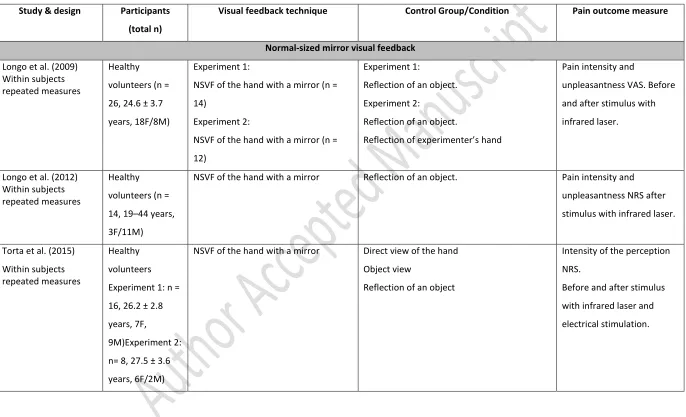
Longo et al. (2009) Within subjects repeated measures
Healthy
volunteers (n =
26, 24.6 ± 3.7
years, 18F/8M)
Experiment 1:
NSVF of the hand with a mirror (n =
14)
Experiment 2:
NSVF of the hand with a mirror (n =
12)
Experiment 1:
Reflection of an object.
Experiment 2:
Reflection of an object.
Reflection of experimenter’s hand
Pain intensity and
unpleasantness VAS. Before
and after stimulus with
infrared laser.
Longo et al. (2012) Within subjects repeated measures
Healthy
volunteers (n =
14, 19–44 years,
3F/11M)
NSVF of the hand with a mirror Reflection of an object. Pain intensity and
unpleasantness NRS after
stimulus with infrared laser.
Torta et al. (2015)
Within subjects repeated measures
Healthy
volunteers
Experiment 1: n =
16, 26.2 ± 2.8
years, 7F,
9M)Experiment 2:
n= 8, 27.5 ± 3.6
years, 6F/2M)
NSVF of the hand with a mirror Direct view of the hand
Object view
Reflection of an object
Intensity of the perception
NRS.
Before and after stimulus
with infrared laser and
Mancini et al. (2013)
Within subjects
repeated measures
Healthy
volunteers (n =
10, mean 25,
range 19– 32
years)
NSVF of the hand with a mirror Reflection of an object Pain intensity NRS during
contact thermal heat
stimulation
Size distorted mirror visual feedback
Johnson and Gohil
(2016)
Within subjects repeated measures
Healthy
volunteers (n =
20, 23.55 ± 4.01
years, 10M/10F)
NSVF of the hand with a mirror Magnified reflection of the hand
Minified reflection of the hand
Direct view of the hand
Pain threshold, tolerance
and intensity VAS. During
cold-pressor task.
Mancini et al. (2011)
Within and between subjects repeated measures
Healthy
volunteers (n =
18, 27,1 ± 4,1
years, 7M/11F
NSVF of the hand with a mirror Within subjects
Magnified reflection of the hand
Minified reflection of the hand
Between subjects
Reflection of an object
Contact heat pain threshold
Osumi et al. (2014) Within and between subjects repeated measures
Healthy
volunteers (n =
44, 21.6 ± years,
17M/27F)
NSVF of the hand with a mirror Magnified reflection of the hand Contact heat pain threshold
Normal-sized visual feedback using other visual feedback techniques
Nierula et al. (2017)
Within subjects
repeated measures
Healthy
volunteers (n =
19,24.1 ± 5.1
years, 19M)
NSVF of the arm of an avatar
co-located with participants’ arm
NSVF of the arm of an avatar displaced 30 cm
away from participants’ body midline
Martini et al. (2014) Within subjects
repeated measures
Healthy
volunteers (n =
24, 25.5 ± 5.8
years, 14F/10M)
NSVF of an avatar index finger moving
in synchrony to participants’ finger.
No visual feedback
Object view
NSVF of an avatar index finger moving in
asynchrony to participants’ finger.
Contact heat pain threshold
Size-distorted visual feedback using other techniques
Romano and Maravita (2014)
Within subjects repeated measures
Healthy
volunteers (n =
38, 24.46 ± 3.88,
30F/8M)
NSVF of the hand looking through a
lens
Magnified view of the hand
Minified view of the hand
Pain intensity VAS, after
noxious stimuli with a
non-invasive needle with a blunt
end.
Romano e al. (2016) Within subjects repeated measures
Healthy
volunteers (n =
21, 23 ± 2 years,
9F/12M)
NSVF of an avatar legs congruent with
the position of participants’ legs
Magnified view of the avatar’s leg
Minified view of the avatar’s leg
Pain intensity VAS, after
noxious stimuli with a
non-invasive needle with a blunt
end.
De Kooning et al. (2017)
Within and between subjects repeated measures
Healthy controls
(n = 34, 44.59
±13.85 years,
11M/23F)
NSVF with real time video of their neck No visual feedback
View of the hand
Magnified view of the neck
Pressure pain threshold
Diers et al. (2013) Within and between subjects repeated measures
Healthy volunteers (n = 18, 54.69 ± 9.09 years, 6M/12F)
NSVF with real time video of their back Within subjects
Magnified view of their back Minified view of their back View of the hand
Pain intensity NRS
Pain induced by pressure
and electrical stimulations.