Open Access
Research article
Magnesium deficiency: effect on bone mineral density in the mouse
appendicular skeleton
Helen E Gruber*
1
, Robert K Rude
2
, L Wei
2
, A Frausto
2
, Barbara G Mills
2
and
H James Norton
3
Address: 1Department of Orthopaedic Surgery, Cannon Bldg, 3rd floor, Carolinas Medical Center, PO Box 32861, Charlotte, N.C. 28203, USA, 2University of Southern California and the Orthopaedic Hospital, 2025 Zonal Ave., Los Angeles, CA 90033, USA and 3Department of Biostatistics, Carolinas Medical Center, PO Box 32861, Charlotte, N.C. 28232, USA
Email: Helen E Gruber* - [email protected]; Robert K Rude - [email protected]; L Wei - [email protected]; A Frausto - [email protected]; Barbara G Mills - ; H James Norton - [email protected]
* Corresponding author
Abstract
Background: Dietary magnesium (Mg) deficiency in the mouse perturbs bone and mineral homeostasis. The objective of the present study was to evaluate bone mineral density of the femur in control and Mg-deficient mice.
Methods: BALB/c mice aged 28 days at study initiation were maintained on a normal or Mg deficient (0.0002% Mg) diet, and at time points 0, 2, 4 or 6 weeks bones were harvested for bone mineral density analysis. Peripheral quantitative computed tomography (pQCT) was used to assess the trabecular metaphyseal compartment and the cortical midshaft.
Results: Although mean total bone density of the femoral midshaft in Mg deficient mice did not differ significantly from controls throughout the study, the trabecular bone compartment showed significantly decreased mineral content after 4 (p < 0.001) and 6 weeks (p < 0.001) of Mg depletion.
Conclusions: This study demonstrates the profound effect of Mg depletion on the trabecular compartment of bone, which, with its greater surface area and turnover, was more responsive to Mg depletion than cortical bone in the appendicular skeleton of the mouse.
Background
Several previous epidemiologic studies have demonstrat-ed a positive correlation between dietary Mg intake and bone density and/or the rate of bone loss during low Mg intake in humans [1–5]. Magnesium depletion has also been strongly indicated as a risk factor for osteoporosis as-sociated with aging and postmenopausal status [6–8]. Since osteoporosis also occurs with greater frequency in certain populations in which Mg depletion is also present, such as diabetes mellitus, alcoholism, and malabsorption syndromes [9], further investigations of animal models in
which dietary Mg intake is decreased are valuable because they can help us to better understand the relationship of Mg inadequacy and bone health.
Previous work by our group has shown that in the Mg-de-pleted mouse hypomagnesemia develops and skeletal Mg content falls significantly [10]. Bone histomorphometry analysis showed that trabecular bone of Mg-depleted mice also shows increased numbers of osteoclasts and reduced numbers of osteoblasts. The present study was conducted in order to compare bone mineral density in the
Published: 17 April 2003
BMC Musculoskeletal Disorders 2003, 4:7
Received: 29 January 2003 Accepted: 17 April 2003
This article is available from: http://www.biomedcentral.com/1471-2474/4/7
trabecular and cortical bone of the mouse femur using pe-ripheral quantitative computed tomography (pQCT). This study demonstrates the profound effect of Mg deple-tion on the trabecular compartment of bone, which, with its greater surface area and turnover, was more quickly re-sponsive to Mg depletion than cortical bone in the appen-dicular skeleton of the mouse.
Methods
The study presented here was approved by the IACUC of Orthopaedic Hospital. Dietary Mg deficiency was induced for up to 6 weeks in BALB/c female mice (Simonsen Lab-oratories, Gilroy, CA) aged 28 days at start of the study. Mice were acclimated to the vivarium for one week, and fed tap water and a control or Mg deficient diet as de-scribed below ad libitum. Animals were housed individu-ally in 7-inch-high plastic cages with 110 inch2 floor area with Sani-chips bedding. The environment was main-tained at 20–25°C, 50% humidity, with 10–15 room air exchanges per hour and a 12:12-hour light: dark cycle. The experimental diets were instituted after a 1-week acclima-tion period. Mice were fed either a normal control Mg diet (0.06% as percent of total diet) or a Mg-deficient diet (0.002% Mg) (both diets obtained from Harlan Teklad, Madison, WI). Dietary calcium intake was as recommend-ed for mice at 0.05%. The complete composition of the diet has been previously published [11]. Pair feeding was performed in order to keep weight gain as close as possi-ble in the Mg-deficient and control groups, as bone mass is closely correlated with body mass. Control mice were pair-fed with the Mg-deficient mice based on weight of food consumed by the Mg-deficient animals accordingly every day. During the study period, distilled deionized water containing <0.003 mg Mg/dl was used for hydration.
Animals were weighed at start of study and at study termi-nations. At baseline, and 2, 4 and 6 weeks of study, ani-mals were euthanized, the right femur removed and the left femur harvested for bone mineral density analysis. Pe-ripheral quantitative computed tomography (pQCT) was performed on the excised left femur using a Stratec XCT-RM and associated software (Stratec Medizintechnik Gm-bh, Pforzheim, Germany. Software version 5.40). The scan was performed at two sites, 12 and 50% from the dis-tal end of the femur. The positions were verified using scout views and one 0.5-mm slice perpendicular to the long axis of the femur shaft was acquired. The scans were analyzed using a threshold for delineation of the external boundary recommended by the software. The bone min-eral density, area and minmin-eral content at each site were re-ported. Quality control of the machine was monitored by scanning a manufacturer-provided phantom on each day of scan. The coefficient of variations for bone mineral con-tent, area and density were 2.2, 3.5 and 2.8%, respectively.
Statistical analysis employed SAS® version 8.2 software and used standard methods of analysis including paired and unpaired t-tests and two-way ANOVA with interac-tion. Data are expressed as means ± S.D.
Results
Pair feeding was used to attempt to keep weight changes as close as possible in control and Mg-deficient animals. As shown in Table 1, there was no difference in the initial vs final mean weights for control animals at 2, 4 or 6 week study timepoints. In the Mg-deficient animals, there was no difference between initial and final mean weights at 2 weeks or at 4 weeks. In the 6 week group, however, the fi-nal weight was significantly less than the mean initial weight of that group (p = 0.002). When mean initial weights were analyzed, the controls and Mg-deficient ani-mals showed no differences in the 4 or 6 week groups. In the 2 week group, however, controls were significantly heavier (p = 0.01). When mean final weights were ana-lyzed, controls and Mg-deficient animals showed no dif-ferences in the 2 and 4 week groups; in the 6 week group, controls were significantly heavier than Mg-deficient ani-mals (p < 0.001). It is unlikely that these body weight dif-ferences influenced bone mineral density since changes seen in trabecular bone mineral content between controls and Mg-deficient animals were already present in the 4 week group, a time at which there was no difference in mean weights of these groups.
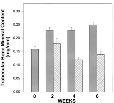
Femurs from mice aged 28 days at start of this study were assessed with pQCT to determine volumetric bone miner-al density (BMD, mg/cm3) of the midshaft and total bone mineral content of the distal femoral metaphyseal (BMC, mg/mm) of control and Mg-deficient animals at baseline (time 0, start of study) and 2, 4 and 6 weeks of study. As shown in Figure 1, BMC was significantly decreased in the metaphysis of Mg-deficient mice. Statistical analysis using ANOVA showed that controls increased significantly over time (with all of the change from baseline to week 2) (p = 0.005). BMC of femurs from Mg-deficient mice did not change significantly over time, and at weeks 4 and 6 were significantly decreased compared to controls at the same time point (p < 0.001). Bone mineral density (mg/cm3) in the metaphysis was similar in controls and Mg-deficient mice at 2 weeks [171.4 ± 25.5 (8) for controls vs 162.1 ± 47.0 (7) for Mg-deficient animals (means ± S.D. (n))]. At 6 weeks, however, the mean bone mineral density in Mg-deficient animals was significantly lower than controls (146.8 ± 37.9 (7) vs control level of 153.3 ± 28 (10), p = 0.02).
significant change over time in either control (p = 0.17) or Mg-deficient mice (p = 0.07). There was no significant dif-ference between controls or Mg-deficient BMD at 2, 4 or 6 weeks (= 0.53, 0.64 and 0.81, respectively). Since there is little trabecular bone in the femoral midshaft, trabecular bone mineral content was not evaluated at this site.
[image:3.612.54.561.102.219.2]Mean cortical thickness was also measured at the midshaft site. Control means increased from 0.316 ± 0.01 (8) (mean ± S.D. (n)) at baseline to 0.323 ± 0.008 (10) but showed no significant difference over 0 – 6 weeks. Mean cortical thickness similarly did not change significantly in the low Mg mice (0.311 ± 0.01 (8) at baseline; 0.321 ± 0.009 (7) at 6 weeks).
Table 1: Body Weights at Study Initiation and Completion a
Time (weeks)
Baseline Initial Baseline Final Control Initial Control Final Low Magnesium Initial Low Magnesium Final
0 25.4 ± 1.6
(n = 8)
25.7 ± 1.7 (n = 8)
2 25.5 ± 1.2 c
(n = 8)
25.5 ± 1.6 (n = 8)
23.9 ± 1.9 (n = 8)
24.4 ± 2.5 (n = 8)
4 25.2 ± 1.5
(n = 8)
23.5 ± 1.5 (n = 8)
25.1 ± 1.5 (n = 8)
24.0 ± 2.7 (n = 8)
6 25.5 ± 1.4
(n = 10)
28.2 ± 3.3 (n = 10)
24.8 ± 1.6 (n = 7)
21.5 ± 2.9 b
(n = 7)
a Data are expressed as means ± S.D. See text for discussion of statistical analysis of data. b Significantly less than corresponding mean Low Mg Initial
weight, p = 0.002, and significantly less than corresponding control mean final weight, p < 0.001. c Significantly greater than corresponding Low Mg
initial mean weight, p = 0.001.
Figure 1
[image:3.612.58.291.289.501.2]Bone mineral content (BMC) of the femoral metaphysis at baseline (time 0) and 2, 4 and 6 weeks of study. Solid bars, controls; hatched bars, Mg-deficient mice. BMC of controls increased significantly over the first 2 weeks (p = 0.005), with no significant change thereafter. Although there was no change in the BMC of femurs from Mg-deficient mice by the second week, BMC decreased significantly by weeks 4 and 6 (p < 0.001). (Numbers/group are as noted in Table 1)
Figure 2
[image:3.612.310.553.291.504.2]Discussion
Although previous studies of the magnesium-deficient mouse had reported that this animal model developed hy-pocalcemia (similar to humans), more recent work from our laboratory showed that the Mg deficient mouse has significantly elevated serum calcium levels at 4 and 6 weeks of study [10]. Our previous work has also shown that Mg depletion has a severe effect on the mouse skele-ton resulting in impaired bone growth, decreased osteob-last numbers, increased osteocosteob-last numbers and stimulation of cytokine activity in bone. In this model of dietary Mg deficiency, ashing studies have shown that the percent ash Mg content decreases significantly by 3 weeks of depletion and further declines by 6 weeks [10]. (Ash calcium and phosphorus contents, however, are not altered).
In the present study, we have further examined bone in the Mg-deficient murine model using peripheral quantita-tive computed tomography. Recent studies by Beamer et al have demonstrated that the pQCT technology can be used with precision on the small skeletal bones of the mouse [12]. These authors have also discussed the fact that pQCT data compare well with other methods of as-sessing bone density such as histomorphometry and dual energy X-ray absorptiometry.
After 4 and 6 weeks of Mg deficiency, BMC of the mouse femur in a site primarily trabecular in nature was signifi-cantly lower than that of control animals at these time points. Femoral BMD, however, and femoral cortical mid-shaft thickness, showed no difference compared to rele-vant control means. These data demonstrate the profound effect of Mg depletion on the trabecular compartment of bone, which, with its greater surface area and turnover, was more responsive to Mg depletion than cortical bone in the appendicular skeleton of the mouse. The general nature of enhanced responsiveness of trabecular bone has been previously noted since trabecular bone not only has greater surface area and greater bone turnover than corti-cal bone, but also the changes of bone as seen in diseases such as osteoporosis have a greater effect in that compart-ment [13]. It is of interest to note that a similar pattern of change – decreased BMC but unchanged midshaft BMD – was seen in the tibia of mice lacking the transforming growth factor-β1 gene studied by Geiser et al [14].
Studies with several animals models subjected to dietary Mg deficiency show that Mg deficiency is a risk factor for osteoporosis. A positive correlation between dietary Mg intake and bone density and/or increased rates of bone loss has been published in several epidemiologic studies [3,5,15–18]. Our understanding of the mechanisms of ac-tion at the bone tissue, cell and hormonal level, however, is still incomplete and much remains to be learned about
the role of dietary Mg intake in the maintenance of bone mass.
Conclusions
This study demonstrates the profound effect of Mg deple-tion on the trabecular compartment of bone, which, with its greater surface area and turnover, was more responsive to Mg depletion than cortical bone in the appendicular skeleton of the mouse.
Competing Interests
None declared.
Authors' Contributions
RKR was the principal investigator and designed the study and supervised the experiments. HEG wrote the manu-script and submitted the data for statistical analysis. HJN performed the statistical analysis. LYW and AF contributed technical assistance. BM assisted with experi-mental design and protocols.
Acknowledgements
This work was supported in part by grant 1 R03 AG16398-01 to RKR from the National Institutes of Health and by funds from Orthopaedic Hospital.
References
1. Angus RM and Sambrook PN Dietary intake and bone mineral densityBone Miner 1988, 4:265-277
2. Freudenheim JL, Johnson NE and Smith EL Relationships between usual nutrient intake and bone-mineral content of women 35–65 years of age: Longitudinal and cross-sectional analysis
Am J Clin Nutr 1986, 44:863-876
3. Yano K, Heilbrun LK, Wasnich RD, Hankin JH and Vogel JM The re-lationship between diet and bone mineral content of multi-ple skeletal sites in elderly women living in HawaiiAm J Clin Nutr 1985, 42:877-888
4. Tucker K, Kiel DP, Hannan MT and Felson DT Magnesium intake is associated with bone mineral density in elderly womenJ Bone Miner Res 1995, 10:S466
5. Tucker KL, Hannan MT, Chen H, Cupples LA, Wilson RWF and Kiel D Potassium, magnesium, and fruit and vegetable intakes are associated with greater bone mineral density in elderly men and womenAm J Clin Nutr 1999, 69:727-736
6. Stendig-Lindberg G, Tepper R and Leichter I Trabecular bone den-sity in a two-year controlled trial of peroral magnesium in osteoporosisMagnesium Research 1993, 6:155-163
7. Sojka JE and Weaver CM Magnesium supplementation and
osteoporosisNutr Rev 1995, 53:71-80
8. Cohen L and Kitzes R Infrared spectroscopy and magnesium content of bone mineral in osteoporotic womenIsr J Med Sci 1981, 17:1123-1125
9. Rude RK Magnesium metabolismIn: Principles of Bone Biology (Ed-ited by: Academic Press, San Diego, CA) Bilezikian JP, Raisz L, Rodan G, Markovac J 1996, 277-293
10. Rude RK, Gruber HE, Wei LY, Frausto A and Mills BG Effect on bone and mineral metabolism in the mouseCalcif Tissue Int 2003, 72:32-41
11. Rude RK, Kirchen ME, Gruber HE, Meyer MH, Luck JS and Crawford DL Magnesium deficiency-induced osteoporosis in the rat: uncoupling of bone formation and bone resorptionMagnesium Research 1999, 12:257-267
12. Beamer WG, Donahue LR, Rosen CJ and Baylink DJ Genetic varia-bility in adult bone density among inbred strains of miceBone 1996, 18:397-403
Publish with BioMed Central and every scientist can read your work free of charge "BioMed Central will be the most significant development for disseminating the results of biomedical researc h in our lifetime."
Sir Paul Nurse, Cancer Research UK
Your research papers will be:
available free of charge to the entire biomedical community
peer reviewed and published immediately upon acceptance
cited in PubMed and archived on PubMed Central
yours — you keep the copyright
Submit your manuscript here:
http://www.biomedcentral.com/info/publishing_adv.asp
BioMedcentral
Treatment(Edited by: Avioli LV) Grune & Stratton, Inc., New York 1983, 85-114
14. Geiser AG, Zeng QQ, Sato M, Helvering LM, Hirano T and Turner CH Decreased bone mass and bone elasticity in mice lacking the transforming growth factor-β1 geneBone 1998, 23:87-93 15. New SA, Bolton-Smith C, Grubb DA and Reid DM Nutritional
in-fluences on bone mineral density: A cross-sectional study in premenopausal womenAm J Clin Nutr 1997, 65:1831-1839 16. New SA, Robins SP, Campbell MK, Martin JCGMJ, Bolton-Smith C,
Brubb DA, Lee SJ and Reid DM Dietary influences on bone mass and bone metabolism: further evidence of a positive link be-tween fruit and vegetable consumption and bone health?Am J Clin Nutr 2000, 71:142-151
17. Carpenter TO, Barton CN and Park YK Usual dietary magnesium intake in NHANES III is associated with femoral bone mass
J Bone Miner Res 2000, 15(Suppl 1):S292(Abstract)
18. Wang MC, Moore EC, Crawford PB, Hudes M, Sabry ZI, Marcus R and Bachrach LK Influence of pre-adolescent diet on quantita-tive ultrasound measurements of the calcaneus in young adult womenOsteoporosis Int 1999, 9:532-535
Pre-publication history
The pre-publication history for this paper can be accessed here: