0095-1137/05/$08.00⫹0 doi:10.1128/JCM.43.12.6073–6085.2005
Copyright © 2005, American Society for Microbiology. All Rights Reserved.
Use of Phylogenetic and Phenotypic Analyses To Identify
Nonhemolytic Streptococci Isolated
from Bacteremic Patients
Tomonori Hoshino,
1,2Taku Fujiwara,
2and Mogens Kilian
1*
Institute of Medical Microbiology and Immunology, Aarhus University, Bartholin Building, DK-8000 Aarhus C, Denmark,1and Division of Pediatric Dentistry, Nagasaki University Graduate School of Biomedical Sciences, Nagasaki 852-8588, Japan2
Received 22 July 2005/Returned for modification 6 September 2005/Accepted 4 October 2005
The aim of this study was to evaluate molecular and phenotypic methods for the identification of nonhe-molytic streptococci. A collection of 148 strains consisting of 115 clinical isolates from cases of infective endocarditis, septicemia, and meningitis and 33 reference strains, including type strains of all relevant
Streptococcus species, were examined. Identification was performed by phylogenetic analysis of nucleotide
sequences of four housekeeping genes,ddl,gdh,rpoB, andsodA; by PCR analysis of the glucosyltransferase (gtf) gene; and by conventional phenotypic characterization and identification using two commercial kits, Rapid ID 32 STREP and STREPTOGRAM and the associated databases. A phylogenetic tree based on concatenated sequences of the four housekeeping genes allowed unequivocal differentiation of recognized species and was used as the reference. Analysis of single gene sequences revealed deviation clustering in eight strains (5.4%) due to homologous recombination with other species. This was particularly evident inS. sanguinis and in members of the anginosus group of streptococci. The rate of correct identification of the strains by both commercial identification kits was below 50% but varied significantly between species. The most significant problems were observed withS. mitisandS. oralisand 11Streptococcusspecies described since 1991. Our data indicate that identification based on multilocus sequence analysis is optimal. As a more practical alternative we recommend identification based onsodAsequences with reference to a comprehensive set of sequences that is available for downloading from our server. An analysis of the species distribution of 107 nonhemolytic streptococci from bacteremic patients showed a predominance of S. oralis and S. anginosus with various underlying infections.
The genusStreptococcuscurrently consists of more than 50
species, most of which belong to one of six phylogenetic clus-ters that are revealed by comparative analysis of 16S rRNA gene sequences. In addition to the pyogenic group, which in-cludes the traditional pathogenic species (i.e., hemolytic strep-tococci), these clusters are the anginosus group, the mitis group, the salivarius group, the bovis group, and the mutans group (30, 34). Many of the species of these five clusters are major constituents of the commensal microbiota of the human oral cavity and upper respiratory tract and are occasionally implicated in various pathologies. The anginosus group,
for-merly called “Streptococcus milleri” in some parts of the world
(16), includes three recognized species (Streptococcus
angino-sus, Streptococcus intermedius, and Streptococcus constellatus) that are primarily associated with suppurative infections of tissues of the mouth and various body sites, including the meninges (9, 37, 44, 54, 56). The mitis group currently includes
12 species, Streptococcus pneumoniae, Streptococcus
pseudo-pneumoniae,Streptococcus mitis, Streptococcus oralis, Strepto-coccus infantis, Streptococcus sanguinis (formerly S. sanguis), Streptococcus gordonii, Streptococcus parasanguinis (formerly S. parasanguis),Streptococcus cristatus(formerlyS. crista), Strep-tococcus peroris,Streptococcus australis, andStreptococcus sinensis.
Although they are commensals of the upper respiratory
tract,S. pneumoniaeis a major cause of both local and systemic
infections and several of the other mitis group streptococci have long been recognized as important etiologic agents of subacute bacterial endocarditis (2, 13); septicemia, particularly in neutropenic cancer patients (5, 6, 29); occasional cases of meningitis (8); and eye infections (1). The two species of the
salivarius group associated with humans (Streptococcus
saliva-riusandStreptococcus vestibularis) are usually considered to be of low virulence, although occasional life-threatening infec-tions such as bacteremia and meningitis have been reported (11, 43). Some species of the bovis group, which is undergoing taxonomic reconstruction, cause endocarditis, particularly as-sociated with colonic neoplasia (4, 20). The mutans group
streptococci (primarilyStreptococcus mutansandStreptococcus
sobrinus) are considered the prime causative agents of human dental caries (22) and also cause subacute endocarditis (3).
Accurate identification of the nonhemolytic streptococci is a prerequisite for understanding the pathogenesis of the men-tioned opportunistic infections and the molecular epidemiol-ogy of the increasing antibiotic resistance among some of these bacteria (47). In clinical laboratories, phenotypic test kits such
as the Rapid ID 32 STREP system (Bio Me´rieux, La Balme les
Grottes, France) and STREPTOGRAM (Wako Pure Chemi-cals, Osaka, Japan) are commonly used for identification of streptococci and related genera (18, 27). The inherent problem of this approach is the large number of species relative to the limited number of biochemical traits that can be analyzed,
* Corresponding author. Mailing address: Institute of Medical Mi-crobiology and Immunology, Aarhus University, Bartholin Building, DK-8000 Aarhus C, Denmark. Phone: 45-8942-1735. Fax: 45-8619-6128. E-mail: [email protected].
6073
on May 15, 2020 by guest
http://jcm.asm.org/
the variability of several traits within species (33, 35, 36, 44), the poor reproducibility of some tests (12, 17, 26, 36, 44), and the lack of sufficient phenotypic data on more recently described species in the underlying databases. The last
prob-lem applies to the speciesS. cristatus(23),S. peroris, S. infantis
(31),S. australis(55),S. sinensis(57),Streptococcus
macedoni-cus (51), Streptococcus infantarius, Streptococcus lutetiensis,
Streptococcus gallolyticus(42), andS. pseudopneumoniae(1). Sequences of the 16S rRNA gene have been widely accepted as the most informative basis for phylogenetic analysis and identification of microorganisms. However, because of signif-icant sequence conservation, the 16S rRNA gene is not
ade-quate for identification of many of theStreptococcus species
such asS. pneumoniae,S. pseudopneumoniae,S. mitis, andS.
oralis, which exhibit more than 99% identity across species and furthermore may show misleading variation between the four rRNA operons (36). As an alternative, sequencing of other housekeeping genes has been used to identify streptococci,
e.g., the genes encodingD-alanine:D-alanine ligase (ddl) (19,
32, 36), glutamate dehydrogenase (gdh) (36, 40), thesubunit
of RNA polymerase (rpoB) (15), and manganese-dependent
superoxide dismutase (sodA) (32, 41, 42). Likewise, the
se-quences of the highly variable spacer region between the 16S
and 23S rRNA genes and thegroESLgenes have been used for
identification of streptococci (10, 48). It is reported that these
methods enable reliable identification ofStreptococcusisolates
to the species level, but they have not been applied to the
whole spectrum of Streptococcus species. Moreover, many
streptococci are naturally competent for genetic transforma-tion (24). To what extent recombinatransforma-tion affects these house-keeping genes and thus the reliability of identification based on sequences of single gene loci has not been analyzed.
Recently, we reported that a PCR-based technique targeting
the streptococcal glucosyltransferase gene (gtf) offers a
conve-nient means of identifying the oral streptococcal species that
produce extracellular polysaccharide, i.e.,S. sanguinis,S.
gor-donii,S. mutans,S. sobrinus,S. salivarius, and some strains of S. oralis (28). The shortcoming of this method is that it is unable to identify other species. However, it has been sug-gested that the glucosyltransferase enzyme (GTF) is an impor-tant virulence factor in systemic infections, being responsible for biosynthesis of the capsule-like extracellular polysaccharide (25) and for adhesion to and invasion and killing of cultured human umbilical endothelial cells (46, 52). For this reason, it
was interesting to investigate the proportion of streptococci from
cases of endocarditis and septicemia that carry thegtfgene.
In the present study, sequencing and phylogenetic analysis of
the four housekeeping genesddl,gdh,rpoB, andsodAand PCR
analysis of thegtfgenes were applied to a collection of
non-hemolytic streptococci isolated from patients with endocardi-tis, septicemia, and meningitis and to relevant type and refer-ence strains to compare the validity of identification based on one or several gene sequences. The results were used to con-struct a DNA sequence database and phenotypic profiles that can facilitate exact identification of nonhemolytic streptococci and furthermore provide an update on the distribution of spe-cies of nonhemolytic streptococci in systemic infections.
MATERIALS AND METHODS
Bacterial strains and culture.The 148 strains included in the study encom-passed 101 consecutive isolates recovered from patients in hospitals in Denmark between 1980 and 1994 and submitted to theStreptococcusreference laboratory at Statens Serum Institut, Copenhagen, for examination. These strains were received from Jørgen Henrichsen (now deceased). In addition, eight Streptococ-cusisolates from bacteremic neutropenic patients in Switzerland were received from Patrick Francioli, Lausanne, Switzerland (6), and six isolates from the human oral cavity (35) were included. For reference purposes, 24 type and 9 reference strains were analyzed:S. sanguinisstrains ATCC 10556T/SK1, SK4,
and SK36 (S. sanguinisstrain whose genome is currently being sequenced at Virginia Commonwealth University; www.sanguinis.mic.vcu.edu); S. oralis
strains NCTC 11427T
/SK23 and SK34;S. gordoniistrains ATCC 10558T
/SK3 and Challis/SK7;S. mitisstrains NCTC 12261T/SK142, NCTC 8029/SK24, ATCC
11843/SK319, and NCTC 8031/SK320; S. pseudopneumoniaestrains CCUG 49455 (ATCC BAA-960T) and CCUG 48465 (ATCC BAA-891);S. parasanguinis
strains ATCC 15912T
and ATCC 15911/SK968; S. australis strain ATCC 700641T/SK956;S. cristatusstrain NCTC 12479T/SK231;S. perorisstrain GTC
848T
/SK958;S. infantisstrain ATCC 27375T
/SK959;S. sinensisstrain CCUG 48488 (DSM 14990T);S. anginosusstrain ATCC 33397T/SK52;S. constellatus
strain ATCC 27823T
/SK53;S. constellatussubsp.pharyngisstrain CCUG 46377 (NCTC 13122T);S. intermediusstrain ATCC 27335T/SK54;S. salivariusstrain
NCTC 8618T
/SK56;S. vestibularisstrain ATCC 49125T
/SK227;S. gallolyticus
subsp.gallolyticusstrain CCUG 35224 (ACM3611T);S. gallolyticussubsp.
mace-donicusstrain CCUG 39970 (ACA-DC 206T
);S. gallolyticussubsp.pasteurianus
strain CCUG 46150 (CIP 107122T);S. infantariussubsp.infantariusstrain CCUG
43820 (NCDO 599T
);S. infantariussubsp.colistrain CCUG 47831 (NCDO
964T);S. lutetiensisstrain CCUG 46149 (CIP 106849T); andS. mutansstrain
NCTC 10449T
/SK28. The strains designated ATCC, NCTC, and CCUG were obtained from the American Type Culture Collection, the National Collection of Type Cultures (Colindale, London, England), and the Culture Collection of the University of Go¨teborg, Go¨teborg, Sweden, respectively, and the SK strains were from our own culture collection (35, 36).
[image:2.585.44.543.81.201.2]The isolates were routinely cultured in Todd-Hewitt broth (TH; Difco Labo-ratories, Detroit, Mich.) and on 5% defibrinated horse blood agar (Statens Serum Institut, Copenhagen, Denmark).
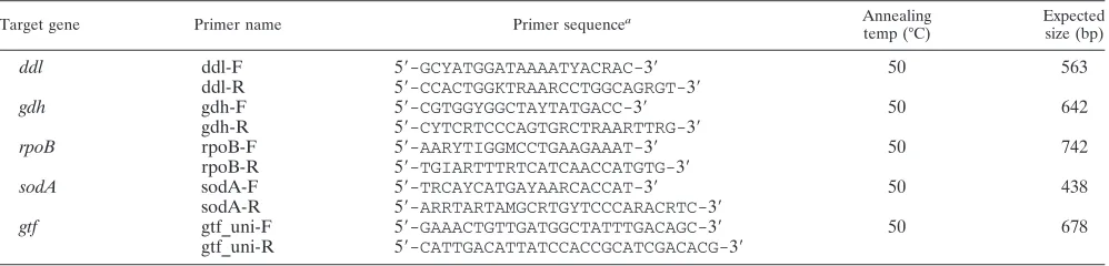
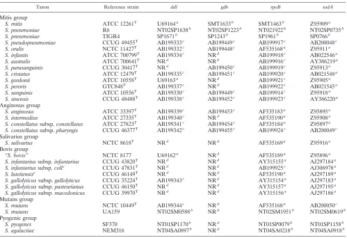
TABLE 1. PCR primers used to amplifyddl,gdh,rpoB,sodA, andgtfgene sequences
Target gene Primer name Primer sequencea Annealing
temp (°C)
Expected size (bp)
ddl ddl-F 5⬘-GCYATGGATAAAATYACRAC-3⬘ 50 563
ddl-R 5⬘-CCACTGGKTRAARCCTGGCAGRGT-3⬘
gdh gdh-F 5⬘-CGTGGYGGCTAYTATGACC-3⬘ 50 642
gdh-R 5⬘-CYTCRTCCCAGTGRCTRAARTTRG-3⬘
rpoB rpoB-F 5⬘-AARYTIGGMCCTGAAGAAAT-3⬘ 50 742
rpoB-R 5⬘-TGIARTTTRTCATCAACCATGTG-3⬘
sodA sodA-F 5⬘-TRCAYCATGAYAARCACCAT-3⬘ 50 438
sodA-R 5⬘-ARRTARTAMGCRTGYTCCCARACRTC-3⬘
gtf gtf_uni-F 5⬘-GAAACTGTTGATGGCTATTTGACAGC-3⬘ 50 678
gtf_uni-R 5⬘-CATTGACATTATCCACCGCATCGACACG-3⬘ aI, inosine; Y, C or T; R, A or G; K, G or T; M, A or C.
on May 15, 2020 by guest
http://jcm.asm.org/
Nomenclature.Tru¨per and De Clari (50) corrected the long-standing namesS.
sanguis,S. parasanguis, andS. cristatoS. sanguinis,S. parasanguinis, andS.
cristatus, respectively, for reasons of Latin grammar. Although we consider these
changes an unnecessary source of confusion and a direct violation of the prin-ciples of the International Code of Bacterial Nomenclature, we have adopted the corrected names in this paper according to the recent ruling of the Judicial Commission of the International Committee of the Taxonomy of Bacteria (53). Preparation of DNA for PCR.DNA was extracted from the bacteria by alka-line lysis (56). Individual strains were inoculated on 5% defibrinated horse blood agar and incubated at 37°C for 18 h in a 5% CO2-enriched atmosphere. The
bacterial colonies were scraped from the agar with a sterile disposable plastic loop (1 mm in diameter) and suspended in 100l of sterile ultrapure water. Then 20l of the bacterial suspension and 80l of 0.05 M sodium hydrate were gently mixed in a microcentrifuge tube. This mixture was incubated at 60°C for 45 min and then 9.2l of 1 M Tris-HCl (pH 7.0) was added to neutralize the pH. The prepared solution was used as the template DNA for the PCR analyses.
PCR primers.Oligonucleotide primers used to amplify fragments of the genes
ddl,gdh,rpoB,sodA, andgtfwere designed on the basis of conserved sequences identified by aligning relevant sequences ofStreptococcusspecies obtained at the GenBank nucleotide database (Table 1). The species-specificgtfprimers used were reported previously (28).
PCR analyses.All PCR mixtures contained 10l of Eppendorf HotmasterMix (Eppendorf AG, Hamburg, Germany), 10l of a 200-fold dilution of the prep-aration of template DNA described above, and a 0.4M concentration of each primer supplemented with sterile ultrapure water to a total volume of 25l. Amplification was performed with a Mastercycler gradient (Eppendorf) using the following parameters: an initial denaturing step at 94°C for 5 min, and 30
cycles of a denaturing step at 94°C for 30 s, a primer-annealing step at the appropriate temperature for 30 s, an extension step at 72°C for 30 s, and a final extension step at 72°C for 5 min. The primer-annealing temperature was opti-mized for each primer set (Table 1) (28). The PCR products were analyzed by 1.5% agarose gel electrophoresis after staining with ethidium bromide.
Sequencing ofddl,gdh,rpoB, andsodA.Amplicons of four housekeeping genes obtained by PCR were purified with Wizard minicolumns (Promega Co., Madison, WI). The gene fragments were sequenced on both strands using the same primers and the Thermo Sequence Dye Terminator cycle sequencing pre-mix kit (Amersham Bioscience AB, Uppsala, Sweden) according to the manu-facturer’s instructions. The following program was used: 30 cycles of denatur-ation at 96°C for 30 s, primer annealing at 45°C for 15 s, and extension at 60°C for 4 min. The products of the sequencing reaction were examined with an auto-matic DNA sequencer (ABI Prism 310 genetic analyzer; Applied Biosystems, Foster City, CA).
Phylogenetic analyses ofddl,gdh,rpoB, andsodA.The Clustal X software (49) downloaded from http://www.ebi.ac.uk was used to align the sequences for each gene separately and as concatenated sequences in the orderrpoB,sodA,ddl, and
gdh. Phylogenetic analysis by the neighbor-joining algorithm (45) was conducted using MEGA version 3 (38). Missing sequences as a result of lack of a PCR amplification product of genes that may be missing or present in a significantly different sequence variant were dealt with as gaps of the same length. The corresponding parameter of the neighbor-joining algorithm was set at “pairwise deletion.” The model used was “nucleotide: Kimura two-parameter.”
[image:3.585.44.543.78.420.2]Criteria for identification based on the sequences of the four housekeeping genes.Unequivocal clustering with a single type strain and other well-described reference strains of a particular species in the unrooted phylogenetic tree based TABLE 2. Accession numbers of housekeeping gene sequences of reference strains extracted from public databases
Taxon Reference strain ddl gdh rpoB sodA
Mitis group
S. mitis ATCC 12261T U69164a SMT1633b SMT1463b Z95909a
S. pneumoniae R6 NT02SP1638b NT02SP1223b NT021922b NT02SP0735b
S. pneumoniae TIGR4 SP1671b SP1243b SP1961b SP0766b
S. pseudopneumoniae CCUG 49455T AB199333c AB199449c AB199917c AB200048c
S. oralis NCTC 11427T AB199332c AB199448c AF535168a Z95911a
S. infantis ATCC 700799T AB199334c NRd AB199918c AB022546a
S. australis ATCC 700641T NRd NRd AB199916c AY386219a
S. parasanguinis CCUG 30417T NRd AB199450c AB199919c Z95913a
S. cristatus ATCC 12479T AB199335c AB199451c AB199920c AB021548a
S. gordonii ATCC 10558T U69163a NRd AB199921c Z95905a
S. peroris GTC848T AB199337c NRd AB199922c AB021545a
S. sanguinis ATCC 10556T AB199330c AB199449c AB199914c Z95918a
S. sinensis CCUG 48488T AB199338c AB199452c AB199923c AY386220a
Anginosus group
S. anginosus ATCC 33397T AB199339c AB199453c AF535183a Z95895a
S. intermedius ATCC 27335T AB199340c NRd AF535190a Z95908a
S. constellatussubsp.constellatus ATCC 27823T AB199341c AB199454c AF535184a Z95897a
S. constellatussubsp.pharyngis CCUG 46377T AB199342c AB199455c AB199924c AB200049c
Salivarius group
S. salivarius NCTC 8618T NRd NRd AF535169a Z95916a
Bovis group
“S. bovis” NCTC 8177 U69162a NRd AF535189a Z95896a
S. infantariussubsp.infantarius CCUG 43820T NRd NRd AY315155a AJ297184a
S. infantariussubsp.colie CCUG 47831T NRd NRd AB199925c AJ306978a
S. lutetiensise CCUG 46149T NRd NRd AF535190a AJ297189a
S. gallolyticussubsp.gallolyticus CCUG 35224T AB199343c NRd AY315154a AJ297183a
S. gallolyticussubsp.pasteurianus CCUG 46150T NRd NRd AY315157a AJ297195a
S. gallolyticussubsp.macedonicus CCUG 39970T NRd NRd AY315156a AJ297186a
Mutans group
S. mutans NCTC 10449T AB199344c NRd AF535168a AB200050c
S. mutans UA159 NT02SM0588b NRd NT02SM1951b NT02SM0619b
Pyogenic group
S. pyogenes SF370 NT01SP1170b NRd NT01SP0079b NT01SP1158b
S. agalactiae NEM316 NT04SA0897b NRd NT04SA0218b NT04SA0918b
a
GenBank accession number. b
TIGR locus name in the genome database of TIGR. c
Accession number of a sequence determined in this study. d
NR, no reference sequence available. e
S. infantariussubsp.coliandS. lutetiensisare synonyms for the same species according to Poyart et al. (42).
on May 15, 2020 by guest
http://jcm.asm.org/
on May 15, 2020 by guest
http://jcm.asm.org/
on concatenated sequences of the four housekeeping genes was taken as evi-dence of conclusive identification of that species. Deviations from this identifi-cation based on phylogenetic analysis of single gene loci were recorded. The single strain ofGemella haemolysans, from which only one of the four house-keeping genes was successfully amplified, was assigned to that species based on a 100% similarity to the corresponding gene sequence (rpoB) of the type strain. PCR analysis ofgtf.As the first screening, PCR analysis with universalgtf
primers, which were able to amplify all streptococcalgtfgenes without regard to species, was carried out. Next, PCR analyses with six sets of species-specificgtf
primers were performed on each isolate that was positive in the first-screen PCR. By the results of this PCR analysis, the isolate was identified according to the species that possessed the targetedgtfgene.
Phenotypic examination and identification.The Rapid ID 32 STREP kit (Bio Me´rieux, La Balme les Grottes, France) was inoculated from a suspension of bacteria grown anaerobically for 2 days on blood agar according to the manu-facturer’s instructions using an ATB1574 automatic dispenser (Vitek Systems, Bio Me´rieux). After incubation for 4 hours at 37°C, the reactions were read and processed by an ATB expression system (Vitek Systems). Phenotypic analysis using the STREPTOGRAM kit (Wako Pure Chemicals, Osaka, Japan) was performed according to the manufacturer’s instructions. Identification was achieved after 20 h by visual comparison of color reactions with the color plate included by the manufacturer.
Identifications obtained by the two kits were classified into three levels: (i) good identification, i.e., the first suggestion supported by a scoreⱖ80% of relative probability; (ii) acceptable identification, the first suggestion supported by a scoreⱕ80% of relative probability; (iii) no identification (“unacceptable profile”) or no suggestion supported by a probability of⬎50%. Each result was compared with the corresponding identification based on phylogenetic analysis of concatenated sequences of the four housekeeping genes.
For comparative purposes all strains were also tested for the ability to hydro-lyze arginine using a traditional tube test as described previously (35).
RESULTS
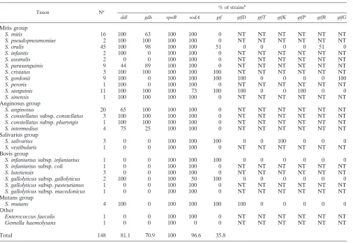
PCR amplification and sequence determination ofddl,gdh, rpoB, andsodAgenes.PCR of theddl,gdh,rpoB,sodA, andgtf genes yielded a single amplified band at approximately 560, 640, 740, 440, and 680 bp, respectively. The rates of successful
amplifications with theddl,gdh,rpoB, andsodAprimers were
80.2, 70.0, 100, and 96.6%, respectively (see below for each species). Sequences covering 292, 431, 517, and 390 bp, respec-tively, of the four amplified genes were used in the analyses. The GenBank accession numbers of sequences determined for the reference strains in this study are shown in Table 2 together with numbers for reference sequences extracted from GenBank. The nucleotide sequences determined for clinical isolates have been deposited in GenBank under accession numbers
AB199330 to AB199446 (ddl), AB199447 to AB199548 (gdh),
AB199914 to AB200046 and AB218984 (rpoB), and AB200047
to AB200167 and AB218985 (sodA). All sequences are also
available for downloading as packages of either single loci or concatenated sequences at http://www.immi.au.dk/service/download /kilian.
Identification of strains based on phylogenetic analysis.Of the 147 strains examined, 145 were identified as belonging to
the genusStreptococcus.The two remaining strains were
iden-tified asEnterococcus faecalisandGemella haemolysansbased
on 100% similarity of the sequences of rpoB and sodA to
sequences of those two species detected in a nucleotide BLAST search (http://www.ncbi.nlm.nih.gov/BLAST/Blast.cgi).
The concatenated sequence of the four housekeeping genes
rpoB, sodA, ddl, andgdhconsisted of 1,630 bp. Phylogenetic
analysis of the concatenated sequences resulted in the tree shown in Fig. 1. The tree revealed clear separation of all currently recognized species supported by significant bootstrap values. Some clusters showed an unexpected degree of se-quence variation. This was most striking in the clusters
con-taining the type and reference strains of S. anginosusand S.
parasanguinis.This situation may reflect the existence of yet unrecognized species within some of the clusters. However, with a few potential exceptions, all strains could be assigned with confidence to currently recognized species. This was con-sidered the conclusive identification of the strains.
The phylogenetic trees based on separate analyses of ddl,
gdh,rpoB, andsodAlikewise showed distinct clusters that could
be correlated with individual species (not shown). However, identifications based on these analyses revealed some devia-tions from the conclusive identification based on analysis of the concatenated sequences. Thus, identification of a total of eight strains (5.5%), including three type strains of the anginosus group of species, deviated from the conclusive identification
(Table 3). The deviating results were based mainly onddland
rpoB gene sequences, whereas no deviation was observed in
results obtained by analysis of thesodA gene. No strain
pos-sessed more than one deviating sequence among the four housekeeping genes examined. Apart from these problems, which can be explained by homologous recombination between
species, the recently describedS. pseudopneumoniaeposed a
problem that was visible in trees based on single loci. In each
of the trees based onrpoB,gdh, andddl, the two strains could
not be distinguished with confidence fromS. mitis, and in the
tree based on sodA sequences, they were indistinguishable
fromS. pneumoniae.
To analyze the nature of the deviating results for some of the anginosus group streptococci further, partial sequencing of 16S rRNA genes was performed for each strain belonging to the
species S. intermedius and S. constellatus using the method
described elsewhere (36). These results confirmed the
identi-fications based on concatenated housekeeping genes byⱖ99%
similarity to the 16S rRNA gene sequences of the relevant type and reference strains in public databases (data not shown).
Rates of positive PCRs. The percentage of PCR-positive
isolates assigned to each of the Streptococcus species as
de-scribed is shown in Table 4. Theddlprimers failed to amplify
a sequence in all strains of the speciesS. australis,S. salivarius,
S. vestibularis,S. infantarius,S. lutetiensis,S. gallolyticus, and in
some strains ofS. parasanguinis,S. anginosus, andS.
interme-dius.ThegdhPCR failed to amplify sequences inS. infantis,S. australis, S. gordonii, S. peroris, and in all members of the
salivarius, bovis, and mutans groups. In contrast, therpoBand
FIG. 1. Phylogenetic tree based on concatenated sequences of four housekeeping genes,rpoB,sodA,ddl, andgdh. The tree was based on the neighbor-joining method. The value on each branch is the estimated confidence limit (expressed as a percentage) for the position of the branch as determined by bootstrap analysis. Only values exceeding 50% are shown. The scale bar (neighbor-joining [NJ] distance) represents a 5% difference in nucleotide sequence.
on May 15, 2020 by guest
http://jcm.asm.org/
sodAprimers yielded amplicons from virtually all strains (100 and 96.6%, respectively) (Table 4).
Identification of strains based on thegtfgene.PCR analysis
with the universalgtfprimer set resulted in an amplicon from
53 strains (35.8%) assigned to the speciesS. sanguinis,S. oralis,
S. gordonii, S. mutans, andS. salivariusin addition toS. infan-tariussubsp. infantarius and S. gallolyticussubsp.gallolyticus. With the exception of strains of the last two taxa, all of these strains also yielded an amplicon with one of the species-specific
gtfprimers. No nonspecific product was observed in any of the
species. The identification based on these results for gtf
-positive strains was in complete agreement with the identi-fication of the strains based on concatenated sequences and,
furthermore, revealed thatgtfsequences were present in all
strains ofS. sanguinis,S. gordonii, S. mutans, andS.
saliva-rius.In contrast, only 51% of strains identified asS. oralis
possessed agtfgene.
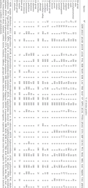
Identification based on phenotypic test kits.A total of 145
strains belonging to the genus Streptococcus as determined by
DNA sequence analysis were examined with the commercial identification kit STREPTOGRAM and the Rapid ID 32 STREP system. The two kits contain 21 and 32 biochemical tests, respec-tively. Eleven tests are shared by the two systems and allow direct comparison of the outcomes (Tables 5 and 6). Although the overall pattern is similar, the proportion of positive reactions in individual tests shared by the two systems shows many differences. It is striking that the test for arginine hydrolysis, which is a
taxo-TABLE 3. Phylogenetically deviating gene sequences in eight
Streptococcusstrains identified according to analysis of
concatenated sequences of four housekeeping genes
Strain Correct identity Deviating gene
Misidentified as Mitis group
SK153 S. oralis ddl S. sanguinis
SK36 S. sanguinis rpoB S. sinensis
SK574 S. sanguinis ddl S. gordonii
1310 S. sanguinis ddl S. gordonii
Anginosus group
SK54T S. intermedius rpoB S. anginosus
1893 S. intermedius gdh S. anginosus
SK53T S. constellatus ddl S. anginosus
CCUG 46377T S. constellatussubsp. pharyngis
[image:6.585.42.283.99.238.2]ddl S. anginosus
TABLE 4. Proportion of successful PCR amplifications of genes from 22 species of nonhemolytic streptococci using the primers listed in Table 1
Taxon Na % of strains
b
ddl gdh rpoB sodA gtf gtfD gtfT gtfK gtfP gtfR gtfG
Mitis group
S. mitis 16 100 63 100 100 0 NT NT NT NT NT NT
S. pseudopneumoniae 2 100 100 100 100 0 NT NT NT NT NT NT
S. oralis 45 100 98 100 100 51 0 0 0 0 51 0
S. infantis 2 100 0 100 100 0 NT NT NT NT NT NT
S. australis 2 0 0 100 100 0 NT NT NT NT NT NT
S. parasanguinis 9 44 89 100 100 0 NT NT NT NT NT NT
S. cristatus 3 100 100 100 100 100 NT NT NT NT NT NT
S. gordonii 9 100 0 100 100 100 100 0 0 0 0 100
S. peroris 1 100 0 100 100 0 NT NT NT NT NT NT
S. sanguinis 11 100 100 100 73 100 100 0 0 100 0 0
S. sinensis 1 100 100 100 100 0 NT NT NT NT NT NT
Anginosus group
S. anginosus 20 65 100 100 100 0 NT NT NT NT NT NT
S. constellatussubsp.constellatus 3 100 100 100 100 0 NT NT NT NT NT NT
S. constellatussubsp.pharyngis 1 100 100 100 100 0 NT NT NT NT NT NT
S. intermedius 4 75 25 100 100 0 NT NT NT NT NT NT
Salivarius group
S. salivarius 3 0 0 100 100 100 0 0 100 0 0 0
S. vestibularis 1 0 0 100 100 0 NT NT NT NT NT NT
Bovis group
S. infantariussubsp.infantarius 1 0 0 100 100 100 0 0 0 0 0 0
S. infantariussubsp.coli 1 0 0 100 100 0 NT NT NT NT NT NT
S. lutetiensis 3 0 0 100 100 0 NT NT NT NT NT NT
S. gallolyticussubsp.gallolyticus 2 100 0 100 50 100 0 0 0 0 0 0
S. gallolyticussubsp.pasteurianus 1 0 0 100 100 0 NT NT NT NT NT NT
S. gallolyticussubsp.macedonicus 1 0 0 100 100 0 NT NT NT NT NT NT
Mutans group
S. mutans 4 100 0 100 100 100 100 0 0 0 0 0
Other
Enterococcus faecalis 1 0 0 100 100 0 NT NT NT NT NT NT
Gemella haemolysans 1 0 0 100 0 0 NT NT NT NT NT NT
Total 148 81.1 70.9 100 96.6 35.8
aN, number of strains. bNT, not tested.
on May 15, 2020 by guest
http://jcm.asm.org/
[image:6.585.44.550.362.710.2]TABLE 5. Physiological characteristics of strains of 22 species of nonhemolytic streptococci determined by the Rapid ID 32 Strep system a Species N b % of strains positive ADH  GLU  GAR  GUR ␣ GAL PAL RIB MAN SOR LAC TRE RAF VP APPA  GAL PYRA  NAG GTA HIP GLYG PUL MAL MEL SAC M  DG TAG  MAN Mitis group S. mitis 16 0 13 69 0 25 50 13 0 0 50 0 19 6 35 4 0 6 94 6 0 56 62 0 63 6 6 0 S. pseudopneumoniae 2 0 50 100 0 0 100 50 50 0 50 50 0 0 100 0 0 0 50 0 0 0 50 0 50 0 50 0 S. oralis 45 2 2 93 0 40 78 0 0 0 29 13 9 6 98 13 0 9 87 0 0 24 38 0 38 0 7 0 S. infantis 2 0 0 100 0 0 0 0 0 0 100 0 0 0 100 100 0 0 100 0 0 100 100 0 100 0 0 0 S. sanguinis 11 36 27 0 0 45 36 0 0 0 82 82 9 9 91 0 0 18 82 0 9 64 91 0 91 27 0 0 S. gordonii 9 44 89 100 0 11 100 11 0 0 78 89 0 0 100 11 0 0 89 11 0 0 100 0 78 67 11 67 S. parasanguinis 9 56 11 78 0 100 100 0 0 11 100 11 68 11 100 11 0 11 33 11 0 0 100 44 100 11 33 0 S. australis 2 50 0 50 0 0 0 0 0 0 100 0 0 0 100 0 0 0 50 0 0 50 100 0 100 0 50 0 S. cristatus 3 33 0 33 0 0 0 0 0 0 33 67 0 0 100 0 0 0 100 0 0 0 67 0 67 0 33 0 S. peroris 1 0 0 100 0 0 100 0 0 0 100 0 0 0 100 0 0 0 100 0 0 0 100 0 100 0 100 0 S. sinensis 1 0 0 100 0 0 0 0 0 0 0 0 0 0 100 0 0 0 0 0 0 0 0 0 0 0 0 0 Anginosus group S. anginosus 20 75 95 0 0 20 100 0 15 0 60 80 0 100 100 0 0 0 0 0 0 60 95 0 100 80 0 15 S. constellatus 3 0 0 0 0 0 100 0 0 0 33 67 0 67 100 0 0 0 0 0 0 0 67 0 67 33 0 0 S. constellatus subsp. pharyngis 1 0 100 100 0 0 100 0 0 0 100 0 0 100 100 0 0 100 0 0 0 100 100 0 100 0 0 0 S. intermedius 4 75 75 100 0 0 100 0 0 0 100 100 0 100 100 25 0 75 75 0 0 100 100 0 100 25 0 25 Bovis group S. infantarius 1 0 0 0 0 100 100 0 0 0 100 0 0 100 100 0 0 0 0 0 0 0 100 0 100 0 0 0 S. lutetiensis 4 0 0 0 0 100 0 0 0 0 25 0 50 75 100 0 0 0 0 0 0 0 50 0 75 0 0 0 S. gallolyticus 2 0 100 0 0 50 0 0 100 0 100 100 100 100 100 50 0 0 0 0 100 100 100 0 100 100 0 0 S. pasteurianus 1 0 100 0 0 100 0 0 0 0 100 100 0 100 100 100 0 0 0 0 0 0 100 0 100 100 0 100 S. macedonicus 1 0 0 100 0 0 0 0 0 0 100 0 0 100 100 0 0 0 0 0 0 0 100 0 100 0 0 0 Mutans group S. mutans 4 0 75 50 0 75 0 25 100 75 100 100 100 100 100 0 0 0 0 25 0 0 100 75 100 50 50 0 Salivarius group S. salivarius 3 0 100 67 0 0 33 0 0 0 33 100 33 100 100 67 0 0 67 0 0 100 100 0 33 33 0 0 a ADH, arginine dihydrolase;  GLU,  -glucosidase;  GAR,  -galactosidase detected with p -nitrophenyl- -D -galactopyranoside as the substrate;  GUR,  -glucuronidase; ␣ GAL, ␣ -galactosidase; PAL, alkaline phosphatase; VP, Voges-Proskauer test; APPA, activity of alanine-phenylalanine-proline-arylamidase;  GAL,  -galactosidase detected with 2-naphthyl- -galactopyranoside as the substrate; PYRA, pyrrolidonyl arylamidase;  NAG, N -acetyl- -glucosaminidase; GTA, glycyl-tryptophan arylamidase; HIP, hydrolysis of hippurate;  MAN,  -mannosidase; RIB, MAN, SOR, LAC, TRE, RAF, GLYG, PUL, MAL, MEL, SAC, and TAG, fermentation of ribose, mannitol, sorbitol, lactose, trehalose, raf finose, glycogen, pullulan, maltose, melibiose, sucrose, and tagatose, respectively. All strains tested were negative for fermentation of melezitose, L -arabinose, D -arabitol, and cyclodextrin. b N, number of strains.
on May 15, 2020 by guest
http://jcm.asm.org/
[image:7.585.139.440.75.720.2]TABLE 6. Physiological characteristics of strains of 22 species of nonhaemolytic streptococci determined by the STREPTOGRAM kit a Species N b % of strains positive AMY LAC NAG TRE MAN FUC ARB RAF ␣ GAL SOR ARA  GAL INU MEL PAL ARG ESC GLN PYRA HIP VP Mitis group S. mitis 16 88 94 75 0 0 56 13 19 31 6 94 19 0 6 43 0 13 0 0 0 0 S. pseudopneumoniae 2 0 100 100 0 0 100 0 0 0 0 50 0 0 0 100 0 0 0 50 0 0 S. oralis 45 0 93 98 24 4 96 91 58 49 9 60 42 0 31 89 2 4 0 0 2 2 S. infantis 2 0 100 100 0 0 100 0 0 0 0 0 50 0 0 0 0 0 0 0 0 0 S. sanguinis 11 0 100 90.9 100 0 82 73 18 55 63 9 0 82 9 0 9 63 0 0 0 0 S. gordonii 9 89 78 100 89 0 78 78 0 0 0 44 22 67 0 89 22 89 0 0 0 0 S. parasanguinis 9 22 89 100 11 0 78 22 100 100 22 11 22 11 56 100 78 33 0 0 0 0 S. australis 2 50 50 100 50 0 50 50 0 0 0 50 50 50 0 50 50 50 0 0 0 0 S. cristatus 3 0 67 33 100 0 0 100 33 0 0 100 0 0 0 0 0 33 0 0 0 0 S. peroris 1 0 100 0 0 0 0 0 0 0 0 0 0 0 0 100 0 0 0 0 0 0 S. sinensis 1 100 100 100 100 0 100 100 0 0 0 100 100 0 0 0 100 100 0 0 0 0 Anginosus group S. anginosus 20 95 95 5 90 15 0 100 20 5 0 5 0 5 15 100 100 100 0 0 5 95 S. constellatus 3 0 67 0 67 0 0 100 0 0 0 67 0 0 0 100 67 67 0 0 0 67 S. constellatus subsp. pharynges 1 0 0 0 0 0 0 0 0 0 0 0 0 0 0 100 100 0 0 0 0 100 S. intermedius 4 75 100 100 100 0 100 100 0 0 0 0 75 0 0 100 100 100 0 0 0 100 Bovis group S. infantarius 1 0 100 0 0 0 0 0 100 100 0 0 0 0 0 0 0 0 0 0 0 100 S. lutetiensis 4 100 100 0 0 0 0 100 100 100 0 0 25 25 0 0 0 100 0 0 0 0 S. gallolyticus 2 100 100 100 100 100 100 100 100 100 0 0 100 0 100 0 0 100 0 0 0 100 S. macedonicus 1 0 100 0 0 0 100 0 0 0 0 0 0 0 0 0 0 0 0 0 0 0 S. pasteurianus 1 100 100 0 100 0 0 100 100 100 0 0 100 0 100 0 0 100 0 0 0 100 Mutans group S. mutans 4 0 100 0 100 100 0 100 75 75 100 25 0 100 75 0 25 75 0 0 0 50 Salivarius group S. salivarius 3 67 67 0 100 0 67 100 100 0 33 67 67 33 0 33 0 100 0 0 0 67 aNAG, N -acetylglucosaminidase detected with p -nitrophenyl-N -acetylglucosaminide as the substrate; FUC, fucopyranosidase detected with p -nitrophenyl- -D -fucopyranoside as the substrate; ␣ GAL, ␣ -galactosidase detected with p -nitrophenyl- -D -galactopyranoside as the substrate;  GAL,  -galactosidase detected with 2-naphthyl- -galactopyranoside as the substrate; PAL, alkaline phosphatase; ARG, arginine dihydrolase; ESC, hydrolysis of esculin; GLN,  -glucuronidase; PYRA, pyrrolidonyl arylamidase; HIP, hydrolysis of hippurate; VP, Voges-Proskauer test; AMY, LAC, TRE, MAN, ARB, RAF, SOR, ARA, IN U, and MEL, fermentation of amygdalin, lactose, trehalose, mannitol, arbutin, raf finose, sorbitol, arabinose, inulin, and melibiose, respectively. bN, number of strains.
on May 15, 2020 by guest
http://jcm.asm.org/
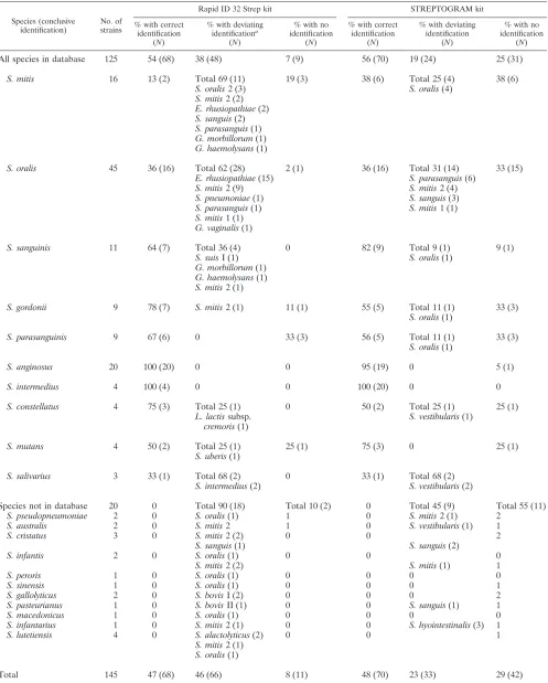
TABLE 7. Comparison of conclusive identification of 145Streptococcusstrains based on phylogenetic analysis of concatenated sequences of four housekeeping genes and identifications obtained with two commercial identification kits
Species (conclusive identification)
No. of strains
Rapid ID 32 Strep kit STREPTOGRAM kit
% with correct identification
(N)
% with deviating identificationa
(N)
% with no identification
(N)
% with correct identification
(N)
% with deviating identification
(N)
% with no identification
(N)
All species in database 125 54 (68) 38 (48) 7 (9) 56 (70) 19 (24) 25 (31)
S. mitis 16 13 (2) Total 69 (11) 19 (3) 38 (6) Total 25 (4) 38 (6)
S. oralis2 (3) S. oralis(4)
S. mitis2 (2)
E. rhusiopathiae(2)
S. sanguis(2)
S. parasanguis(1)
G. morbillorum(1)
G. haemolysans(1)
S. oralis 45 36 (16) Total 62 (28) 2 (1) 36 (16) Total 31 (14) 33 (15)
E. rhusiopathiae(15) S. parasanguis(6)
S. mitis2 (9) S. mitis2 (4)
S. pneumoniae(1) S. sanguis(3)
S. parasanguis(1) S. mitis1 (1)
S. mitis1 (1)
G. vaginalis(1)
S. sanguinis 11 64 (7) Total 36 (4) 0 82 (9) Total 9 (1) 9 (1)
S. suisI (1) S. oralis(1)
G. morbillorum(1)
G. haemolysans(1)
S. mitis2 (1)
S. gordonii 9 78 (7) S. mitis2 (1) 11 (1) 55 (5) Total 11 (1) 33 (3)
S. oralis(1)
S. parasanguinis 9 67 (6) 0 33 (3) 56 (5) Total 11 (1) 33 (3)
S. oralis(1)
S. anginosus 20 100 (20) 0 0 95 (19) 0 5 (1)
S. intermedius 4 100 (4) 0 0 100 (20) 0 0
S. constellatus 4 75 (3) Total 25 (1) 0 50 (2) Total 25 (1) 25 (1)
L. lactissubsp.
cremoris(1)
S. vestibularis(1)
S. mutans 4 50 (2) Total 25 (1) 25 (1) 75 (3) 0 25 (1)
S. uberis(1)
S. salivarius 3 33 (1) Total 68 (2) 0 33 (1) Total 68 (2)
S. intermedius(2) S. vestibularis(2)
Species not in database 20 0 Total 90 (18) Total 10 (2) 0 Total 45 (9) Total 55 (11)
S. pseudopneumoniae 2 0 S. oralis(1) 1 0 S. mitis2 (1) 2
S. australis 2 0 S. mitis2 1 0 S. vestibularis(1) 1
S. cristatus 3 0 S. mitis2 (2) 0 0 2
S. sanguis(1) S. sanguis(2)
S. infantis 2 0 S. oralis(1) 0 0 0
S. mitis2 (2) S. mitis(1) 1
S. peroris 1 0 S. oralis(1) 0 0 0 0
S. sinensis 1 0 S. oralis(1) 0 0 0 1
S. gallolyticus 2 0 S. bovisI (2) 0 0 0 2
S. pasteurianus 1 0 S. bovisII (1) 0 0 S. sanguis(1) 1
S. macedonicus 1 0 S. oralis(1) 0 0 0 0
S. infantarius 1 0 S. mitis2 (1) 0 0 S. hyointestinalis(3) 1
S. lutetiensis 4 0 S. alactolyticus(2) 0 0 1
S. mitis2 (1)
S. oralis(1)
Total 145 47 (68) 46 (66) 8 (11) 48 (70) 23 (33) 29 (42)
aOther species includedErysipelothrix rhusiopathiae,Gemella morbillorum,Gemella haemolysans,Gardnerella vaginalis, andLactococcus lactissubsp.cremoris.
on May 15, 2020 by guest
http://jcm.asm.org/
nomically very informative property when determined by a stan-dard tube test (35), shows significant differences. Less than 50% of the strains belonging to species that are invariably positive in
standard tests (S. sanguinisandS. gordonii) show positive
reac-tions with the two kits.
With reference to the conclusive identification based on phylogenetic analysis of the concatenated sequences, the rate of correct identification obtained in the two kits was calculated (Table 7). Of all 145 strains examined, 68 (47%) and 70 (48%) were correctly identified by Rapid ID 32 STREP and the STREPTOGRAM kit, respectively. The rate of incorrect iden-tification in the two systems was 46% and 23%, respectively. STREPTOGRAM gave a “no code” response rather than an incorrect identification significantly more often than Rapid ID
32 STREP. Among 16 strains ofS. mitis, only 2 (13%) and 6
(38%) strains were correctly identified by Rapid ID 32 STREP and STREPTOGRAM, respectively. Both kits correctly
iden-tified 16 (36%) of 45 strains ofS. oralis.The rate of correct
identification of strains of other species varied between 33 and 100%.
The best results were obtained with members of the
an-ginosus group of streptococci, i.e.,S. anginosus, S.
interme-dius, andS. constellatus. Eleven species described since 1991
(Table 7) are not included in the databases associated with the two identification kits. Eighteen of the 20 strains representing these species were assigned to other species by Rapid ID 32
STREP. Among these were strains of S. gallolyticus and S.
pasteurianus, which were “correctly” identified as “S. bovis,” a species name that, however, is now considered illegitimate (42). STREPTOGRAM assigned incorrect names to nine (35%) of the 20 strains, whereas 11 (55%) remained uniden-tified (Table 7).
Species distribution and properties of blood isolates.A total of 107 strains were isolated from human blood. Further infor-mation on the underlying disease was not available for 80 of these isolates. The remaining 27 strains were from patients
with subacute bacterial endocarditis (n ⫽14) and meningitis
(n ⫽ 1) and from patients suffering from neutropenia (n ⫽
12). Figure 2 shows the species distribution of the 107 isolates according to the conclusive identification based on
phyloge-netic analysis of concatenated sequences of the four
house-keeping genes. As shown in the figure,S. oralisaccounted for
40% of all blood isolates of nonhemolytic streptococci, exclud-ingS. pneumoniae.The second most frequently isolated species was S. anginosus (18%). Isolates from endocarditis patients
belonged toS. oralis(n⫽6),S. mitis(n⫽2),S. gordonii(n⫽
2), andS. sanguinis,S. parasanguinis,S. salivarius, andS.
aus-tralis(each represented by one isolate). The predominant
spe-cies isolated from neutropenic patients wereS. oralis(n⫽6)
andS. mitis(n⫽5). The only other species represented in this
group of patients wasS. sanguinis(n⫽1).
Among the 107 blood isolates, 42 (39%) produced
extracel-lular polysaccharide as suggested by the presence of agtfgene.
Among 43 blood isolates of S. oralis, in which gtfis variably
present, 22 (51%) had thegtfgene. Eight (57%) of 14 isolates
from patients with subacute bacterial endocarditis produced extracellular polysaccharide.
DISCUSSION
Several factors contribute to the difficulties that are encoun-tered in the identification of nonhemolytic streptococci by both molecular and biochemical methods. The taxonomy of this group of bacteria has been revised frequently, and many new species have been described, often without sufficient informa-tion about differential characteristics and the extent of pheno-typic and genetic variation within proposed species (16, 44). Satisfactory classification of bacteria reflects their phylogenetic diversification within distinct boundaries. It has sometimes been doubted that distinct subpopulations that can be equated with species exist in the population of nonhemolytic strepto-cocci. Nonhemolytic streptococci are naturally competent for genetic transformation (24), and there is direct evidence of interspecies homologous recombination in genes encoding
transpeptidases (“penicillin-binding proteins”) in S.
pneu-moniae,S. mitis, andS. oralis(14, 21). However, the frequency of detectable recombination affecting penicillin-binding pro-teins in the natural populations of these species is undoubt-edly influenced by an extraordinary selection pressure ex-erted by beta-lactam antibiotics. To what extent interspecies homologous recombination affects housekeeping genes in which functionally acceptable mutations are selectively neu-tral is not known.
The data summarized in Table 3 show that in 5.5% of the 145 Streptococcus strains examined, the phylogeny of one of the four housekeeping genes deviated from that of the other genes and revealed clear evidence of recombination with
an-otherStreptococcusspecies. The practical consequence of this
is that these eight strains would have been misidentified if identification were based on the sequence of the affected gene alone. In five of eight strains the gene affected by
recombina-tion wasddl, in two strains it wasrpoB, and in one strain it was
gdh. The superoxide dismutase gene,sodA, was not affected in
any of the strains examined in spite of the fact that sequences of this gene were successfully determined for all but five strains (Table 4). Thus, for unknown reasons, the frequency of
devi-ating sequences in sodA was significantly lower than in ddl
(P ⬍ 0.05).
The data in Table 3 furthermore demonstrate that recom-bination always occurred between members of the same
phy-FIG. 2. Species distribution of 111 blood isolates of nonhemolytic streptococci.
on May 15, 2020 by guest
http://jcm.asm.org/
[image:10.585.43.283.67.236.2]logenetic group, i.e., either within the mitis group or within the anginosus group of species, probably determined by the re-quirement for sufficient sequence similarity to allow efficient
integration (39). It is remarkable that 14% (n⫽4) of the 28
strains belonging to the anginosus group of streptococci showed evidence of recombination affecting one of the four
housekeeping genes. By comparison, only 4% (n ⫽4) of the
102 mitis group streptococci were affected, but with significant differences between individual species of the group. Thus, only
1 (2%) of 45S. oralisstrains showed evidence of recombination
with another species, while a remarkable 27% (n⫽3) of 11S.
sanguinis strains possessed deviating gene sequences
appar-ently originating in S. gordonii or S. sinensis. These figures
clearly demonstrate differences in the frequency of transfer of housekeeping gene sequences between species of nonhemo-lytic streptococci. Of particular practical significance is the finding that three of the four anginosus group strains with deviating sequences were type strains, which usually serve as references for gene sequence-based identification.
Recombination may also be partly responsible for the diffi-culties encountered in finding phenotypic traits that allow un-equivocal differentiation of species of nonhemolytic strepto-cocci. As shown in Tables 5 and 6 and by previous studies (5, 35, 36), very few characters are of differential value. Compar-ison of the results presented in Tables 5 and 6 furthermore demonstrates how different versions of a test for the same target character may give different results. This is even more signifi-cant when results obtained in traditional tube tests are com-pared with results obtained in commercially available kits such as Rapid ID 32 STREP (36). For example, the ability to
hy-drolyze arginine is a characteristic of all members ofS.
san-guinis,S. gordonii,S. parasanguinis, andS. cristatusand is there-fore valuable for differentiating this group of closely related
species fromS. mitis,S. oralis, S. pneumoniae, andS. infantis
(36). Strains previously referred to as “S. mitisbiovar 2,” which
are arginine hydrolase positive (35), belong to the speciesS.
parasanguinisand to a yet unnamed taxon according to recent taxonomic studies (36). As shown in Tables 5 and 6, only a small proportion of strains (31% and 44%, respectively)
be-longing to S. sanguinis, S. gordonii, S. parasanguinis, and S.
cristatusgave a positive result in the test for arginine hydrolysis/ dihydrolase in the two commercial kits. Although these results do not necessarily invalidate the ability of the commercial kits to identify clinical isolates of these species, it is clear that the results obtained with these kits do not adequately reflect the presence or absence of particular genes and associated prop-erties. Thus, examination of all strains by a traditional tube test (35) showed arginine hydrolase activity in all strains assigned
to the speciesS. australis,S. parasanguinis,S. gordonii,S.
san-guinis, andS. intermediusin 18 of 20S. anginosusand in two of fourS. constellatusstrains.
Another problem of direct practical relevance is that the databases associated with the two commercial kits evaluated in this study are not adjusted according to the frequently chang-ing taxonomy of this group of bacteria. As a consequence, members of species described after 1991 are not correctly identified by either of the two commercial kits (Table 7).
Combined, the problems mentioned above contributed to a rate of correct identification of the present strain collection that was below 50% for both kits. The rate of correct
identi-fication varied significantly between species. Not unexpectedly,
the most significant problems were observed forS. mitisandS.
oralis, of which only 13 and 36%, respectively, were correctly identified by Rapid ID 32 STREP and 38 and 36%, respec-tively, by STREPTOGRAM. Among all 145 strains examined 46.9% and 22.1% were incorrectly identified by the API Rapid ID 32 STREP and the STREPTOGRAM kits, respectively. The remaining 6.2% and 29.6% of the strains were unidenti-fied by these systems, respectively (Table 7).
The results obtained in this study and in our recent taxo-nomic study (36) indicate that phenotypic characterization is of limited value for identification of many species of nonhemo-lytic streptococci.
Correct identification of nonhemolytic streptococci is to some extent achievable by partial sequencing of the 16S rRNA gene and searching for homologous sequences in public data-bases (7, 30). However, there are two problems with this
ap-proach. First, the method does not allow differentiation ofS.
mitis, S. oralis, S. pseudopneumoniae, and S. pneumoniae be-cause of significant sequence conservation of the 16S rRNA genes in this group of bacteria and because of misleading recombinatory sequences in some versions of that gene in some strains (36). Second, many sequences in the public data-bases are mislabeled, either because of incorrect identification of the source strain or because of nonrecorded revised classi-fication of the strain subsequent to deposition of the sequence. It is therefore important to restrict searches to the type strains of the species. This is an option at the database of the Ribo-somal Database Project II (http://rdp.cme.msu.edu/index.jsp). Our study demonstrates that unequivocal identification of clinical isolates of nonhemolytic streptococci can be achieved by phylogenetic analysis of concatenated sequences of four
housekeeping genes,ddl,gdh,rpoB, andsodA, amplified by the
primer sets listed in Table 1. As the resulting identification is based on the phylogeny of the isolates as reflected in several loci distributed along the entire genome, we assume that this is as close as one can get to a correct identification, although yet unrecognized taxa may still occur, as suggested by the signifi-cant sequence divergence in some of the clusters (Fig. 1). This method can be an important tool in taxonomic studies of strep-tococci and may eventually replace the technically more de-manding and less reproducible DNA-DNA hybridization. However, an important question is if it is feasible and biolog-ically meaningful to define species limits according to particu-lar thresholds of sequence simiparticu-larities. Comprehensive studies of additional groups of bacteria are required to obtain an appropriate basis for such deliberations.
Multilocus sequence analysis is at present unrealistic as a general identification tool for clinical microbiology laborato-ries. As an alternative, our findings support the suggestion by Poyart and Kawamura and their coworkers (32, 41, 42) that
sodA would be the best choice as a single informative gene.
This is based on the observation that the gene could be am-plified in the whole range of species, including members of related genera, and that the frequency of phylogenetically de-viating sequences in this gene appears to be low. The only shortcomings of this choice are that the primers curiously failed
to amplify a sequence in someS. sanguinisstrains (Table 4) and
that thesodAsequences ofS. pseudopneumoniaeandS.
pneu-moniaewere indistinguishable.
on May 15, 2020 by guest
http://jcm.asm.org/
The phylogenetic identification methods, whether based on single or multiple genes, rely on the availability of comprehen-sive sets of reference sequences that adequately reflect the genetic polymorphism in the respective gene loci. The se-quences generated by this study may be downloaded as com-plete packages of sequences, either concatenated sequences or individual sequences, from www.immi.au.dk/service/download /kilian. By adding a sequence determined for a clinical isolate in the correct format and length, a phylogenetic tree showing the location of the target strain may easily be performed with the MEGA3 software (38), which can be downloaded from http://www.megasoftware.net/.
The species distribution of 107 blood isolates, the majority of which were consecutive isolates from one laboratory in
Denmark, showed a clear predominance of S. oralis and S.
anginosus(Fig. 2). Unfortunately, detailed clinical information about underlying diseases is lacking for the majority of the
isolates. However, it is clear that S. oralis was predominant
among isolates from cases of subacute bacterial endocarditis, although the number of strains is relatively limited. The species S. mitisand S. oralis were the most frequent among strains from patients with neutropenia, in agreement with previous reports (5, 29). Although these two species are among the predominant streptococci in dental plaque, their relative pro-portions in plaque do not adequately explain their almost ex-clusive occurrence in neutropenic patients. Likewise, the high
proportion of S. anginosusfrom cases of bacteremia and its
absence among isolates from known cases of endocarditis and neutropenia are hardly a coincidence. Based on the existing literature (9, 44, 54) it is conceivable that many bacteremias caused by anginosus group streptococci were associated with local suppurative infections.
There is only limited information about potential virulence factors that may explain the differential distribution of the species in various extraoral pathologies. It has been suggested that the glucosyltransferase enzyme is an important virulence factor in systemic infections due to its ability to synthesize capsule-like extracellular polysaccharide (25) and to enhance adhesion to and invasion and killing of endothelial cells in vitro (46, 52). In this study, 39% of the 107 isolates from blood
possessed thegtf gene. More specifically, 51% of the blood
isolates ofS. oralis, in which the property is variably present,
weregtfpositive. The fact that a similar proportion was found
in a collection ofS. oralisstrains mainly isolated from the oral
cavity (36) does not support the hypothesis that GTF activity constitutes an advantage to the survival of viridans streptococci in the bloodstream. Our data do not exclude that GTF may be a determinant of the severity of disease, but it is unrelated to extracellular polysaccharide production, as sucrose, which is the necessary substrate, is absent in blood.
The results shown in Table 4 indicate that S. infantarius
subsp.infantariusandS. gallolyticussubsp.gallolyticuspossess
the gtf gene and produce extracellular polysaccharide from
sucrose. These taxa were previously included in the now
for-mally illegitimate “S. bovis,” some strains of which are known
to produce extracellular polysaccharide. However, it has not been clear which of the more recently described taxa produce extracellular polysaccharide (16). This property may help in identifying clinical isolates that belong to the bovis group of streptococci.
We conclude that identification of nonhemolytic strepto-cocci using the two commercial kits tested is not a valid ap-proach at the present time. The shortcomings of the kits reflect the general problem of identifying these bacteria on the basis of phenotypic characterization combined with the lack of up-dating of the associated databases. Phylogenetic analysis of the sequences of several housekeeping genes or carefully selected
single genes (e.g.,sodA) with reference to a set of
representa-tive sequences will secure correct identification of clinical iso-lates. The principle proved its usefulness in an updated analysis of the distribution of species of nonhemolytic streptococci from bacteremic patients.
ACKNOWLEDGMENTS
The technical help provided by Tove Findahl and Lise Hald is gratefully acknowledged.
This study was supported by the Danish Medical Research Council and by a visiting scientist stipend to T.H. within the framework of the bilateral agreement between the Danish Rectors’ Conference and the Japan Society for the Promotion of Science (no. 0413101).
REFERENCES
1.Arbique, J. C., C. Poyart, P. Trieu-Cuot, G. Quesne, G. C. Mda, A. G. Steigerwalt, R. E. Morey, D. Jackson, R. J. Davidson, and R. R. Facklam. 2004. Accuracy of phenotypic and genotypic testing for identification of
Streptococcus pneumoniae and description of Streptococcus
pseudopneu-moniaesp. nov. J. Clin. Microbiol.42:4686–4696.
2.Baddour, L. M.1994. Virulence factors among gram-positive bacteria in experimental endocarditis. Infect. Immun.62:2143–2148.
3.Banas, J. A.2004. Virulence properties of Streptococcus mutans. Front. Biosci.9:1267–1277.
4.Bassetti, M., G. Secchi, S. Borziani, G. Melica, A. Cassottana, L. Martinelli, S. Chierchia, and D. Bassetti.2004. Successful treatment of four-valve native endocarditis caused byStreptococcus bovis. Int. J. Cardiol.97:159–160. 5.Beighton, D., A. D. Carr, and B. A. Oppenheim.1994. Identification of
viridans streptococci associated with bacteraemia in neutropenic cancer pa-tients. J. Med. Microbiol.40:202–204.
6.Bochud, P. Y., P. Eggiman, T. Calandra, G. Van Melle, L. Saghafi, and P. Francioli.1994. Bacteremia due to viridans streptococcus in neutropenic patients with cancer: clinical spectrum and risk factors. Clin. Infect. Dis. 18:25–31.
7.Bosshard, P. P., S. Abels, M. Altwegg, E. C. Bo¨ttger, and R. Zbinden.2004. Comparison of conventional and molecular methods for identification of aerobic catalase-negative gram-positive cocci in the clinical laboratory. J. Clin. Microbiol.42:2065–2073.
8.Cabellos, C., P. F. Viladrich, J. Corredoira, R. Verdaguer, J. Ariza, and F. Gudiol.1999. Streptococcal meningitis in adult patients: current epidemiol-ogy and clinical spectrum. Clin. Infect. Dis.28:1104–1108.
9.Chang, W. N., J. J. Wu, C. R. Huang, Y. C. Tsai, C. C. Chien, and C. H. Lu. 2002. Identification of viridans streptococcal species causing bacterial men-ingitis in adults in Taiwan. Eur. J. Clin. Microbiol. Infect. Dis.21:393–396. 10.Chen, C. C., L. J. Teng, S. Kaiung, and T. C. Chang.2005. Identification of clinically relevant viridans streptococci by sequence analysis of the 16S-23S ribosomal DNA spacer region. J. Clin. Microbiol.42:2651–2657. 11.Conangla, G., L. Rodriguez, C. Alonso-Tarres, A. Avila, and A. G. de la
Campa. 2004.Streptococcus salivarius meningitis after spinal anesthesia. Neurologia19:331–333. (In Spanish.)
12.Coykendall, A. L.1989. Classification and identification of the viridans strep-tococci. Clin. Microbiol. Rev.2:315–328.
13.Douglas, C. W. I., J. Heath, K. K. Hampton, and F. E. Preston.1993. Identity of viridans streptococci isolated from cases of infective endocarditis. J. Med. Microbiol.39:179–182.
14.Dowson, C. G., T. J. Coffey, C. Kell, and R. A. Whiley.1993. Evolution of penicillin resistance inStreptococcus pneumoniae; the role ofStreptococcus
mitis in the formation of a low affinity PBP2B inS. pneumoniae. Mol.
Microbiol.9:635–643.
15.Drancourt, M., V. Roux, P. E. Fournier, and D. Raoult.2004.rpoBgene sequence-based identification of aerobic gram-positive cocci of the genera
Streptococcus, Enterococcus, Gemella, Abiotrophia, and Granulicatella.
J. Clin. Microbiol.42:497–504.
16.Facklam, R.2002. What happened to the streptococci: overview of taxo-nomic and nomenclature changes. Clin. Microbiol. Rev.15:613–630. 17.Facklam, R., G. S. Bosley, D. Rhoden, A. R. Franklin, N. Weaver, and R.
Schulman.1985. Comparative evaluation of the API 20S and AutoMicrobic gram-positive identification systems for non-beta-hemolytic streptococci and aerococci. J. Clin. Microbiol.21:535–541.