0095-1137/05/$08.00⫹0 doi:10.1128/JCM.43.2.791–795.2005
Copyright © 2005, American Society for Microbiology. All Rights Reserved.
Simple Method for Determination of the Number of
Helicobacter pylori
CagA Variable-Region EPIYA Tyrosine Phosphorylation Motifs
by PCR
Richard H. Argent,
1* Youli Zhang,
2and John C. Atherton
1Institute of Infection, Immunity, and Inflammation, and the Wolfson Digestive Diseases Centre, Queen’s Medical Centre,
University of Nottingham, Nottingham, United Kingdom,1and Division of Gastroenterology, The Affiliated
Hospital of Jiangsu University, Zhenjiang, Jiangsu, People’s Republic of China2
Received 30 June 2004/Returned for modification 17 August 2004/Accepted 8 October 2004
Helicobacter pylori strains possessing thecagpathogenicity island are associated with the development of
gastric cancer. The CagA protein is translocated into epithelial cells and becomes phosphorylated on tyrosine residues within EPIYA motifs, which may be repeated within the variable region of the protein. Strains possessing CagA with greater numbers of these repeats have been more closely associated with gastric carcinogenesis. Phosphorylated CagA leads to epithelial cell elongation, which is dependent on the number of variable-region EPIYA motifs. Thus, determination of the degree of CagA phosphorylation and the number of EPIYA motifs appears to be more important than detection ofcagAalone. Determination of the number of EPIYA motifs by nucleotide sequencing, however, is a laborious and expensive process. We describe here a novel and rapid PCR method for determination of the pattern of repeats containing the EPIYA motif. This will aid in the identification of those strains that may be more likely to cause disease.
Helicobacter pyloriis estimated to infect the stomachs of half
the world’s population and is associated with the development of gastroduodenal diseases, including peptic ulceration and gastric cancer. Strains possessing thecagpathogenicity island (PaI) are more likely to cause disease than those lacking this locus (17–19, 21). ThecagPaI comprises 27 to 31 genes and encodes a type IV secretory system (11) that forms a syringe-like structure that penetrates epithelial cells, facilitating the translocation of CagA (1, 11) but also inducing the secretion of proinflammatory cytokines and chemokines such as interleu-kin-8 (31). Within the cytosol, CagA may become phosphory-lated (4, 10, 22, 25, 28) by Src family kinases (4, 27, 29) and can then interact with the phosphatase SHP-2 (15, 16, 33), leading to epithelial cell elongation and formation of the “humming-bird” phenotype (9, 25, 26).
Phosphorylation of CagA occurs within tyrosine phosphor-ylation motifs (TPMs) containing the EPIYA sequence (5, 15, 20, 28, 30). CagA proteins show size variation due to the presence of repeat sequences containing the EPIYA motif within the C-ter-minal variable region (7, 12, 29, 32). We and others have shown that CagA proteins possessing greater numbers of EPIYA repeats increase phosphorylation of the protein (3, 15, 29), increase the extent of hummingbird phenotype formation (3, 15), and are more likely to be associated with the development of gastric cancer (7, 32). Determination of the number of TPMs within the CagA variable region may therefore be more important than determination of the presence ofcagAalone.
Nucleotide sequencing of thecagAvariable region is a time-consuming and expensive procedure. Owen et al. recently
de-veloped a restriction fragment length polymorphism-PCR ap-proach to determine the potential number ofcagATPMs (23), based on the TPM motif (R/K)X2–3(D/E)X2–3Y, which was
con-sidered to be the motif phosphorylated within CagA (9, 22). However, using mutagenesis and mass spectrometry, it has re-cently been shown that all EPIYA motifs may undergo phosphor-ylation (5, 15, 20, 29, 30). We have therefore developed a simple and rapid PCR-based method to determine the number and type of EPIYA motifs present within thecagA3⬘variable region.
MATERIALS AND METHODS
H.pyloristrains (Table 1) were grown on blood-agar plates (Oxoid, Basing-stoke, United Kingdom) in a microaerobic environment within a MACS-500
[image:1.585.301.542.527.678.2]* Corresponding author. Mailing address: Institute of Infection, Im-munity, and Inflammation, C Floor West Block, Queen’s Medical Centre, University of Nottingham, Nottingham NG7 2UH, United Kingdom. Phone: 44-115-924-9924, ext. 41063. Fax: 44-115-970-9923. E-mail: [email protected].
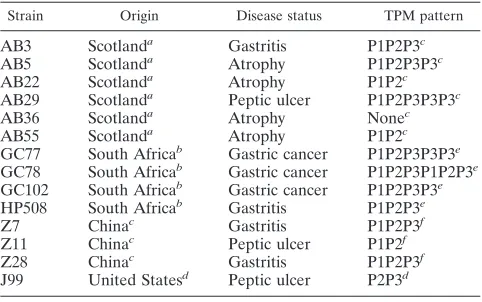
TABLE 1. H.pyloristrains used in this study
Strain Origin Disease status TPM pattern
AB3 Scotlanda Gastritis P1P2P3c
AB5 Scotlanda Atrophy P1P2P3P3c
AB22 Scotlanda Atrophy P1P2c
AB29 Scotlanda Peptic ulcer P1P2P3P3P3c
AB36 Scotlanda Atrophy Nonec
AB55 Scotlanda Atrophy P1P2c
GC77 South Africab Gastric cancer P1P2P3P3P3e
GC78 South Africab Gastric cancer P1P2P3P1P2P3e
GC102 South Africab Gastric cancer P1P2P3P3e
HP508 South Africab Gastritis P1P2P3e
Z7 Chinac Gastritis P1P2P3f
Z11 Chinac Peptic ulcer P1P2f
Z28 Chinac Gastritis P1P2P3f
J99 United Statesd Peptic ulcer P2P3d
aEl-Omar et al. (14). bKidd et al. (18). cThis study. dAlm et al. (2). eArgent et al. (3).
fAccession numbers AJ832141, AJ832142, and AJ832145.
791
on May 16, 2020 by guest
http://jcm.asm.org/
workstation (Don Whitley Scientific, Shipley, United Kingdom) for three pas-sages before extraction of genomic DNA as previously described (6). PCR amplification of thecagA3⬘variable region was performed as previously de-scribed by using the primers cag2 and cag4 (24), and sequencing of the 550- to 800-bp products was performed by the Biopolymer Synthesis and Analysis Unit (Queen’s Medical Centre, University of Nottingham, Nottingham, United King-dom). Determination of the number of nucleotide sequences encoding the EPIYA motifs, and the types of motifs encoded, was carried out by using the forward primer cag2 (or cagA28F) and the reverse primers P1C, cagA-P2CG, cagA-P2TA, and cagA-P3E (Table 2). A reaction mixture containing 0.2 mM concentrations of each deoxynucleoside triphosphate, a 0.4 nM concentra-tion of the forward primer, a 0.08 nM concentraconcentra-tion of the reverse primer, 0.05 U ofTaqDNA polymerase (Roche Diagnostics, Penzberg, Germany)/l, and 1
l of genomic DNA in buffer (10 mM Tris-HCl [pH 8.3], 1.5 mM magnesium chloride, 50 mM potassium chloride) was incubated at 95°C for 90 s, followed by 35 cycles at 95°C for 30 s, 57°C for 60 s, and 72°C for 30 s and a final extension at 72°C for 5 min. PCR products were separated on a 1.5% (wt/vol) agarose gel.
RESULTS AND DISCUSSION
CagA proteins differ in size due to the presence of repeat regions containing EPIYA motifs, and we have previously shown thatH.pyloristrains isolated from South Africa possess three to six TPMs within this variable region (3). PCR ampli-fication of the entire cagA variable region by using primers cag2 and cag4 (24) can give a size-based estimation of the likely number of TPMs. However, someH.pyloristrains possesscagA
with insertions or deletions between TPM repeats, thereby generating size variations without affecting the number of EPIYA motifs. Determination of the exact number of EPIYA motifs has therefore previously involved sequencing the entire
cagAvariable region, a laborious and generally expensive pro-cess.
To simplify determination of the number and type of EPIYA motifs present, we developed a PCR-based approach to iden-tify individual EPIYA motifs, using a single forward primer and multiple reverse primers. Analysis of the sequences of
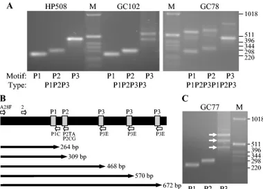
[image:2.585.43.284.81.173.2]cagA variable regions showed that there are three types of repeat region containing the EPIYA motif present: the first (P1) position has the consensus sequence EPIYA(Q/ K)VNKKK(T/A)GQ, the second (P2) has the consensus se-quence EPIY(A/T)QVAKKV, and the third (P3) has the con-sensus sequence EPIYATIDDLGGPFPL. The third (P3) position, however, is different inH.pyloristrains isolated from East Asia, having the sequence EPIYATIDFDEANQAG. In
[image:2.585.102.482.391.664.2]FIG. 1. PCR amplification ofcagAvariable-region TPMs from South AfricanH.pyloristrains. (A and C) Genomic DNA fromH.pyloristrains HP508, GC102, and GC78 (A) and GC77 (C) were used to PCR amplify thecagAvariable-region EPIYA motifs, using the forward primer cagA28F and the reverse primers cagA-P1C (P1), cagA-P2CG and cagA-P2TA (equimolar mixture; P2), or cagA-P3E (P3). M, size markers (in base pairs). (B) Schematic representation of thecagAvariable region fromH.pyloristrain GC77 showing the EPIYA motifs (gray), the annealing positions of the cagA28F (A28F) and cag2 (2) forward primers and the cagA-P1C (P1C), cagA-P2TA (P2TA), cagA-P2CG (P2CG), and cagA-P3E (P3E) reverse primers, and the expected sizes of the amplified products.
TABLE 2. Primers used for the amplification ofcagATPMs
Primer Sequence (5⬘-3⬘) Motif
amplified
cag2a GGAACCCTAGTCGGTAATG
cag4a ATCTTTGAGCTTGTCTATCG
cagA28F TTCTCAAAGGAGCAATTGGC
cagA-P1C GTCCTGCTTTCTTTTTATTAACTTKAGC P1 cagA-P2CG TTTAGCAACTTGAGCGTAAATGGG P2 cagA-P2TA TTTAGCAACTTGAGTATAAATGGG P2
cagA-P3E ATCAATTGTAGCGTAAATGGG P3
a
Data from Rudi et al. (24).
792 ARGENT ET AL. J. CLIN. MICROBIOL.
on May 16, 2020 by guest
http://jcm.asm.org/
order to amplify the three types of TPM motif, we designed primers complementary to the nucleotide sequences encoding these repeats (Table 2). The original P1 reverse primer (cagA-P1) was found to preferentially amplify the P2 repeat, due to high identity between the primers, and so was redesigned to amplify the A(Q/K)VNKKK(T/A)G repeat instead (cagA-P1C). The P2 primers (cagA-P2CG and cagA-P2TA) were designed to amplify the PIY(A/T)QVAK repeat, and the P3 primer (cagA-P3E) was designed to amplify the PIYATID repeat (Table 2).
Figure 1 shows the amplification of TPM repeats within the
cagAvariable regions of South AfricanH.pyloristrains known to possess three to six EPIYA motifs by nucleotide sequencing (3). From the sequences we expected that the strains would have the EPIYA motif patterns P1P2P3 (HP508), P1P2P3P3 (GC102), P1P2P3P3P3 (GC77), and P1P2P3P1P2P3 (GC78), which are the exact patterns obtained by PCR (Fig. 1). We predicted that, with the forward primer cagA28F, which an-neals to nucleotide positions 546053 to 546072 in theH.pylori
strain J99 chromosome, the amplified fragment sizes would be 264 to 291 bp for the first P1 site, 309 to 336 bp for the first P2 site, and 465 to 498 bp for the first P3 site. ForcagAfrom strain GC77, which has three copies of the P3 motif, we predicted that amplification of the P1 and P2 sites would generate frag-ments of 264 and 309 bp, respectively, and that amplification of the P3 sites would produce products of 468, 570, and 672 bp (Fig. 1B), which are the approximate sizes generated in the PCR amplifications (Fig. 1C). PCR with the forward primer cag2 in place of cagA28F generates the same products except that, as predicted, they are ca. 99 bp shorter in length.
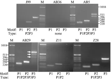
We next looked at cagA variable regions from H. pylori
strains from other populations, along with a control from ge-nome sequence strain J99 (ATCC 700824) and from a strain lackingcagA(Fig. 2). ThecagAfrom strain J99 has the EPIYA motif pattern P2P3 and therefore lacks a P1 motif, and this can be seen in the PCR data. There are no products generated for thecagA-negative sample (AB36), whereascagAfrom strains AB3, AB5, AB22, AB29, and AB55 predict the EPIYA motif patterns P1P2P3, P1P2P3P3, P1P2, P1P2P3P3P3, and P1P2, respectively (Fig. 2). To confirm that our PCR-based predic-tion of TPMs was correct, we performed nucleotide sequenc-ing of the entire cagA 3⬘ variable region from strains AB3, AB5, AB22, AB29, and AB55 after amplification of this region with primers cag2 and cag4 (Table 3). This showed that the
cagA variable region from these strains possessed the exact patterns of EPIYA motifs expected from the PCR data.
Although the PCR prediction of EPIYA motifs proved re-liable with “Western” isolates ofH.pylori, we also wanted to determine whether this method could be used to type thecagA
[image:3.585.117.474.70.326.2]variable regions of H. pylori strains from East Asia that are known to possess differences in sequence. To do this, we PCR amplified thecagA EPIYA motifs from H. pyloristrains iso-lated from China. Strain Z11 has a nucleotide sequence that encodes a Western type of CagA, with a P1P2 motif pattern, whereas strains Z7 and Z28 have an “East Asian” type ofcagA, with a P1P2P3 pattern. The reverse primer cagA-P3E was designed to amplify the P3 motif within all types ofcagA by annealing to the nucleotide sequence encoding the EPIYATID motif (Table 2). PCR amplification ofcagAfrom ChineseH.pyloristrains, carried out with the forward primer
FIG. 2. PCR amplification ofcagAvariable-region TPMs fromH.pyloristrains. Genomic DNA fromH.pyloristrains J99, AB36, AB5, AB29, Z11, and Z28 were used to amplify thecagAvariable-region EPIYA motifs, using the forward primers cagA28F (J99, AB36, AB5, and AB29) or cag2 (Z11 and Z28) and the reverse primers cagA-P1C (P1), cagA-P2CG and cagA-P2TA (equimolar mixture; P2), or cagA-P3E (P3).H.pylori strain J99 lacks the P1 motif, and strain AB36 does not possesscagA. M, size markers (in base pairs).
on May 16, 2020 by guest
http://jcm.asm.org/
cag2, again revealed the predicted patterns (Fig. 2). Strain Z11 has the EPIYA motif pattern P1P2, whereas strains Z7 and Z28 have the pattern P1P2P3. Amplification with the forward primer cagA28F gave the same patterns, although we found that, with East Asian types ofcagA, PCRs with the cag2 primer gave clearer results.
We have therefore shown that PCR can be used to deter-mine the number of cagA variable-region EPIYA motifs in strains from Africa, Britain, and China. Although we have not testedcagAvariable regions from other populations, we expect that this method would be compatible for use with almost all
H. pyloristrains. The methodology we describe is simple and
inexpensive; after genomic DNA extraction, PCRs and analysis can be carried out within a few hours, and this method allows the rapid screening of multiple strains. Although most strains
ofH.pyloripossess the three types of TPM analyzed here, or
minor variations in these motifs, we cannot exclude the possi-bility that some strains may rarely possess alternative se-quences that would not be recognized by our primers.
Our new method will help in further work to elucidate the biological and clinical importance of multiple tyrosine phos-phorylation sites. In particular, it is important that we sepa-rately identify P1, P2, and P3 sites since the relative importance of each type of TPM containing the EPIYA repeat remains unclear. Although Higashi et al. (15) showed that the P3 motif of CagA from strain NCTC 11637 (which has a P1P2P3P3P3, or ABCCC, TPM pattern) became more phosphorylated within epithelial cells than the other motifs and induced the hummingbird phenotype in more cells, we (3) showed that CagA fromH.pyloristrain GC78 (which has a P1P2P3P1P2P3 pattern; Table 1) became more phosphorylated and induced the hummingbird phenotype more cell than CagA from strain GC77 (which has a P1P2P3P3P3 pattern; Table 1). Finally, we point out that aspects of CagA other than its level of tyrosine phosphorylation may impact its cellular effects. CagA from East Asian strains interacts with SHP-2 phosphatase via the binding motif pY-(S/T/A/V/I)-X-(V/I/L)-X-(W/F) (13), which is present in CagA from East AsianH.pyloristrains but not in Western strains and leads to greater activation of the phospha-tase (8, 15). However, despite the absence of this consensus sequence in non-East Asian CagA proteins, it is clear that these strains can still induce formation of the hummingbird phenotype (3, 8). Whether strains with an East Asian SHP-2 binding motif are associated with more severe disease is a further question worth addressing.
ACKNOWLEDGMENTS
This study was funded by a grant from Cancer Research UK. Y.Z. was funded by a scholarship from the Jiangsu Provincial Department of Education of the People’s Republic of China to conduct research in the United Kingdom. J.C.A. is funded by a Senior Clinical Fellowship from the Medical Research Council (United Kingdom).
REFERENCES
1.Akopyants, N. S., S. W. Clifton, D. Kersulyte, J. E. Crabtree, B. E. Youree, C. A. Reece, N. O. Bukanov, E. S. Drazek, B. A. Roe, and D. E. Berg.1998. Analyses of thecagpathogenicity island ofHelicobacter pylori. Mol. Micro-biol.28:37–53.
2.Alm, R. A., L.-S. L. Ling, D. T. Moir, B. L. King, E. D. Brown, P. C. Doig, D. R. Smith, B. Noonan, B. C. Guild, B. L. deJonge, G. Carmel, P. J. Tummino, A. Caruso, M. Uria-Nickelsen, D. M. Mills, C. Ives, R. Gibson, D. Merberg, S. D. Mills, Q. Jiang, D. E. Taylor, G. F. Vovis, and T. J. Trust.
1999. Genomic-sequence comparison of two unrelated isolates of the human gastric pathogenHelicobacter pylori. Nature397:176–180.
3.Argent, R. H., M. Kidd, R. J. Owen, R. J. Thomas, M. C. Limb, and J. C. Atherton.2004. Determinants and consequences of different levels of CagA phosphorylation for clinical isolates ofHelicobacter pylori. Gastroenterology
127:514–523.
4.Asahi, M., T. Azuma, S. Ito, Y. Ito, H. Suto, Y. Nagai, M. Tsubokawa, Y. Tohyama, S. Maeda, M. Omata, T. Suzuki, and C. Sasakawa.2000. Helico-bacter pyloriCagA protein can be tyrosine phosphorylated in gastric epithe-lial cells. J. Exp. Med.191:593–602.
5.Asahi, M., Y. Tanaka, T. Izumi, Y. Ito, H. Naiki, D. Kersulyte, K. Tsujikawa, M. Saito, K. Sada, S. Yanagi, A. Fujikawa, M. Noda, and Y. Itokawa.2003.
Helicobacter pyloriCagA containing ITAM-like sequences localized to lipid rafts negatively regulates VacA-induced signaling in vivo. Helicobacter8:1– 14.
6.Atherton, J. C.1997. Molecular methods for detecting ulcerogenic strains of
H.pylori, p. 133–143.InC. L. Clayton and H. L. T. Mobley (ed.), Methods in molecular medicine,Helicobacter pyloriprotocols. Humana Press, Totowa, N.J.
7.Azuma, T., A. Yamakawa, S. Yamazaki, K. Fukuta, M. Ohtani, Y. Ito, M. Dojo, Y. Yamazaki, and M. Kuriyama.2002. Correlation between variation of the 3⬘region of thecagAgene inHelicobacter pyloriand disease outcome in Japan. J. Infect. Dis.186:1621–1630.
8.Azuma, T., S. Yamazaki, A. Yamakawa, M. Ohtani, A. Muramatsu, H. Suto, Y. Ito, M. Dojo, Y. Yamazaki, Y. Keida, H. Higashi, and M. Hatakeyama.
2004. Association between diversity in the Src homology 2 domain-contain-ing tyrosine phosphatase binddomain-contain-ing site ofHelicobacter pyloriCagA protein and gastric atrophy and cancer. J. Infect. Dis.189:820–827.
9.Backert, S., S. Moese, M. Selbach, V. Brinkmann, and T. F. Meyer.2001. Phosphorylation of tyrosine 972 of theHelicobacter pyloriCagA protein is essential for induction of a scattering phenotype in gastric epithelial cells. Mol. Microbiol.42:631–644.
10.Backert, S., E. Ziska, V. Brinkmann, U. Zimny-Arndt, A. Fauconnier, P. R. Jungblut, M. Naumann, and T. F. Meyer.2000. Translocation of the Heli-cobacter pyloriCagA protein in gastric epithelial cells by a type IV secretion apparatus. Cell. Microbiol.2:155–164.
11.Censini, S., C. Lange, Z. Xiang, J. E. Crabtree, P. Ghiara, M. Borodovsky, R. Rappuoli, and A. Covacci.1996.cag, a pathogenicity island ofHelicobacter pylori, encodes type I-specific and disease-associated virulence factors. Proc. Natl. Acad. Sci. USA93:14648–14653.
12.Covacci, A., S. Censini, M. Bugnoli, R. Petracca, D. Burroni, G. Macchia, A. Massone, E. Papini, Z. Xiang, N. Figura, and R. Rappuoli.1993. Molecular characterization of the 128-kDa immunodominant antigen ofHelicobacter pyloriassociated with cytotoxicity and duodenal ulcer. Proc. Natl. Acad. Sci. USA90:5791–5795.
13.de Sousa, D., L. J. Fabri, A. Nash, D. J. Hilton, N. A. Nicola, and M. Baca.
2002. SH2 domains from suppressor of cytokine signaling-3 and protein tyrosine phosphatase SHP-2 have similar binding specificities. Biochemistry
41:9229–9236.
14.El-Omar, E. M., K. Oien, L. S. Murray, A. El-Nujumi, A. Wirz., D. Gillen, C. Williams, G. Fullarton, and K. E. L. McColl.2000. Increased prevalence of precancerous changes in relatives of gastric cancer patients: critical role of
Helicobacter pylori. Gastroenterology118:22–30.
15.Higashi, H., R. Tsutsumi, A. Fujita, S. Yamazaki, M. Asaka, T. Azuma, and M. Hatakeyama.2002. Biological activity of theHelicobacter pylorivirulence factor CagA is determined by variation in the tyrosine phosphorylation sites. Proc. Natl. Acad. Sci. USA99:14428–14433.
16.Higashi, H., R. Tsutsumi, S. Muto, T. Sugiyama, T. Azuma, M. Asake, and M. Hatakeyama.2002. SHP-2 tyrosine phosphatase as an intracellular target ofHelicobacter pyloriCagA protein. Science295:683–686.
17.Jenks, P. J., F. Me´graud, and A. Labigne. 1998. Clinical outcome after infection withHelicobacter pyloridoes not appear to be reliably predicted by the presence of any of the genes of thecagpathogenicity island. Gut43:
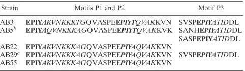
[image:4.585.44.285.80.150.2]752–758. TABLE 3. Nucleotide sequence analysis ofH.pyloristrainsa
Strain Motifs P1 and P2 Motif P3
AB3 EPIYAKVNKKKTGQVASPEEPIYTQVAKKVN SVSPEPIYATIDDL AB5b EPIYAQVNKKKAGQVASPEEPIYTQVAKKVK SANHEPIYATIDDL
SASPEPIYATIDDL AB22 EPIYAKVNKKKAGQVASPEEPIYAQVAKKVN
AB29c EPIYAKVNKKKAGQVASPEEPIYAQVAKKVN SVSPEPIYATIDDL AB55 EPIYAKVNKKKAGQVASPEEPIYAQVAKKVN
aP1, P2, and P3 EPIY(A/T) motifs are shown in boldface. The annealing regions for the reverse primers are shown in italics.
bStrain AB5 has two P3 motifs, as shown in the final column.
cStrain AB29 has three identical P3 motifs, as shown in the final column.
794 ARGENT ET AL. J. CLIN. MICROBIOL.
on May 16, 2020 by guest
http://jcm.asm.org/
18.Kidd, M., A. J. Lastovica, J. C. Atherton, and J. A. Louw.2001. Conservation of thecagpathogenicity island is associated withvacAalleles and gastrodu-odenal disease is South AfricanHelicobacter pyloriisolates. Gut49:11–17. 19.Maeda, S., H. Yoshida, T. Ikenoue, K. Ogura, F. Kanai, N. Kato, Y.
Shira-tori, and M. Omata.1999. Structure ofcagpathogenicity island in Japanese
Helicobacter pyloriisolates. Gut44:336–341.
20.Mimuro, H., T. Suzuki, J. Tanaka, M. Asahi, R. Haas, and C. Sasakawa.
2002. Grb2 is a key mediator ofHelicobacter pyloriCagA protein activities. Mol. Cell10:745–755.
21.Nilsson, C., A. Sille´n, L. Eriksson, M.-L. Strand, H. Enroth, S. Normark, P. Falk, and L. Engstrand.2003. Correlation betweencagpathogenicity island composition andHelicobacter pylori-associated gastroduodenal disease. In-fect. Immun.71:6573–6581.
22.Odenbreit, S., J. Pu¨ls, B. Sedlmaier, E. Gerland, W. Fischer, and R. Haas.
2000. Translocation ofHelicobacter pyloriCagA into gastric epithelial cells by type IV secretion. Science287:1497–1500.
23.Owen, R. J., S. I. Sharp, S. A. Chisholm, and S. Rijpkema.2003. Identifi-cation ofcagAtyrosine phosphorylation DNA motifs inHelicobacter pylori
isolates from peptic ulcer patients by novel PCR-restriction fragment length polymorphism and real-time fluorescence PCR assays. J. Clin. Microbiol.
41:3112–3118.
24.Rudi, J., C. Kolb, M. Maiwald, D. Kuck, A. Sieg, P. R. Galle, and W. Stremmel.1998. Diversity ofHelicobacter pylori vacAandcagAgenes and relationship to VacA and CagA protein expression, cytotoxin production, and associated diseases. J. Clin. Microbiol.36:944–948.
25.Segal, E. D., J. Cha, J. Lo, S. Falkow, and L. S. Tompkins.1999. Altered states: involvement of phosphorylated CagA in the induction of host cellular growth changes byHelicobacter pylori. Proc. Natl. Acad. Sci. USA96:14559– 14564.
26.Segal, E. D., S. Falkow, and L. S. Tompkins.1996.Helicobacter pylori at-tachment to gastric cells induces cytoskeletal rearrangements and tyrosine phosphorylation of host cell proteins. Proc. Natl. Acad. Sci. USA93:1259– 1264.
27.Selbach, M., S. Moese, C. R. Hauck, T. F. Meyer, and S. Backert.2002. Src is the kinase of theHelicobacter pyloriCagA protein in vitro and in vivo. J. Biol. Chem.277:6775–6778.
28.Stein, M., R. Rappuoli, and A. Covacci.2000. Tyrosine phosphorylation of theHelicobacter pyloriCagA antigen aftercag-driven host cell translocation. Proc. Natl. Acad. Sci. USA97:1263–1268.
29.Stein, M., F. Bagnoli, R. Halenbeck, R. Rappuoli, W. J. Fantl, and A. Covacci.2002. c-Src/Lyn kinases activateHelicobacter pyloriCagA through tyrosine phosphorylation of the EPIYA motifs. Mol. Microbiol.43:971–980. 30.Tsutsumi, R., H. Higashi, M. Higuchi, M. Okada, and M. Hatakeyama.2003. Attenuation of Helicobacter pyloriCagA䡠SHP-2 signaling by interaction between CagA and C-terminal Src kinase. J. Biol. Chem.278:3664–3670. 31.Tummuru, M. K. R., S. A. Sharma, and M. J. Blaser.1995.Helicobacter
pylori picB, a homologue of theBordetella pertussistoxin secretion protein, is required for induction of IL-8 in gastric epithelial cells. Mol. Microbiol.
18:867–876.
32.Yamaoka, Y., T. Kodama, K. Kashima, D. Y. Graham, and A. R. Sepulveda.
1998. Variants of the 3⬘region of thecagAgene inHelicobacter pyloriisolates from patients with differentH.pylori-associated diseases. J. Clin. Microbiol.
36:2258–2263.
33.Yamazaki, S., A. Yamakawa, Y. Ito, M. Ohtani, H. Higashi, M. Hatakeyama, and T. Azuma.2003. The CagA protein ofHelicobacter pyloriis translocated into epithelial cells and binds to SHP-2 in human gastric mucosa. J. Infect. Dis.187:334–337.