Investigating long noncoding RNAs using
animal models
Michael Feyder, Loyal A. Goff
J Clin Invest.
2016;
126(8)
:2783-2791.
https://doi.org/10.1172/JCI84422
.
The number of long noncoding RNAs (lncRNAs) has grown rapidly; however, our
understanding of their function remains limited. Although cultured cells have facilitated
investigations of lncRNA function at the molecular level, the use of animal models provides
a rich context in which to investigate the phenotypic impact of these molecules. Promising
initial studies using animal models demonstrated that lncRNAs influence a diverse number
of phenotypes, ranging from subtle dysmorphia to viability. Here, we highlight the diversity
of animal models and their unique advantages, discuss the use of animal models to profile
lncRNA expression, evaluate experimental strategies to manipulate lncRNA function in
vivo, and review the phenotypes attributable to lncRNAs. Despite a limited number of
studies leveraging animal models, lncRNAs are already recognized as a notable class of
molecules with important implications for health and disease.
Review
Find the latest version:
Introduction
Approximately three-quarters of the mammalian genome is tran-scribed into RNA (1–3); however, only a fraction of this transcrip-tion produces mRNA, whose mature nucleotide sequence serves as a template for protein synthesis (3). The function of this nonpro-tein-coding RNA (or noncoding RNA) is mostly obscure, despite a larger number of noncoding genes than protein-coding genes. Given the broad functional repertoire derived from protein-cod-ing RNA, it is perhaps not surprisprotein-cod-ing that the relatively few known functions for noncoding RNAs also span diverse cellular process-es, resulting in an array of noncoding RNA subclassifications (4). One particular subclass, long noncoding RNAs (lncRNAs), rep-resents a large family of noncoding RNA molecules with potential-ly broad implications for basic science, health, and disease. In this Review, we focus on the use of animal models to discover novel lncRNAs and to investigate their significance in vivo.
lncRNA definitions and mechanisms
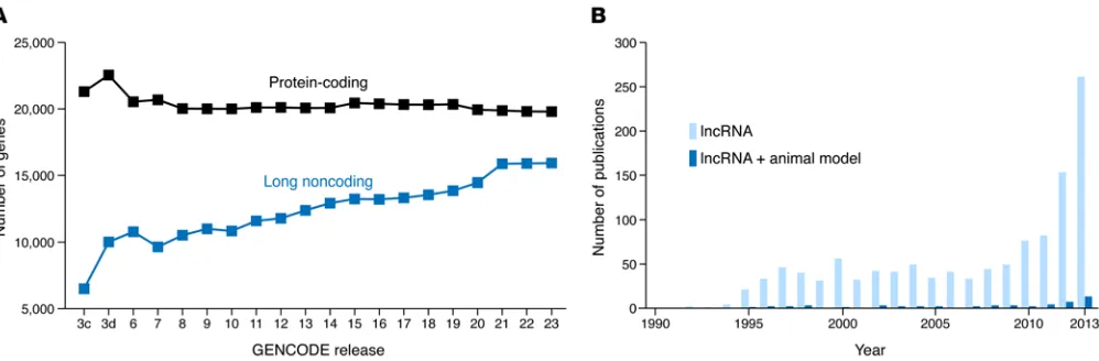
lncRNAs represent a burgeoning class of molecules broadly defined as RNA transcripts longer than 200 nucleotides, with no protein-coding potential. This length is somewhat arbitrary, but it serves to distinguish them from shorter, biologically distinct non-coding RNAs, such as microRNAs. In humans, high-throughput experimental approaches have led to the rapid identification of approximately 16,000 lncRNA genes thus far, rivaling the approx-imately 20,000 protein-coding genes (5–10) (Figure 1A). These lncRNAs are informatically predicted to lack protein-coding potential, although notable cases of presumed lncRNAs
encod-ing micropeptides (11, 12) or actencod-ing as precursors to microRNAs (13, 14) exist. Therefore, current definitions attempt to corral this emerging class of molecules, whose validation and function in vivo remain to be fully elucidated (Figure 1B).
lncRNAs are found in both the nucleus and the cytoplasm. The majority of lncRNAs reside in the nucleus (10), where they can act proximally or distally to their site of transcription, functioning in cis or trans, respectively. For example, during X chromosome inactivation in mice, the lncRNA Xist functions in cis to initiate silencing of genes across the same X chromosome from which it was originally transcribed (15). Conversely, the mouse lncRNA Trp53cor1 (also known as lincRNA-p21) acts in trans to globally repress the expression of hundreds of genes distant from its site of transcription (16). Additionally, the transcription of a cis-acting lncRNA, per se, rather than its resulting RNA product, can also have a biological effect (17). As discussed below, this possibility raises important considerations when designing experiments to manipulate lncRNA expression and function.
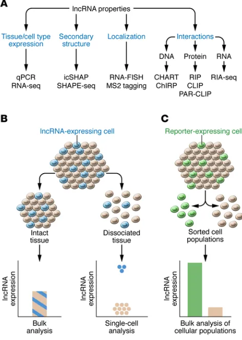
The molecular mechanisms for most lncRNAs remain large-ly unknown. They can bind to DNA, RNA, or proteins, and many techniques have been developed to assay these interactions (Fig-ure 2A). Techniques exploring lncRNA-DNA interactions, such as capture hybridization analysis of RNA targets (CHART) (18) and chromatin isolation by RNA purification (ChIRP) (19), utilize com-plementary oligonucleotides that hybridize to a lncRNA of interest and serve as an affinity handle to enrich for bound DNA. Similar hybridization approaches, such as radioimmunoassay sequencing (RIA-seq), can also be used to assay lncRNA-RNA interactions (20). Other techniques, such as RNA immunoprecipitation (RIP) or its variants, cross-linking immunoprecipitation (CLIP) (21, 22), and photoactivatable ribonucleoside–enhanced cross-link-ing immunoprecipitation (PAR-CLIP) (23), utilize antibodies to purify lncRNA-protein complexes. Additionally, high-through-put approaches to investigate RNA secondary structure, such as in vivo click selective 2′hydroxyl acylation and profiling experi-ment (icSHAPE) (24) or selective 2′-hydroxyl acylation analyzed
The number of long noncoding RNAs (lncRNAs) has grown rapidly; however, our understanding of their function remains limited. Although cultured cells have facilitated investigations of lncRNA function at the molecular level, the use of animal models provides a rich context in which to investigate the phenotypic impact of these molecules. Promising initial studies using animal models demonstrated that lncRNAs influence a diverse number of phenotypes, ranging from subtle dysmorphia to viability. Here, we highlight the diversity of animal models and their unique advantages, discuss the use of animal models to profile lncRNA expression, evaluate experimental strategies to manipulate lncRNA function in vivo, and review the phenotypes attributable to lncRNAs. Despite a limited number of studies leveraging animal models, lncRNAs are already recognized as a notable class of molecules with important implications for health and disease.
Investigating long noncoding RNAs using
animal models
Michael Feyder1,2 and Loyal A. Goff1,2,3
1McKusick-Nathans Institute for Genetic Medicine, 2Department of Neuroscience, and 3Johns Hopkins University School of Medicine, Baltimore, Maryland, USA.
Conflict of interest: L.A. Goff is a co-inventor on two patents, neither of which is directly related to the manuscript: (a) “High-throughput Methodology for Identifying RNA-Protein Interactions Transcriptome-wide,” B.D. Gregory, J.L. Rinn, F. Li, L.A. Goff, and C. Trapnell (U.S. Patent no. 9,097,708 B2); and (b) “Rational Probe Optimization for Microarray Detection of MicroRNAs,” R. Getts, R.P. Hart, and L.A. Goff (U.S. Patent Application no. PCT/US2005/038261).
drogenesis-associated transcript (CISTR) and the spatially prox-imal protein-coding gene parathyroid hormone–like hormone (PTHLH), diminishing the expression of PTHLH mRNA (32). These findings highlight how lncRNA dysfunction impacts cellu-lar processes with clinically relevant consequences.
Notably, lncRNA expression in human tumors is associated with clinical outcomes for a variety of cancers, and xenograft stud-ies in mice have been instrumental when extending these findings in vivo. For example, more than 100 lncRNAs correlate with over-all or progression-free survival for ovarian cancer, prostate cancer, glioblastomas, and lung squamous cell carcinomas (33). Nine of these lncRNAs consistently correlate across these cancer subtypes (33). Other examples include the lncRNA HOTAIR, which is over-expressed in primary breast tumors, and elevated expression of
HOTAIR in these tumors correlates with an increased probability of
metastasis and a decreased overall rate of survival (34). Addition-ally, xenografts overexpressing HOTAIR in a cell line derived from metastatic breast tissue have an increased propensity for metastasis in mice (34). Furthermore, approximately 1,900 lncRNAs are dif-ferentially expressed in T cell acute lymphoblastic leukemia (35). This finding is further supported in the same study by demonstrat-ing in mice that one of these lncRNAs, LUNAR1, is necessary for xenograft tumor growth of a human T cell lymphoma cell line (35). Finally, xenograft studies can also support the in vivo significance of lncRNAs implicated in biological processes such as hypoxia that are known to promote cancer progression (36). For example, the lncRNAs NPTN-IT1 (also known as lncRNA-LET) (37), TP53COR1 (38), and LINC-ROR (39) all regulate hypoxia-induced signaling and affect xenograft growth. In summary, accumulating evidence implicates lncRNAs in cancer development, a finding supported by the use of laboratory animals for xenograft studies.
Diversity of animal models
Animal models vary in biological complexity, which can be lever-aged depending on the area of investigation. Commonly used ani-mal models more evolutionarily divergent from humans include the nematode (Caenorhabditis elegans), the fruit fly (Drosophila
melanogaster), and the zebrafish (Danio rerio). The reduced
com-by primer extension sequencing (SHAPE-seq) (25), have also been developed. Collectively, these techniques demonstrate that lncRNAs interact with diverse macromolecules, potentially impacting a wide range of biological functions.
lncRNAs and disease
Accumulating evidence suggests that lncRNA dysfunction pro-motes disease (26). Notably, approximately 40% of disease- or trait-associated SNPs are found within the noncoding regions flanking protein-coding genes, where a subclass of lncRNAs, termed long intergenic noncoding RNAs, reside (27). Although the functional consequences, if any, for many of these SNPs remain to be experimentally evaluated, they may promote disease by affecting lncRNA function and/or expression (28). For example, an SNP within the human lncRNA myocardial infarction–asso-ciated transcript (MIAT), which is assoinfarction–asso-ciated with an increased risk for myocardial infarction, also increases the binding affinity of MIAT for nuclear proteins (29). This same SNP also increases expression levels of MIAT, although the precise molecular mech-anisms remain unknown (29). Additionally, in a human cell line, the binding of lncRNAs to the protein complex Mediator promotes permissive chromatin states necessary for gene expression, and mutations in Mediator that result in intellectual disability have been shown to impair its interaction with lncRNAs (30). An alter-native scenario, whereby an alteration of a lncRNA disrupts its interaction with Mediator, could conceivably produce similar, yet less pronounced, effects. These results contribute to a growing body of evidence implicating lncRNAs in disease.
Genetic linkage studies have uncovered new lncRNAs, pro-viding additional evidence to suggest that lncRNA dysfunction promotes disease. For example, HELPP syndrome, which occurs in mothers during pregnancy and is characterized by hemolysis, elevated liver enzymes, and low platelet counts, is associated with a previously unknown placental lncRNA (31). Mutations within this lncRNA increase the in vitro proliferation of human placenta cells in a model of this syndrome (31). Additionally, brachydac-tyly type E can result from a chromosomal rearrangement that disrupts local interactions between the human lncRNA
chon-Figure 1. lncRNA biology is a burgeoning field. (A) The number of genes designated as lncRNAs in humans has steadily increased over successive
[image:3.585.42.541.57.221.2]or humans, despite diverging primary sequences (49). These results suggest that the higher-order structure of a lncRNA, rather than its primary sequence, may be conserved. Given the approxi-mately 500 computationally predicted, intergenic RNA secondary structures conserved across vertebrates (57), zebrafish may be a robust model for probing a conserved lncRNA function that is not readily apparent from the primary sequence.
Species-specific lncRNAs may broadly highlight the involve-ment of lncRNAs in certain conditions in which certain animal models have afforded advantages. For example, both the fruit fly and the nematode are preferentially used to model aging, as their life span is shorter than that of the mouse or the rat (58). Using a genetic model of aging in the nematode, it has been demonstrat-ed that binding of the lncRNA tts-1 to the ribosome suppresses ribosomal levels and promotes longevity, implicating lncRNAs in this phenotype (59).
Other advantages of animal models more evolutionarily dis-tant from humans include the ability to conduct genetic screens (60). For example, expanded nucleotide repeats within the human lncRNA ATXN8OS (also known as SCA8) result in the neurode-generative disorder spinocerebellar ataxia (61). Using the retina of the fruit fly as a model system, the expression of ATXN8OS, either with or without expanded repeats, results in neurodegeneration (62). However, a genetic screen in this same study demonstrated that this phenotype is differentially modified by RNA-binding pro-teins (62), in agreement with accumulating evidence implicating aberrant RNA-protein interactions in neurodegenerative disorders (63). Therefore, the use of animal models more evolutionarily dis-tant from humans to study lncRNA function may serve to uncover fundamental roles of lncRNAs and their dysfunction in disease.
Animal models and lncRNA profiling
Animal models generate a variety of tissue and cell types from which to profile lncRNA expression. This feature renders animal models well suited for the study of lncRNAs, given that lncRNA expression is more tissue and cell type specific than is protein-cod-ing gene expression (5, 10, 64). When compared with cultured cells, material derived from animals is generally less abundant and more time-consuming to generate. Nonetheless, animal mod-els provide direct, in vivo evidence of endogenous lncRNA expres-sion and dramatically narrow the relevant search space when investigating phenotypes.
Modern techniques available for genomics research, such as RNA-seq, provide an unbiased, high-throughput approach to investigating endogenous lncRNA expression and to discovering and assembling novel lncRNAs de novo (64). lncRNA expres-plexity of these species can be advantageous for investigating the
role of lncRNAs in highly conserved biological processes, whose dysfunction may contribute to human disease. For example, stud-ies of protein-coding genes utilizing C. elegans and D. melanogaster have aided in understanding the molecular mechanisms of apop-tosis, whose dysfunction contributes to cancer development and progression (40). Less evolutionarily divergent animal models include the rat (Rattus norvegicus), the mouse (Mus musculus), and the nonhuman primate. The greater biological complexity and physiological similarity of these organisms to humans may better model the complex biology of diseases, such as that underlying tumor growth and metastasis (41, 42). Additional considerations when choosing an appropriate animal model include financial cost, ethical considerations, potential throughput, and ease of experimental manipulation.
Evolutionary conservation of lncRNAs
[image:4.585.46.282.60.392.2]In addition to humans, numerous lncRNAs are also found in the fruit fly (43–46), the nematode (47), the zebrafish (48, 49), and the mouse (1, 6). Cross-species comparisons have demonstrated that lncRNAs can contain short, conserved regions (9, 50) and also show evidence of purifying selection (51). In general, how-ever, their primary sequence is weakly conserved across species (49, 52–56). For example, only 29 lncRNAs are conserved between zebrafish and humans (49). Strikingly, in the same study, the phe-notype in zebrafish following functional inactivation of conserved lncRNAs can be rescued by an orthologous gene from either mice
Figure 2. Techniques to investigate lncRNA properties and tissue
expres-sion. (A) These techniques include quantitative PCR (qPCR) and RNA-seq
development of the cerebral cortex (64, 68). Profiling endogenous lncRNA expression may therefore highlight the diversity of genet-ically defined cell types and their potential to promote disease.
Additionally, environmental stimuli may affect lncRNA expression, making animal models an attractive resource for certain areas of research, such as neuroscience. Interestingly, lncRNAs are overrepresented in the human brain (10), where they may be fundamental to synaptic transmission (56). Additionally, both neuronal activation (69) and drugs of abuse (70, 71) promote lncRNA expression. The diversity of cell types within the brain, combined with myriad environmental stimuli, has the potential to reveal numerous unrecognized lncRNAs. In sum, animal models are a rich source of material from which lncRNA expression can be profiled across tissues or cell types, providing insight into their potential function in vivo.
Animal models and lncRNA function
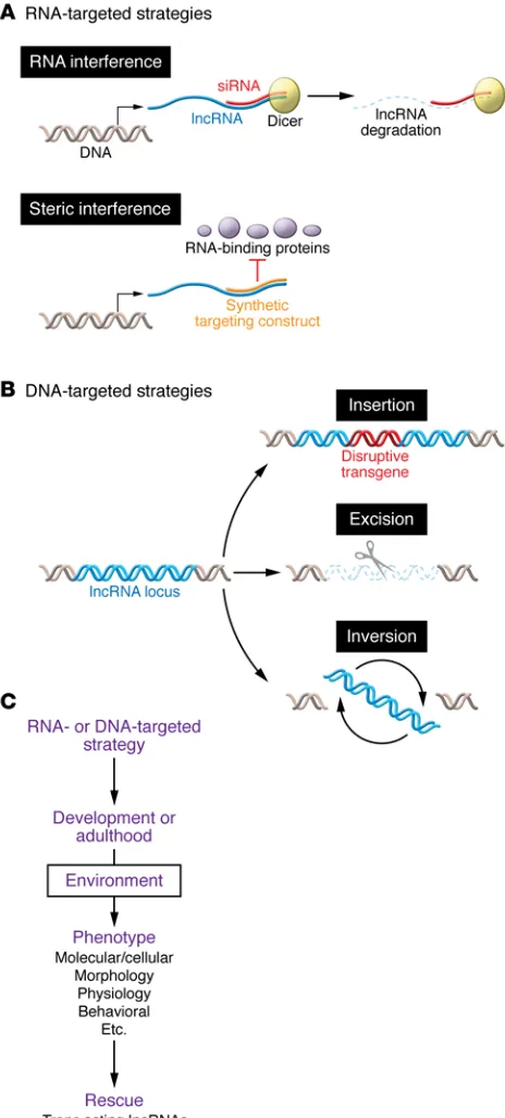
The function of a gene is commonly inferred by attenuating or ablat-ing its expression. Strategies to do so include physically interactablat-ing with its RNA (RNA-targeted approaches) or altering the underly-ing genetic locus (DNA-targeted approaches) (Figure 3, A and B). In animal models, these approaches may be applied at the zygotic stage, thereby modeling congenital conditions. They may also be applied to a specific cell population or during a defined time during development. Precise spatial and/or temporal control can refine the conclusions of a study or can circumvent deleterious developmen-tal effects, such as embryonic lethality. Although these approaches have been successfully applied to protein-coding genes, their use requires caution. We discuss the advantages and limitations of these approaches below, highlighting examples from the literature to illustrate the diverse functions attributed to lncRNAs in vivo.
RNA-targeted approaches and animal models. RNA-targeted
strategies introduce an exogenous RNA or an RNA analog that specifically binds to and functionally inactivates an endogenous lncRNA through complementary base pairing. Commonly used RNA-targeted approaches include RNAi and the use of morpholi-nos. These two approaches differ mechanistically. RNAi promotes lncRNA degradation, while morpholinos sterically hinder cellular processes, such as splicing, and prevent the formation or function of a mature lncRNA. Steric interference can also disrupt the bind-ing of a lncRNA to other macromolecules, preventbind-ing the forma-tion of funcforma-tional complexes (49).
[image:5.585.43.275.57.571.2]sion within grossly dissected tissue can reveal tissue specificity, although lncRNAs expressed in only a minority of cells may be undetectable. This limitation can be circumvented by assessing gene expression within individual cells (Figure 2B). This strate-gy may uncover rare or novel cells types contributing to diseases such as cancer, in which tumor relapses are thought to arise from a genetically transformed subpopulation of cells (65–67). Addition-ally, cellular populations labeled with a fluorescent reporter pro-tein can be dissociated into individual cells, which are then sorted on the basis of fluorescence detection and separately analyzed (Figure 2C). This strategy can increase the sensitivity of lncRNA detection and has revealed novel lncRNAs necessary for the
Figure 3. DNA- and RNA-targeted strategies and general workflow.
(also known as Neat2) can attenuate its expression by 80% (82). Although this approach was principally used to attenuate a patho-logical protein-coding RNA in a mouse model of myotonic dystro-phy type 1, the attenuation of Malat1 suggests that lncRNAs are potential targets for this condition and possibly other muscle dis-orders. However, only RNAs retained in the nucleus are sensitive to this approach (82), a feature well suited for targeting the abun-dance of nuclear lncRNAs (10). This work not only highlights the value of animal models in advancing novel therapeutic approach-es but also the feasibility of correcting pathological gene exprapproach-es- expres-sion, including that of lncRNAs.
DNA-targeted approaches and animal models. Strategies to
manipulate the genetic locus encoding a lncRNA, many of which have recently been reviewed (83), represent an alternative to RNA-targeted approaches. Transcription of both cis- and trans-act-ing lncRNAs can be eliminated, in contrast to the dose-dependent, posttranscriptional effects of RNA-targeted approaches. Further-more, if this ablation occurs in the germline, it is possible to gener-ate transgenic animals whose offspring constitutively lack a given lncRNA. These aspects of DNA-targeted approaches confer sever-al advantages compared with RNA-targeted approaches.
DNA-targeted approaches can facilitate expression profiling of a lncRNA if its locus is replaced with a reporter gene. This strat-egy was used in mice for 18 different lncRNA gene loci (84). For example, the lncRNA Peril was found to be expressed in discrete regions of the mouse brain and spinal cord, while Mdgt expression in mice appears restricted to the testes, brain, thymus, and colon (84). This methodology may also detect lncRNAs that are found only in a minority of cells. For example, the same study revealed that mouse Pantr2 (also known as linc-Brn1b) is expressed in select brain regions throughout cortical development and is selectively expressed in upper layers of the cortex in adulthood (84). This reporter strategy, therefore, provides sufficient resolu-tion to assess lncRNA expression, not only between tissues, but also within a tissue.
A spectrum of phenotypes has been reported following the ablation of a lncRNA by DNA-targeted approaches. For example, mice lacking Peril or Mdgt die perinatally with varying degrees of penetrance, demonstrating an essential, life-supporting role for both genes (84). Surviving Peril- and Mdgt-deficient mice are developmentally stunted, having smaller body sizes and reduced body weight compared with WT animals (84). A similar develop-mental phenotype was observed in mice lacking the lncRNA Pint (84). lncRNA ablation can also result in more subtle phenotypes. For example, deletion of Sra1 in mice improves obesity-related measures when animals are fed a high-fat diet (85), and deletion of Hotair in mice results in skeletal abnormalities of the vertebrae and wrist (86). Thus, DNA-targeted approaches have demonstrat-ed that lncRNAs may affect not only dramatic phenotypes but also more nuanced ones.
DNA-targeted approaches and genomic imprinting. Animal
mod-els of lncRNA function using DNA-targeted approaches have been instrumental in investigating the phenotypic effects of genomic imprinting, whereby gene expression is derived from a parent-spe-cific allele, while transcription from the other allele is epigeneti-cally repressed. Both viability and body weight are influenced by imprinted lncRNA expression. For example, deletion of maternal Zebrafish are an attractive animal model for investigating
lncRNAs, because morpholinos can be readily microinjected at the embryonic, one-cell stage to attenuate lncRNA function throughout development and into adulthood (72). In these ani-mals, the lncRNAs terminator, alien, and punisher are necessary for cardiovascular development, complementing similar inferences derived from cultured human and mouse cells (73). Additionally, attenuation of megamind (also known as tuna) in zebrafish impairs locomotion and disrupts brain development, while attenuation of
cyrano in zebrafish results in neural tube defects and dysmorphic
head and eyes (49). These results highlight the pronounced roles of lncRNAs in zebrafish development.
Mouse models are another valuable resource for under-standing lncRNA function in vivo. Transgenic mice engineered to constitutively overexpress an antisense RNA targeted to Sfta3 (also known as Nanci) have abnormal epithelial morphogenesis in their developing lungs (74). A more localized, tissue-specific interference of lncRNAs using RNAi can also have phenotypic consequences. shRNA-mediated downregulation of Miat (also known as Gomafu) in the medial prefrontal cortex of the brain promotes anxiety-like behaviors (75), while local siRNA-medi-ated downregulation of Munc, which is specifically expressed in skeletal muscle, impairs myogenesis (76). Additionally, the use of an shRNA to revert the upregulation of Arid2-IR in the mouse kidney following renal inflammation also reverts the biochemical signatures of this condition (77). Future work using mutant- and virus-mediated strategies will likely reveal additional phenotypes directly attributable to lncRNAs.
Limitations of RNA-targeted approaches. While informative,
these RNA-targeted approaches have important caveats. RNAi is effective for RNA exported to the cytoplasm but is relatively inef-ficient for RNAs residing in the nucleus (78), where many lncRNAs are found (10). This inefficiency may be particularly apparent for
cis-acting lncRNAs, which act near their site of origin and
there-fore may necessitate rapid binding for inactivation. This limitation may be circumvented by using RNA analogs, such as locked nucle-ic acids, whnucle-ich are better suited for nuclear targets because of their faster kinetics (79). However, sterically interfering RNA analogs, including both locked nucleic acids and morpholinos, cannot be genetically encoded, precluding the generation of transgenic animals. Furthermore, both strategies need sufficient expression levels to be effective, potentially inducing off-target effects and general toxicity within the cell. Conversely, insufficient levels for either strategy may only attenuate, rather than eliminate, lncRNA function and may not be sufficient to produce a phenotypic effect (80). Finally, binding of an exogenous RNA after transcription may not affect lncRNA function, because its transcription, per se, may have a biological effect. Despite these limitations, success-fully applied RNA-targeted approaches have revealed significant phenotypes following lncRNA dysfunction.
Clinical application of RNA-targeted approaches. Findings from
Meg-3 (also known as Gtl2) in mice results in perinatal death, an
effect not observed following paternal deletion (87). Similar-ly, ablation of maternal H19 in mice results in greater offspring body mass compared with that of offspring with paternal deletion (88, 89). Additionally, female mice lacking a single Tsix allele, a lncRNA implicated in X chromosome inactivation, produce fewer surviving offspring than do males lacking the same gene (90, 91). Finally, deletion of the X-linked Tsx in male mice results in smaller testes, reduced fear-related behaviors, and enhanced short-term memory (92). Collectively, these studies demonstrate diverse in vivo functions for imprinted lncRNAs.
Animal models of lncRNA function have also been instru-mental when investigating the complementary molecular mecha-nisms underlying genomic imprinting (93). For example, deletion of paternal Kcnq1ot1 (also known as KvDMR1) in mice results in offspring with reduced body mass and de-repression of proximal genes, an effect not observed after maternal deletion (94). These results were further refined following a more targeted ablation strategy in mice, in which a premature termination sequence was inserted downstream of the Kcnq1ot1 promoter (95). This alter-ation also resulted in gene de-repression, demonstrating that Kcn-q1ot1 transcription is necessary for gene silencing (95). Finally, complementary molecular studies have demonstrated that mouse
Kcnq1ot1 interacts with chromatin and epigenetic modifiers with
tissue specificity (96). This complementary use of animal mod-els to investigate lncRNA function highlights the utility of animal models when exploring lncRNA function in vivo.
Limitations of DNA-targeted approaches. DNA-targeted
approaches also have important limitations. Oftentimes, a large region encompassing the majority, if not all, of a lncRNA gene is removed, although smaller domain-specific (97) and promoter- specific (90, 94, 95) deletions may be possible. Insertion of a pre-mature termination sequence in the gene body may also ablate lncRNA function (98). These strategies differ from those used for protein-coding genes, whereby a single nucleotide deletion or insertion is often sufficient to abolish a protein product. These larg-er genomic altlarg-erations may introduce unintended and confound-ing consequences by removconfound-ing regulatory elements within the deleted region to affect the expression of neighboring genes (83, 99). For example, during embryonic stem cell differentiation in mice, overexpression of the lncRNA Haunt diminishes the expres-sion of the neighboring HOXA gene (100). Conversely, enhancer elements within the Haunt locus facilitate HOXA expression (100). These opposing influences within a locus may complicate efforts to alter a lncRNA locus without also perturbing regulatory elements embedded within that locus. Because of the extensive number of regulatory elements within both the human (101) and mouse (102) genomes, these concerns may be particularly acute and represent the norm rather than the exception. Rescue strat-egies in which the disrupted gene is experimentally reintroduced may control for these potentially confounding effects. However, this experimental design can only apply to trans-acting lncRNAs for which the integration site of the transgene is independent of its function. These limitations are important caveats to consider when interpreting data generated by DNA-targeted approaches.
Another important consideration is that different DNA-tar-geted approaches may result in different phenotypes. In mice,
replacing the lncRNA gene Fendrr with a reporter gene results in lung defects and perinatal death (84), a phenotype in agreement with clinical studies demonstrating that deletions within this locus in humans results in abnormal lung development and neo-natal death (103). In contrast, a second study demonstrated that insertion of a premature transcriptional termination sequence within the same mouse locus results in prenatal death, body wall abnormalities, and heart malfunction (80). These contrasting phenotypes exemplify how different DNA-targeting strategies may produce inconsistent phenotypes and highlight the fact that caution should be exercised in making premature functional con-clusions based on a single approach. Notably, this second study (80) also reported an attempted RNA-targeted strategy, in which a constitutively expressed antisense oligonucleotide resulted in a 60% reduction of Fendrr expression levels in mice that lacked any abnormal phenotype. Thus, three different strategies to manip-ulate lncRNA expression produced three different phenotypes, highlighting the challenges encountered when designing and interpreting lncRNA studies.
Interpretation of an absent phenotype. Loss of a lncRNA can
result in no discernible phenotype. Despite being highly con-served throughout mammalian evolution, ablation of the brain- expressed Linc0046 (also known as Visc-2) in mice does not result in any overt anatomical or behavioral phenotype (104). Similarly, three independent mouse strains lacking Malat1, which is high-ly expressed in the brain and liver, appear to develop normalhigh-ly (105–107). Finally, the loss of Neat1 in mice does not result in any overt phenotype, except for the loss of mammalian-specific nucle-ar subregions termed pnucle-araspeckles, where Neat1 is usually local-ized (108). Because paraspeckles are thought to reflect higher- order compartmentalization within the nucleus that is necessary for intricate regulation of mammalian gene expression, the absence of any obvious phenotype following the loss of Neat1 prompt-ed a critical reassessment of their function (109). Paraspeckles are induced following exposure to infectious diseases or cellular stressors, and it is possible that a phenotype in mice lacking Neat1 may only be apparent following an environmental manipulation, such as viral infection or exposure to microbes, that would normal-ly induce paraspeckles (108).
This possibility extends to all studies using genetic ablation in animal models. Gene-environment interactions may unmask unrecognized phenotypes that are important in understanding complex diseases such as psychiatric disorders with known envi-ronmental risk factors (110, 111). The mechanism of risk confer-ral for many of these environmental factors, such as exposure to environmental pathogens, maternal stress during pregnancy, or maternal substance abuse during pregnancy, may be investigated by using animal models in a controlled laboratory setting to reveal latent phenotypes and resolve gene-environment interactions (Figure 3C) (112–116).
pheno-types. Compensatory effects may further mitigate the appear-ance of a phenotype.
Alternative approaches. Overexpression of a lncRNA is an
alter-native strategy to assess its phenotypic effects. This approach has been successfully applied to animal models using protein-coding genes to study neuropsychiatric disorders that may result from a failure to maintain homeostasis after a gain or loss of gene expres-sion (117). When applied to lncRNAs, overexpresexpres-sion experiments may produce a phenotype opposite that occurring after lncRNA ablation, indicating bidirectional effects of lncRNA expression lev-els. Alternatively, they may induce a phenotype that is absent or seemingly unrelated to those observed following lncRNA ablation. However, this approach is only applicable for trans-acting lncRNAs, whose function is independent of their site of genomic integration.
Genome-editing technology. Major technological advances in
genome editing (118) open the possibility of altering the genomes not only of conventional model organisms, but also of less wide-ly used animal models that have historicalwide-ly been less amenable to alterations. TALENs or clustered regularly interspaced palin-dromic repeat (CRISPR/Cas9) methods have been widely used to excise or invert regions of a lncRNA locus in zebrafish (119, 120), mice (121, 122), and rats (123). These technologies may be extended to other animal models, adopted for specialized areas of investigation, such as the pig used to model cystic fibrosis (124), the songbird used to model language acquisition (125), and the nonhuman primate used to model the neurodegenerative disorder Huntington’s disease (126, 127). However, caveats and concerns similar to those previously discussed for standard DNA-targeted approaches still apply, given the comparatively large segments of
the genome that may need to be altered in order to affect lncRNA expression and function.
Conclusions
Animal models are promising tools for aiding in the discovery of novel lncRNAs and investigating the phenotypic significance of these molecules. However, lncRNAs inherently differ from their protein-coding counterparts, and hence their investigation requires overcoming a host of important new challenges and addressing new considerations with respect to experimental design and data interpretation. Despite these caveats, it has been possible to con-vincingly demonstrate that lncRNA dysfunction in animal models results in diverse phenotypes, ranging from lethality to subtle dys-morphia. So far, only a fraction of lncRNAs have been assessed, and this substantial gap in knowledge highlights a pressing need to progress beyond the initial cataloging of lncRNAs and to under-stand their impact in vivo. We anticipate that these initial find-ings represent a promising beginning to the diverse functions of lncRNAs in vivo and in understanding their relevance to disease.
Acknowledgments
We thank Andrew Holmes, Caroline Siebald, and members of the Goff laboratory for their helpful comments during the preparation of this manuscript.
Address correspondence to: Loyal A. Goff, Institute of Genetic Medicine, Miller Research Building, Room 449, 733 N. Broadway Ave., Baltimore, Maryland 21205, USA. Phone: 443.287.0251; E-mail: [email protected].
1. Okazaki Y, et al. Analysis of the mouse transcriptome based on functional annota-tion of 60,770 full-length cDNAs. Nature. 2002;420(6915):563–573.
2. Kapranov P, et al. RNA maps reveal new RNA classes and a possible function for pervasive tran-scription. Science. 2007;316(5830):1484–1488. 3. Djebali S, et al. Landscape of transcription in
human cells. Nature. 2012;489(7414):101–108. 4. St Laurent G, Wahlestedt C, Kapranov P. The
landscape of long noncoding RNA classification.
Trends Genet. 2015;31(5):239–251.
5. Cabili MN, et al. Integrative annotation of human large intergenic noncoding RNAs reveals global properties and specific subclasses. Genes Dev. 2011;25(18):1915–1927.
6. Guttman M, et al. Chromatin signature reveals over a thousand highly conserved large non-coding RNAs in mammals. Nature. 2009;458(7235):223–227.
7. Bono H, Kasukawa T, Furuno M, Hayashizaki Y, Okazaki Y. FANTOM DB: database of Functional Annotation of RIKEN Mouse cDNA Clones.
Nucleic Acids Res. 2002;30(1):116–118.
8. Harrow J, et al. GENCODE: the reference human genome annotation for The ENCODE Project.
Genome Res. 2012;22(9):1760–1774.
9. Iyer MK, et al. The landscape of long noncoding RNAs in the human transcriptome. Nat Genet. 2015;47(3):199–208.
10. Derrien T, et al. The GENCODE v7 catalog of
human long noncoding RNAs: analysis of their gene structure, evolution, and expression.
Genome Res. 2012;22(9):1775–1789.
11. Anderson DM, et al. A micropeptide encoded by a putative long noncoding RNA regulates muscle performance. Cell. 2015;160(4):595–606. 12. Nelson BR, et al. A peptide encoded by a
transcript annotated as long noncoding RNA enhances SERCA activity in muscle. Science. 2016;351(6270):271–275.
13. Keniry A, et al. The H19 lincRNA is a developmen-tal reservoir of miR-675 that suppresses growth and Igf1r. Nat Cell Biol. 2012;14(7):659–665. 14. Dey BK, Pfeifer K, Dutta A. The H19 long
non-coding RNA gives rise to microRNAs miR-675-3p and miR-675-5p to promote skeletal muscle differentiation and regeneration. Genes Dev. 2014;28(5):491–501.
15. Froberg JE, Yang L, Lee JT. Guided by RNAs: X-inactivation as a model for lncRNA function.
J Mol Biol. 2013;425(19):3698–3706.
16. Huarte M, et al. A large intergenic noncod-ing RNA induced by p53 mediates global gene repression in the p53 response. Cell. 2010;142(3):409–419.
17. Latos PA, et al. Airn transcriptional overlap, but not its lncRNA products, induces imprinted Igf2r silencing. Science. 2012;338(6113):1469–1472. 18. Simon MD, et al. The genomic binding sites
of a noncoding RNA. Proc Natl Acad Sci USA. 2011;108(51):20497–20502.
19. Chu C, Qu K, Zhong FL, Artandi SE, Chang HY. Genomic maps of long noncoding RNA occupan-cy reveal principles of RNA-chromatin interac-tions. Mol Cell. 2011;44(4):667–678. 20. Kretz M, et al. Control of somatic tissue
differ-entiation by the long non-coding RNA TINCR.
Nature. 2013;493(7431):231–235.
21. Wang Z, Tollervey J, Briese M, Turner D, Ule J. CLIP: construction of cDNA libraries for high-throughput sequencing from RNAs cross-linked to proteins in vivo. Methods. 2009;48(3):287–293.
22. Ule J, Jensen K, Mele A, Darnell RB. CLIP: a method for identifying protein-RNA interaction sites in living cells. Methods. 2005;37(4):376–386. 23. Hafner M, et al. Transcriptome-wide
identifica-tion of RNA-binding protein and microRNA tar-get sites by PAR-CLIP. Cell. 2010;141(1):129–141. 24. Spitale RC, et al. Structural imprints in vivo
decode RNA regulatory mechanisms. Nature. 2015;519(7544):486–490.
25. Lucks JB, et al. Multiplexed RNA structure characterization with selective 2’-hydroxyl acylation analyzed by primer extension sequenc-ing (SHAPE-Seq). Proc Natl Acad Sci USA. 2011;108(27):11063–11068.
26. Wapinski O, Chang HY. Long noncoding RNAs and human disease. Trends Cell Biol. 2011;21(6):354–361.
loci for human diseases and traits. Proc Natl Acad
Sci USA. 2009;106(23):9362–9367.
28. Gong J, Liu W, Zhang J, Miao X, Guo AY. lncRNASNP: a database of SNPs in lncRNAs and their potential functions in human and mouse. Nucleic Acids Res. 2015;43(Database issue):D181–D186.
29. Ishii N, et al. Identification of a novel non-cod-ing RNA, MIAT, that confers risk of myocardial infarction. J Hum Genet. 2006;51(12):1087–1099. 30. Lai F, et al. Activating RNAs associate with
Mediator to enhance chromatin architecture and transcription. Nature. 2013;494(7438):497–501. 31. van Dijk M, et al. HELLP babies link a novel lin-cRNA to the trophoblast cell cycle. J Clin Invest. 2012;122(11):4003–4011.
32. Maass PG, et al. A misplaced lncRNA caus-es brachydactyly in humans. J Clin Invcaus-est. 2012;122(11):3990–4002.
33. Du Z, et al. Integrative genomic analyses reveal clinically relevant long noncoding RNAs in human cancer. Nat Struct Mol Biol. 2013;20(7):908–913.
34. Gupta RA, et al. Long non-coding RNA HOTAIR reprograms chromatin state to promote cancer metastasis. Nature. 2010;464(7291):1071–1076. 35. Trimarchi T, et al. Genome-wide mapping
and characterization of Notch-regulated long noncoding RNAs in acute leukemia. Cell. 2014;158(3):593–606.
36. Wilson WR, Hay MP. Targeting hypoxia in cancer therapy. Nat Rev Cancer. 2011;11(6):393–410. 37. Yang F, et al. Repression of the long noncoding
RNA-LET by histone deacetylase 3 contributes to hypoxia-mediated metastasis. Mol Cell. 2013;49(6):1083–1096.
38. Yang F, Zhang H, Mei Y, Wu M. Reciprocal regu-lation of HIF-1α and lincRNA-p21 modulates the Warburg effect. Mol Cell. 2014;53(1):88–100. 39. Takahashi K, Yan IK, Haga H, Patel T.
Modula-tion of hypoxia-signaling pathways by extracellu-lar linc-RoR. J Cell Sci. 2014;127(Pt 7):1585–1594. 40. Naora H, Montell DJ. Ovarian cancer metastasis: integrating insights from disparate model organ-isms. Nat Rev Cancer. 2005;5(5):355–366. 41. Sharpless NE, Depinho RA. The mighty mouse:
genetically engineered mouse models in can-cer drug development. Nat Rev Drug Discov. 2006;5(9):741–754.
42. Khanna C, Hunter K. Modeling metastasis in vivo. Carcinogenesis. 2005;26(3):513–523. 43. Tupy JL, et al. Identification of putative
non-coding polyadenylated transcripts in Dro-sophila melanogaster. Proc Natl Acad Sci USA. 2005;102(15):5495–5500.
44. Young RS, et al. Identification and properties of 1,119 candidate lincRNA loci in the Drosoph-ila melanogaster genome. Genome Biol Evol. 2012;4(4):427–442.
45. Graveley BR, et al. The developmental tran-scriptome of Drosophila melanogaster. Nature. 2011;471(7339):473–479.
46. Brown JB, et al. Diversity and dynamics of the Drosophila transcriptome. Nature. 2014;512(7515):393–399.
47. Nam J-W, Bartel DP. Long noncoding RNAs in C. elegans. Genome Res. 2012;22 (12):2529–2540. 48. Pauli A, et al. Systematic identification of long
noncoding RNAs expressed during zebrafish embryogenesis. Genome Res. 2012;22(3):577–591. 49. Ulitsky I, Shkumatava A, Jan CH, Sive H, Bartel
DP. Conserved function of lincRNAs in verte-brate embryonic development despite rapid sequence evolution. Cell. 2011;147(7):1537–1550. 50. Hezroni H, Koppstein D, Schwartz MG, Avrutin
A, Bartel DP, Ulitsky I. Principles of long non-coding RNA evolution derived from direct com-parison of transcriptomes in 17 species. Cell Rep. 2015;11(7):1110–1122.
51. Ponjavic J, Ponting CP, Lunter G. Functionality or transcriptional noise? Evidence for selection within long noncoding RNAs. Genome Res. 2007;17(5):556–565.
52. Washietl S, Kellis M, Garber M. Evolutionary dynamics and tissue specificity of human long noncoding RNAs in six mammals. Genome Res. 2014;24(4):616–628.
53. Kutter C, et al. Rapid turnover of long noncoding RNAs and the evolution of gene expression. PLoS
Genet. 2012;8(7):e1002841.
54. Babak T, Blencowe BJ, Hughes TR. A systematic search for new mammalian noncoding RNAs indicates little conserved intergenic transcrip-tion. BMC Genomics. 2005;6:104.
55. Wang J, et al. Mouse transcriptome: neutral evolution of ‘non-coding’ complementary DNAs. Nature. 2004;431(7010):doi:10.1038/ nature03016.
56. Necsulea A, et al. The evolution of lncRNA rep-ertoires and expression patterns in tetrapods.
Nature. 2014;505(7485):635–640.
57. Washietl S, Hofacker IL, Lukasser M, Hütten-hofer A, Stadler PF. Mapping of conserved RNA secondary structures predicts thousands of func-tional noncoding RNAs in the human genome.
Nat Biotechnol. 2005;23(11):1383–1390.
58. Fontana L, Partridge L, Longo VD. Extending healthy life span--from yeast to humans. Science. 2010;328(5976):321–326.
59. Essers PB, et al. A long noncoding rna on the ribo-some is required for lifespan extension. Cell Rep. 2015;10(3):339–345.
60. St Johnston D. The art and design of genetic screens: Drosophila melanogaster. Nat Rev Genet. 2002;3(3):176–188.
61. Koob MD, et al. An untranslated CTG expansion causes a novel form of spinocerebellar ataxia (SCA8). Nat Genet. 1999;21(4):379–384. 62. Mutsuddi M, Marshall CM, Benzow KA, Koob
MD, Rebay I. The spinocerebellar ataxia 8 noncoding RNA causes neurodegeneration and associates with staufen in Drosophila. Curr Biol. 2004;14(4):302–308.
63. Ramaswami M, Taylor JP, Parker R. Altered ribostasis: RNA-protein granules in degenerative disorders. Cell. 2013;154(4):727–736.
64. Molyneaux BJ, et al. DeCoN: genome-wide anal-ysis of in vivo transcriptional dynamics during pyramidal neuron fate selection in neocortex.
Neuron. 2015;85(2):275–288.
65. Shapiro E, Biezuner T, Linnarsson S. Single-cell sequencing-based technologies will revolu-tionize whole-organism science. Nat Rev Genet. 2013;14(9):618–630.
66. Ding L, et al. Clonal evolution in relapsed acute myeloid leukaemia revealed by whole-genome
sequencing. Nature. 2012;481(7382):506–510. 67. Magrangeas F, et al. Minor clone provides a
reser-voir for relapse in multiple myeloma. Leukemia. 2013;27(2):473–481.
68. Aprea J, et al. Transcriptome sequencing during mouse brain development identifies long non-cod-ing RNAs functionally involved in neurogenic commitment. EMBO J. 2013;32(24):3145–3160. 69. Barry G, et al. The long non-coding RNA
Goma-fu is acutely regulated in response to neuronal activation and involved in schizophrenia-as-sociated alternative splicing. Mol Psychiatry. 2014;19(4):486–494.
70. Albertson DN, Schmidt CJ, Kapatos G, Bannon MJ. Distinctive profiles of gene expression in the human nucleus accumbens associated with cocaine and heroin abuse.
Neuropsychopharma-cology. 2006;31(10):2304–2312.
71. Michelhaugh SK, Lipovich L, Blythe J, Jia H, Kapa-tos G, Bannon MJ. Mining Affymetrix microarray data for long non-coding RNAs: altered expres-sion in the nucleus accumbens of heroin abusers.
J Neurochem. 2011;116(3):459–466.
72. Nasevicius A, Ekker SC. Effective targeted gene ‘knockdown’ in zebrafish. Nat Genet. 2000;26(2):216–220.
73. Kurian L, et al. Identification of novel long noncod-ing RNAs underlynoncod-ing vertebrate cardiovascular development. Circulation. 2015;131(14):1278–1290. 74. Herriges MJ, et al. Long noncoding RNAs are
spatially correlated with transcription factors and regulate lung development. Genes Dev. 2014;28(12):1363–1379.
75. Spadaro PA, et al. Long noncoding RNA-directed epigenetic regulation of gene expression is asso-ciated with anxiety-like behavior in mice. Biol
Psychiatry. 2015;78(12):848–859.
76. Mueller AC, et al. MUNC, a long noncoding RNA that facilitates the function of MyoD in skeletal myogenesis. Mol Cell Biol. 2015;35(3):498–513. 77. Zhou Q, Huang XR, Yu J, Yu X, Lan HY. Long
noncoding RNA Arid2-IR is a novel therapeu-tic target for renal inflammation. Mol Ther. 2015;23(6):1034–1043.
78. Zeng Y, Cullen BR. RNA interference in human cells is restricted to the cytoplasm. RNA. 2002;8(7):855–860.
79. Sarma K, Levasseur P, Aristarkhov A, Lee JT. Locked nucleic acids (LNAs) reveal sequence requirements and kinetics of Xist RNA localiza-tion to the X chromosome. Proc Natl Acad Sci
USA. 2010;107(51):22196–22201.
80. Grote P, et al. The tissue-specific lncRNA Fendrr is an essential regulator of heart and body wall development in the mouse. Dev Cell. 2013;24(2):206–214.
81. Bennett CF, Swayze EE. RNA targeting therapeu-tics: molecular mechanisms of antisense oligo-nucleotides as a therapeutic platform. Annu Rev
Pharmacol Toxicol. 2010;50:259–293.
82. Wheeler TM, et al. Targeting nuclear RNA for in vivo correction of myotonic dystrophy. Nature. 2012;488(7409):111–115.
83. Li L, Chang HY. Physiological roles of long non-coding RNAs: insight from knockout mice. Trends
Cell Biol. 2014;24(10):594–602.
brain development. Elife. 2013;2:e01749. 85. Liu S, et al. SRA gene knockout protects against
diet-induced obesity and improves glucose toler-ance. J Biol Chem. 2014;289(19):13000–13009. 86. Li L, et al. Targeted disruption of Hotair leads to
homeotic transformation and gene derepression.
Cell Rep. 2013;5(1):3–12.
87. Zhou Y, et al. Activation of paternally expressed genes and perinatal death caused by deletion of the Gtl2 gene. Development. 2010;137(16):2643–2652. 88. Leighton PA, Ingram RS, Eggenschwiler J,
Efstra-tiadis A, Tilghman SM. Disruption of imprinting caused by deletion of the H19 gene region in mice. Nature. 1995;375(6526):34–39.
89. Ripoche MA, Kress C, Poirier F, Dandolo L. Dele-tion of the H19 transcripDele-tion unit reveals the exis-tence of a putative imprinting control element.
Genes Dev. 1997;11(12):1596–1604.
90. Lee JT. Disruption of imprinted X inactiva-tion by parent-of-origin effects at Tsix. Cell. 2000;103(1):17–27.
91. Sado T, Wang Z, Sasaki H, Li E. Regulation of imprinted X-chromosome inactivation in mice by Tsix. Development. 2001;128(8):1275–1286. 92. Anguera MC, Ma W, Clift D, Namekawa S, Kelleher RJ, Lee JT. Tsx produces a long non-coding RNA and has general functions in the germline, stem cells, and brain. PLoS Genet. 2011;7(9):e1002248.
93. Kanduri C. Long noncoding RNAs: lessons from genomic imprinting. Biochim Biophys Acta. 2016;1859(1):102–111.
94. Fitzpatrick GV, Soloway PD, Higgins MJ. Regional loss of imprinting and growth deficiency in mice with a targeted deletion of KvDMR1. Nat Genet. 2002;32(3):426–431.
95. Mancini-Dinardo D, Steele SJ, Levorse JM, Ingram RS, Tilghman SM. Elongation of the Kcnq1ot1 transcript is required for genomic imprinting of neighboring genes. Genes Dev. 2006;20(10):1268–1282.
96. Pandey RR, et al. Kcnq1ot1 antisense noncoding RNA mediates lineage-specific transcriptional silencing through chromatin-level regulation.
Mol Cell. 2008;32(2):232–246.
97. Mohammad F, Mondal T, Guseva N, Pandey GK, Kanduri C. Kcnq1ot1 noncoding RNA mediates transcriptional gene silencing by interacting with Dnmt1. Development. 2010;137(15):2493–2499. 98. Sleutels F, Zwart R, Barlow DP. The non-coding Air
RNA is required for silencing autosomal imprinted genes. Nature. 2002;415(6873):810–813. 99. Bassett AR, et al. Considerations when
investigating lncRNA function in vivo. Elife. 2014;3:e03058.
100. Yin Y, et al. Opposing roles for the lncRNA haunt and its genomic locus in regulating HOXA gene activation during embryonic stem cell differenti-ation. Cell Stem Cell. 2015;16(5):504–516. 101. ENCODE Project Consortium. An integrated
encyclopedia of DNA elements in the human genome. Nature. 2012;489(7414):57–74. 102. Yue F, et al. A comparative encyclopedia of
DNA elements in the mouse genome. Nature. 2014;515(7527):355–364.
103. Szafranski P, et al. Small noncoding differentially methylated copy-number variants, including lncRNA genes, cause a lethal lung developmental disorder. Genome Res. 2013;23(1):23–33. 104. Oliver PL, et al. Disruption of Visc-2, a brain-
expressed conserved long noncoding RNA, does not elicit an overt anatomical or behavioral phe-notype. Cereb Cortex. 2015;25(10):3572–3585. 105. Eißmann M, et al. Loss of the abundant
nuclear non-coding RNA MALAT1 is com-patible with life and development. RNA Biol. 2012;9(8):1076–1087.
106. Zhang B, et al. The lncRNA Malat1 is dispensable for mouse development but its transcription plays a cis-regulatory role in the adult. Cell Rep. 2012;2(1):111–123.
107. Nakagawa S, et al. Malat1 is not an essential component of nuclear speckles in mice. RNA. 2012;18(8):1487–1499.
108. Nakagawa S, Naganuma T, Shioi G, Hirose T. Paraspeckles are subpopulation-specific nuclear bodies that are not essential in mice. J Cell Biol. 2011;193(1):31–39.
109. Nakagawa S, Hirose T. Paraspeckle nuclear bodies — useful uselessness? Cell Mol Life Sci. 2012;69(18):3027–3036.
110. Rutter M. Environmentally mediated risks for psychopathology: research strategies and findings. J Am Acad Child Adolesc Psychiatry. 2005;44(1):3–18.
111. Caspi A, Moffitt TE. Gene-environment interac-tions in psychiatry: joining forces with neurosci-ence. Nat Rev Neurosci. 2006;7(7):583–590. 112. Nestler EJ, Hyman SE. Animal models of
neuropsychiatric disorders. Nat Neurosci. 2010;13(10):1161–1169.
113. Cryan JF, Holmes A. The ascent of mouse: advances in modelling human depression and anxiety. Nat Rev Drug Discov. 2005;4(9):775–790. 114. Holmes A, Wellman CL. Stress-induced
prefrontal reorganization and executive
dys-function in rodents. Neurosci Biobehav Rev. 2009;33(6):773–783.
115. Adamec R, Holmes A, Blundell J. Vulnerability to lasting anxiogenic effects of brief exposure to predator stimuli: sex, serotonin and other fac-tors-relevance to PTSD. Neurosci Biobehav Rev. 2008;32(7):1287–1292.
116. Millstein RA, Holmes A. Effects of repeated maternal separation on anxiety- and depres-sion-related phenotypes in different mouse strains. Neurosci Biobehav Rev. 2007;31(1):3–17. 117. Ramocki MB, Zoghbi HY. Failure of neuronal
homeostasis results in common neuropsychiatric phenotypes. Nature. 2008;455(7215):912–918. 118. Hsu PD, Lander ES, Zhang F. Development and
applications of CRISPR-Cas9 for genome engi-neering. Cell. 2014;157(6):1262–1278. 119. Liu Y, Luo D, Zhao H, Zhu Z, Hu W, Cheng CH.
Inheritable and precise large genomic deletions of non-coding RNA genes in zebrafish using TALENs. PLoS ONE. 2013;8(10):e76387. 120. Xiao A, et al. Chromosomal deletions and
inver-sions mediated by TALENs and CRISPR/Cas in zebrafish. Nucleic Acids Res. 2013;41(14):e141. 121. Han J, et al. Efficient in vivo deletion of a large
imprinted lncRNA by CRISPR/Cas9. RNA Biol. 2014;11(7):829–835.
122. Welsh IC, et al. Chromatin architecture of the Pitx2 locus requires CTCF- and Pitx2-dependent asymmetry that mirrors embryonic gut laterality.
Cell Rep. 2015;13(2):337–349.
123. Cheng X, et al. A CRISPR/Cas9 system based targeted disruption of a novel rat long non-cod-ing RNA in a rat genetic model of hypertension.
FASEB J. 2015;29(1 Supplement). http://www.
fasebj.org/content/29/1_Supplement/814.1. 124. Rogers CS, et al. Disruption of the CFTR gene
produces a model of cystic fibrosis in newborn pigs. Science. 2008;321(5897):1837–1841. 125. Agate RJ, Scott BB, Haripal B, Lois C, Nottebohm
F. Transgenic songbirds offer an opportunity to develop a genetic model for vocal learning. Proc
Natl Acad Sci USA. 2009;106(42):17963–17967.
126. Yang SH, et al. Towards a transgenic model of Huntington’s disease in a non-human primate.
Nature. 2008;453(7197):921–924.
127. Sasaki E, et al. Generation of transgenic non- human primates with germline transmission.
Nature. 2009;459(7246):523–527.