Journal of Chemical and Pharmaceutical Research, 2015, 7(7):564-569
Research Article
ISSN : 0975-7384
CODEN(USA) : JCPRC5
Expression and immunogenicity of H glycoprotein gene of canine distemper
virus cloned in eukaryotic expression vector
Nitin Sharma*, Usha Tiwari, Manoj Kalakoti, Neha K. Mehra and Pragya Pandey
Division of Animal Cell Culture Laboratory, Institute of Biotechnology, G.B. Pant University of Agriculture & Technology, Patwadangar (Nainital), UK, India
_____________________________________________________________________________________________
ABSTRACT
Canine distemper virus H gene is immunogenic and induces neutralizing antibodies against Canine distemper virus (CDV). In the present study the recombinant plasmid pSin.cdv.h transfected HeLa cell expressed H protein which was confirmed by immunoperoxidase test. The in vivo immunological response was studied in dogs immunized through IM route. The immune response on 30th day post vaccination induced by 4µg dose of rplasmid revealed protective serum neutralizing antibody titer 120, while control group did not show any antibody titer against CDV. Stimulation Index (SI) was found higher in vaccinated group as compared to healthy control animals. The T-cell response in the blood of vaccinated dogs suggested the role of cell mediated immunity in protection apart from neutralizing antibodies in serum. The challenge test revealed 100% protection of vaccinated dogs while all control dogs died of cdv infection. Therefore, this study demonstrates that DNA vaccination with 4µg dose of pSin.cdv.h can provide protective immunity 30 days post vaccination.
Keywords: Canine distemper virus, L.B. medium, E.coli (DH5α) strain, Ampicilline antibiotic, Dogs
_____________________________________________________________________________________________
INTRODUCTION
Canine distemper virus is 150 to 300 nm in diameter. CDV wild type isolates from (seals, a German dog a ferret and large fields) are significantly different from CDV vaccine strains [1, 2]. Canine distemper is one among the infectious diseases of dogs caused by virus canine distemper (CDV) which affects pups of age group mostly between 2 to 6 months old. The mortality is between 30 to 80% and the recovery from the disease depends on the immune status of the animal [3]. Many killed and live modified vaccines against CDV are in use [4]. CDV belongs to family Paramyxoviridae, under genus Morbillivirus [5]. CDV genome is 15,690 nucleotides in length and consists of a short 3’ leader region and six genes encoding the N, P, M, F, H, and L proteins [6-11].
EXPERIMENTAL SECTION
1.1.Recombinant plasmid (pSin.cdvh)
The recombinant plasmid pSin.cdvh was available in the Biotechnology Laboratory, IBIT, Bareilly. pSin vector contains replicase gene which produces thousand copies of gene insert mRNA.
1.2. Cell Line
______________________________________________________________________________
used for peroxidase based serum neutralization test. HeLa cell line was used in the study for in vitro expression analysis of recombinant plasmids (pSin.cdvh) and was maintained in GMEM (Micro lab), supplemented with 10% new born calf serum (Gibco, NY),100 µg/ml penicillin and streptomycin.
1.3. Virus
Virulent canine distemper virus as well as BHK21 cell culture adapted CDV were available in the laboratory.
1.4. Conjugates and Hyperimmune serum
Direct polyclonal antibody conjugate for CDV was purchased from VMRD, Pullman, USA. Hyperimmune sera against H gene were raised in mice using recombinant plasmid. The recombinant plasmid (50 µg/mouse) was administered four times at interval of one week, to six mice. The blood was collected from eye of the mouse and serum was harvested.
1.5. In vitro expression analysis 1.5.1. Transfection of HeLa cells
Cells were trypsinised using trypsin-versenate solution (TVS) and then 4 ml of GMEM containing 10% FCS and penicillin and streptomycin (50 µl/ml) was added, to make cell suspension of 1X10⁵ cells/ml. Harvested exponentially growing cells by trypsinization and prepared cell suspension in growth medium. Prepared the calcium phosphate-DNA coprecipitate as follows: combined 50 µl of 2.5M CaCl2 with 10 µl of plasmid DNA in a sterile
microfuge tube, Added 40 µl DW, kept at room temperature.
Immediately transferred the calcium phosphate-DNA suspension using 20 µl suspension for each wells of microtitre plate. Added 100 µl of cell culture suspension in each wells of 96 wells microtitter plate. Rocked the plate gently to mix the medium, which will become yellow-orange and turbid. Carried out this step as quickly as possible because the efficiency of transfection declines rapidly once the DNA precipitate is formed. Kept control wells without transfection. Incubated at 370C in a humidified incubator with an atmosphere of 5% CO2 for 72 hours. Examined for
gene expression by IPT.
1.5.2. Raising primary antibody against pSin.cdvh in mice
Primary polyclonal antibody against the CDV H gene was raised in mouse by hyper immunization of six mice with pSin.cdvh plasmid. 50µl of plasmid DNA was injected IM rout in lateral region of thigh muscles of each mouse and repeated every week for four weeks consecutively. Mice were bled through inner canthus of eyes with a capillary and serum was prepared.
1.5.3. Immunoperoxidase test (IPT)
After 72 hours, transfected HeLa cells were washed with 1XPBS twice and fixed with 80% chilled acetone at 40C for 10 min and air-dried. Put a few drops of CDV H hyperimmune serum and incubated at 370C for 1 hr and again washed with PBS, added a few drops of horseradish peroxidase (HRPO) conjugated rabbit anti-mouse antibody to wells and incubated at 370C for 1 hr in humid chamber. The cells were again washed with PBS thrice and incubated with 2-3 drops of Nadi reagent for 5 min. After the development of color, cells were washed with PBS, dried in air and observed under microscope and photographed, protocol was carried out as per [12].
1.6. Immune response studies 1.6.1. Immunization of dogs
The in vivo experimental setup was designed with sixteen one month age old pups. One week before the start of immunization trial, all pups were treated with antihelminthics (Albendazole). These dogs were then tested for seronegativity against canine distemper virus. Dogs of group 1 were injected interamuscularly with 4 µg recombinant plasmid (pSin.cdv.H) in nuclease free water, and group 2 were injected intramuscularly 6 µg vector alone (pSin) in nuclease free water, while group 3 were kept as health control.
1.6.2. Collection of blood and serum samples
______________________________________________________________________________
1.6.3. Serum neutralization test (SNT) using NPLA
The neutralizing antibodies in the blood were measured through neutralizing peroxidase- linked assay (NPLA) [13].
1.6.4. Lymphocyte proliferation assay
Whole blood was collected as per the method described earlier in EDTA coated vial. 4 ml of lymphocyte separation medium (LPM 1077, PAA) was taken in a 15 ml conical tube. Equal volume of whole blood sample was carefully layered over the LSM. The tubes were centrifuged at 1800 rpm for 30 min at 40C. Lymphocytes were collected from the plasma-LSM interface and washed twice with PBS at 1200 rpm for 10 min and finally the cell pellet was resuspended in 2 ml of RPMI-1640 growth medium (Sigma) without having phenol red and supplemented with 10% FCS. The cell count and viability was determined by Trypan-blue dye exclusion method [14]. The blastogenic response of lymphocytes was assessed by MTT colorimetry method [15].
RESULTS AND DISCUSSION
Canine distemper (CD) is highly contagious disease of dogs characterized by fever, conjunctivitis, discharge from the eyes, bronchitis with coughing nasal discharge and difficult breathing, gastroenteritis with diarrhea, vomiting and slight rash. When the disease progresses to central nervous system it results in neurological disorders with the signs of in-coordinated gait, head pressing, chorea (muscles tremors), seizures and nystagmus (uncontrolled eye movements).
Among the six proteins of canine distemper namely H, N, P, M, L and F, the highest antigenic variation is found in the H protein, whereas F and P proteins are affected to a much lower extent [16]. As the H protein’s primary function is cell receptor binding, its characteristic is therefore most closely related to the effective infectivity and host specificity of the virus. The protective efficacy of anti H monoclonal antibodies is higher than that of anti F mAbs [17]. So in our study we have used CDVH gene of Lederle strain to induce immunity against canine distemper.
1.7. Expression of recombinant plasmid
The expression ability of recombinant plasmids was checked by immunoperoxidase test (IPT) in HeLa cell line and cells were found to express the protein by development of purple color. Intense purple coloration of cells was observed in which HeLa cells were transfected with pSin.cdv.h (Fig.1), while healthy cell control did not show any color change.
Fig.1. HeLa cells transfected with recombinant plasmid pSin.cdv.H and analysed with IPT showing purple color
1.8. Immunological response of pSin.CDV.H in dogs 1.8.1. Serum neutralizing antibody response
______________________________________________________________________________
Table 1. Antibody titer of dogs after immunization
Sl. No. Groups No. of dogs SN* antibody titer on 30th day post immunization 1. Vaccinated (pSin.cdv.H) (4 µg) 4 120
2. Vector alone (pSin) (6 µg) 4 0 3. Healthy control 4 0
*SN titer was calculated as reciprocal of highest dilution of serum able to neutralize 50% of cdv in culture replicates.
1.8.2. Lymphoproliferative assay on PBMCs of immunized dogs
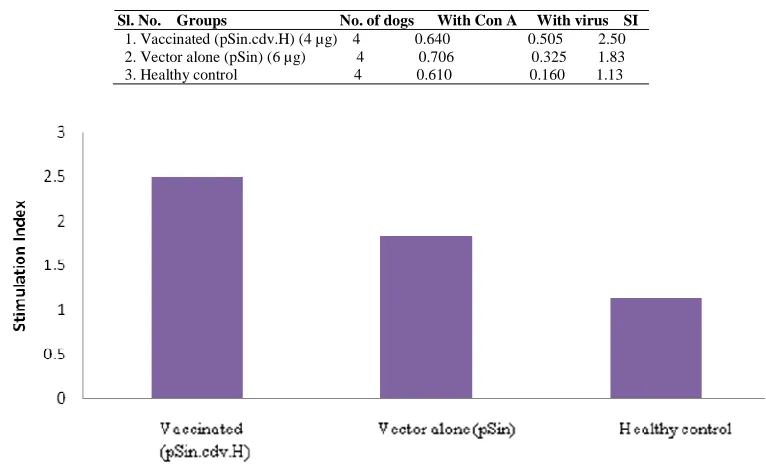
Dogs immunized with 4 µg of pSin.cdv.H shown stimulation index (SI) of 2.50 at 30th days of immunization with cell culture adapted CDV. Unstimulated samples from each group maintained maximum absorbance (A550) near
[image:4.595.107.490.259.492.2]about 0.6. The SI (with virus) showed an increasing trend from control groups to vaccinated groups. The results obtained are presented in the (Table 2) and (Fig.2).
Table 2. Antigen specific response of lymphocytes at 30th day post immunization as assessed by MTT assay
Sl. No. Groups No. of dogs With Con A With virus SI 1. Vaccinated (pSin.cdv.H) (4 µg) 4 0.640 0.505 2.50 2. Vector alone (pSin) (6 µg) 4 0.706 0.325 1.83 3. Healthy control 4 0.610 0.160 1.13
Fig. 2. In vitro lymphoproliferative responses of dogs on 30th days post immunization
1.9. Protection of dogs against CDV challenge
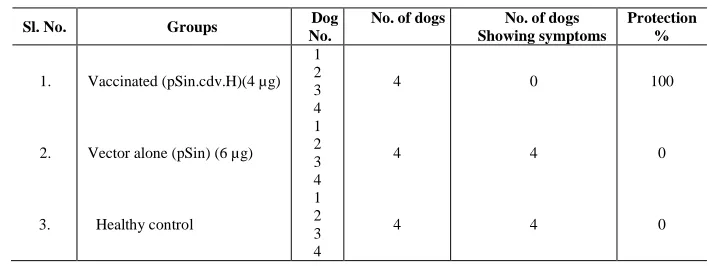
Thirty days after immunization with pSin.cdv.H, all 4 vaccinated dogs belonging to group no. A completely resisted the challenge of 105 ID50 virulent CDV.
The 4 dogs of vector control and also 4 dogs of healthy control group showed severe symptoms of disease such as fever, gastroenteritis with diarrhea, vomiting and conjunctivitis observed between 10-15 days post challenge (Table 3).
______________________________________________________________________________
Table 3. Protection test of dogs challenged with 105ID50 of CDV
Sl. No. Groups Dog
No.
No. of dogs No. of dogs Showing symptoms Protection % 1. 2. 3.
Vaccinated (pSin.cdv.H)(4 µg)
Vector alone (pSin) (6 µg)
Healthy control 1 2 3 4 1 2 3 4 1 2 3 4 4 4 4 0 4 4 100 0 0
The result of the present study would be useful for the development of DNA vaccine against CDV disease. Studies are required to test pSin.CDV.H developed in inducing protective immune response in dogs. The 4 µg dose of pSin.cdv.H with regard to their immune response as assessed on 30th day post vaccination by lymphoproliferative test and stimulation index, SN antibody titer and was found to confer total protection 30th days post vaccination through intramuscular route. Thus, study confirmed that 4 µg recombinant plasmid was optimum for induction of immunity and protection in dogs.
Acknowledgement
Authors thank Director, IVRI and also thank Director, IBIT Izatnagar, Bareilly for providing facilities to carry out this work.
REFERENCES
[1] Mamaev L, Denikina N, Belikov S, Volchkov V, Visser I, Fleming M, Kai C, Harder T, Liess B, Osterhaus A. Vet. Microbiol., 1995, 44, 251-259.
[2] Harder T, Kenter M, Vos H, Siebelink K, Huisman W, van Amerongen G, Orvell C, Barrett T, Appel M, Osterhaus A. J. Gen. Virol., 1996, 77, 397-405.
[3] Fenner F, Bachmann PA, Gibbs EPJ, Murphy FA, Studdert MJ and White DO. Veterinary Virology. edition F. Fenner, Academic Press, Inc., New York, 1987, 488-503.
[4] Bolt G, Jensen T, Gottschalck E, Arctander P, Appel M, Buckland R and Blixenkrone-Moller M. J Gen Virol, 1997, 78( 2), 367-372.
[5] Appel M and Summers BA. Canine distemper: current status in L.E. Carmichael edition. Recent advances in canine infectious diseases. International Veterinary Information Service, Ithaca, N.Y, 1999.
[6] Barrett T, Clarke DK, Evans SA and Rima BK. Virus Res., 1987, 8, 373-386.
[7] Barrett T, Subbarao M, Belsham G, Mahy B. In The Paramyxoviruses edition by D. Kingsbury New York Plenum Press, New York, 1991, 82-102.
[8] Bellini WJ, Englund G, Richardson CD, Rozenblatt S, Lazzarini RA. J. Virol., 1986, 58, 408-416. [9] Curran MD, Clarke DK and Rima BK. J. Gen. Virol., 1991, 72, 443-447.
[10] Rozenblatt S, Eizenberg O, Euglund G and Bellini WJ. J. Virol., 1985, 53, 691-694. [11] Sidhu MS, Husar W, Cook SD, Dowling PC and Udem SA. Virology, 1993, 193, 66-72. [12] Nakane PK, Kawaoi A. J. Histochem. Cytochem., 1974, 22, 1084.
[13] Terpstra C, Bloemraad M, Gielkens AJL. Veterinary Microbiology, 1984, 9, 113-120.
[14] Freshney R, IAN. A manual of basic technique 5th edition John Wiley and Sons, New Jersey, 2006, 360-362. [15] Mosmann T J. Immunology Method, 1983, 65, 55-63.
[16] Blixenkrone-Moeller M, Svansson V, Have P, Orvell C, Appel MJG, Pedersen IR, Dietz HH and Henriksen P. Vet. Microbiol., 1993, 37, 163-173.
[17] Hirayama N, Senda M, Nakashima N, Takagi M, Sugiyama M, Yoahikawa Y and Yamanouchi K. J. Gen. Virol., 1991, 72, 2827-2830.
[18] Xiong C, Levis R, Shen P, Schlesinger S, Rice CM and Huang HV. Science, 1989, 243, 1188-1191.
[19] Hariharan MJ, Driver DA, Townsend K, Brumm D, Polo JM, Belli BA, Catton DJ, Hsu D, Mittelstaedt D, McCormack JE, Karavodin J, Dubensky TW, Chang SMJr. and Banks TA. J. Virol., 1998, 72, 950-958.
______________________________________________________________________________
[21] Sharma N, Kaushik P, Rai A, Maurya R, Pal P, Tiwari U. International J. Applied Bio. Pharma. Technol., 2010, 1, 339-348.