0095-1137/09/$08.00
⫹
0
doi:10.1128/JCM.01899-08
Copyright © 2009, American Society for Microbiology. All Rights Reserved.
Development and Validation of a Microsphere-Based Luminex Assay
for Rapid Identification of Clinically Relevant Aspergilli
䌤
Kizee A. Etienne,
1Rui Kano,
1,2and S. Arunmozhi Balajee
1*
Mycotic Disease Branch, Centers for Disease Control and Prevention, Atlanta, Georgia,
1and Department of Pathobiology,
Nihon University School of Veterinary Medicine, 1866 Kameino, Fuzisawa-city, Kanagawa 252-8510, Japan
2Received 1 October 2008/Returned for modification 23 January 2009/Accepted 13 February 2009
A Luminex-based assay for the rapid identification of
Aspergillus
species was designed, optimized, and
validated with 131 clinical isolates of
Aspergillus fumigatus
,
A. flavus
,
A
.
niger
,
A
.
terreus
,
A. ustus
, and
A.
versicolor
. The six species-specific probes were directed toward the internal transcribed spacer 1 (ITS-1) region
and tested in a multiplex format with results generated within 6 h. Species identifications generated by the
Aspergillus
Luminex assay were 100% concordant with results from comparative sequence analyses of the ITS-1
region and showed excellent specificity. The
Aspergillus
Luminex assay is a rapid, relatively simple method that
may prove to be a useful diagnostic tool for rapid
Aspergillus
identification in clinical laboratory settings.
Correct
Aspergillus
species identification may impact
thera-peutic decision making since previous studies have clearly
demonstrated species-specific differences in antifungal
suscep-tibilities (1, 8, 15, 17, 18, 21, 24). Such identification strategies,
if available in a multiplexed format, can reduce time and labor
in clinical microbiology laboratories. Luminex xMAP
(Lumi-nex Corp., Austin, TX) is a microsphere-based multiplexing
system where microspheres are internally dyed with various
proportions of red and infrared fluorescent dyes, producing
different spectral addresses detected by two lasers.
User-de-signed species-specific probes can then be bound to these
mi-crospheres and tested in a 96-well format using the biotinylated
PCR amplicons with hybridization reactions quantified by the
fluorescence of the reporter molecule
streptavidin-R-phyco-erythrin (SAPE; Molecular Probes, Carlsbad, CA) (13).
The Luminex xMAP technology has been previously
em-ployed for genotyping a wide range of microorganisms,
includ-ing fungi. Bovers et al. developed a Luminex assay based on
the intergenic spacer 1 region for the identification of clinically
relevant
Cryptococcus spp.
(6). In studies by Diaz et al., the
Luminex platform was used to differentiate between clinically
relevant
Cryptococcus,
Malassezia, and
Trichosporon
species,
and in addition, the investigators employed a mini-cluster
probe for identification of new species in these genera based
on the intergenic spacer 1, D1/D2, and internal transcribed
spacer 1 (ITS-1) regions (10–12). Similarly, Das et al.
em-ployed the Luminex assay for the identification and
differen-tiation of six clinically relevant
Candida
species based on the
ITS-2 region (9). In another study, the nucleotide variation in
the RNA polymerase II second largest subunit B2 was
ex-ploited to design a Luminex assay for genotyping human
pathogenic fusaria (20).
In the present study, we designed and validated a rapid
identification method using the Luminex xMAP technology to
identify six clinically important
Aspergillus
species:
A.
fumiga-tus,
A. flavus,
A. niger,
A. terreus,
A. ustus, and
A. versicolor. The
Aspergillus
Luminex assay displayed good specificity and, as
designed, can be used for multiplexed and high-throughput
detection of clinically relevant aspergilli.
MATERIALS AND METHODS
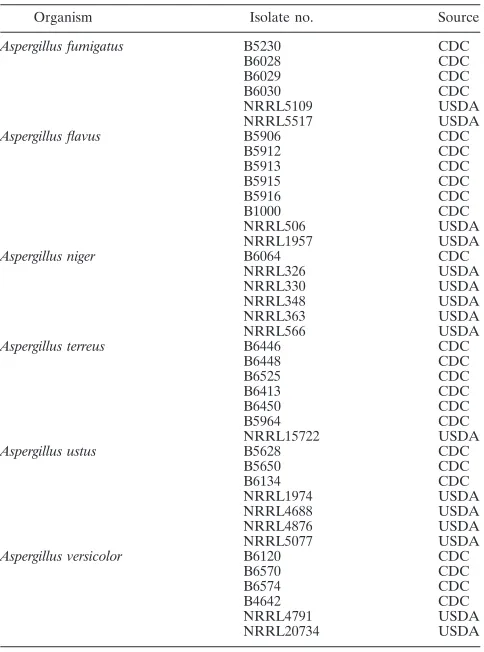
Aspergillus isolates.Two different panels of aspergilli were used in this study. The first panel consisted of 44Aspergillusisolates that represented previously validated and type isolates from the culture collections of the Centers for Disease Control and Prevention (CDC), Atlanta, GA, and the National Center for Agricultural Utilization Research, U.S. Department of Agriculture, Peoria, IL, respectively (Table 1). This panel of isolates (denoted as reference isolates for the purposes of this study) was used for the initial Luminex assay development. Once the assay conditions were established using the reference aspergilli, the
AspergillusLuminex assay was tested on an additional set of 131 clinical Aspergil-lusisolates that included 89A. fumigatus, 17A. flavus, 12A. niger, 4A. terreus, 3
A. ustus, and 6A. versicolorisolates. All the clinical aspergilli were obtained from the Mycotic Diseases Branch Culture Collection, CDC, Atlanta, GA.
Genomic DNA, PCR, and sequencing of the ITS-1 region.All isolates were stored frozen and subcultured on Sabouraud’s dextrose agar plates before DNA extraction was performed. Genomic DNA extraction was performed as described previously (16), and the PCRs were performed with the ITS primers ITS 600 F (5⬘-GGAAGTAAAAGTCGTAACAAGG-3⬘) and ITS 600 R (5⬘-TCCTCCGC TTATTGATATGC-3⬘). The cycling conditions were as follows: 95°C for 3 min, 35 cycles of 95°C for 45 s, an annealing step at 57°C for 30 s, an extension step at 72°C for 3 min, and a final extension step at 72°C for 10 min. The presence and size of amplicons were verified on a gel, fragments were purified using the ExoSAP-IT PCR purification kit (USB Corporation, Cleveland, OH), and the forward and reverse fragments were sequenced with the PCR primer sets as described elsewhere (5).
The identities of all 175Aspergillusisolates were confirmed by comparative sequence analysis of the ITS-1 and ITS-2 regions.
Aspergillusspecies-specific Luminex probe design.Sequences generated from the ITS-1 region of the 44 referenceAspergillusisolates were aligned using the software ClustalW, and candidate regions specific to each species were identified for the Luminex probe design. Each species-specific Luminex probe was de-signed to have at least a 2-nucleotide difference compared to other probes and was 21 to 25 mer in length. The six species-specific probes were AF (A. fumiga-tus), AL (A. flavus), AN (A. niger), AT (A. terreus), AU (A. ustus), and AV (A. versicolor) (Table 2). The stability, melting temperature, and other factors for each probe and ITS-1 complement were evaluated using the software Oligo (Molecular Biology Insights and BioMath, Cascade, CO).
Probe coupling to microspheres.The species-specific Luminex probes AF, AL, AN, AT, AU, and AV (designed as detailed above) were covalently coupled to carboxylated microspheres 130, 131, 132, 133, 134, and 135, respectively, as previously described with some modifications (9). In brief, 2.5⫻106of each
* Corresponding author. Mailing address: Mycotic Diseases Branch,
Centers for Disease Control and Prevention, Mail Stop-G 11, 1600
Clifton Road, Atlanta, GA 30333. Phone: (404) 639-3337. Fax: (404)
639-3546. E-mail: [email protected].
䌤
Published ahead of print on 25 February 2008.
1096
on May 16, 2020 by guest
http://jcm.asm.org/
microsphere set was transferred to a low-binding microcentrifuge tube (Eppen-dorf, Westbury, NY). The microspheres were centrifuged (Eppendorf) for 3 min atⱖ8,000⫻g. After the supernatant was removed, avoiding the pellet, 25l of 2-(N-morpholino) ethanesulfonic acid (MES) (Sigma, St. Louis, MO), 30 mg/ml of EDC [1-ethyl-3-(3-dimethylaminopropyl)carbodiimide hydrochloride] (Pierce, Milwaukee WI), and 500 picomoles of each species-specific probe were coupled to the designated bead region. The solution(s) was shaken in the dark at room temperature for 30 min and 30 mg/ml EDC was added again, followed by a second 30-min incubation period. The microspheres were washed with 500l of 0.02% Tween (Sigma, St. Louis, MO) and centrifuged for 3 min atⱖ8,000⫻g, and the supernatant was removed and 500l 0.1% lauryl sulfate added (Sigma, St. Louis, MO). Finally, this solution was centrifuged for 3 min atⱖ8,000⫻g, the supernatant was removed, and 50l of Tris-EDTA buffer (TE) was added (Sigma, St. Louis, MO). The microspheres coupled with probes were stored at 4°C in the dark until ready for use.
Assay to confirm the binding of Aspergillusprobes to microspheres.The species-specific Luminex probes that were bound to designated microspheres were tested to confirm that the probes were bound to the respective micro-spheres. First, a biotinylated reverse probe that complemented the species-specific Luminex probes was designed. A working solution containing 3l of each species-specific microsphere set was diluted to 1 ml with 1.5⫻TMAC (5 M tetramethyl ammonium chloride–20% Sarkosyl–1 M Tris-HCl [pH 8.0]–0.5 M EDTA [pH 8.0]–dH2O) (Sigma, St. Louis, MO). In a 96-well conical plate
(Corning, Corning, NY), 10l of the probe complement of each Luminex species-specific probe was added to each well, followed by 33l of bead solution and 7l of TE buffer to bring the final solution volume to 50l per well; the appropriate negative controls, consisting of wells containing TE buffer and mi-crosphere solution, were included. The plates were sealed with microseal film (Bio-Rad, Hercules, CA) and heated to 94°C for 5 min for initial denaturation, followed by hybridization at 52°C for 30 min. After 30 min, the plate was centrifuged atⱖ8,000⫻gfor 2 min, the supernatant was carefully removed to avoid the pelleted product, and 75l of SAPE (4 mg/ml) in 1⫻TMAC was
added. For the final hybridization of SAPE, the plate was heated at 52°C for 10 min, read on the Luminex200 using MasterPlexCT (Miraibio, San Francisco, CA), and analyzed using MasterPlex GT analysis software (Miraibio, San Fran-cisco, CA) as detailed below in the section on data analysis.
PCR primer design forAspergillusisolates.After the Luminex species-specific probes were designed and bound to the microspheres, three sets of PCR primers that would yield amplicon lengths amenable to the Luminex assay were designed to amplify 100-bp, 250-bp, and 600-bp portions of the ITS regions using the following respective sequences: ITS 100 F (5⬘GGAAGTAAAAGTCGTAAC AAGG 3⬘) and ITS 100 R (5⬘GAGATCCA/GTTGTTGAAAGTTT-3⬘); ITS 250 F (5⬘GGAAGTAAAAGTCGTAACAAGG-3⬘) and ITS 250 R (5⬘-GCTGCGT TCTTCATCGATGC-3⬘); and ITS 600 F (5⬘-GGAAGTAAAAGTCGTAACA AGG-3⬘) and ITS 600 R (5⬘-TCCTCCGCTTATTGATATGC-3⬘). Each reverse primer was labeled with a biotin molecule, and PCR amplicons for the reference aspergilli were generated using the primer pairs. The PCR conditions were as described in the section on genomic DNA. After PCR, 10 microliters of the PCR product was added to a 96-well conical plate (Corning), and the Luminex assay was performed as described earlier (in the section on coupling confirmation) to determine the appropriate amplicon length that yields efficient hybridization with the species-specific Luminex probes.
The reproducibility of the Luminex assay was assessed as follows. PCR prod-ucts obtained from the reference aspergilli (n⫽44) were tested on the Luminex platform, and aliquots of the PCR amplicons were frozen on day 1. On days 2 and 3, the frozen aliquots were thawed and tested in the Luminex assay in indepen-dent runs. Additionally, on day 4, genomic DNA was extracted again from the referenceAspergillusisolates and subjected to PCR and Luminex analyses. The results from all four experiments were analyzed to evaluate the hybridization efficiency of the biotin-labeled PCR products after storage and to compare the results of two independent Luminex assays performed on the same batch of isolates.
Validation of theAspergillusLuminex assay.Once the assay parameters (in-cluding amplicon size, PCR conditions, and coupling confirmation) were estab-lished, theAspergillusLuminex assay was employed to genotype a panel of 131
Aspergillusclinical isolates. The species identifications of the 131 aspergilli were also derived employing comparative sequence analyses of the ITS regions. For the Luminex assay, the genomic DNA of all aspergilli was subjected to PCR using primer pairs ITS 250 F and ITS 250 R; 10l of the PCR product, 33l of bead solution containing the sixAspergillusspecies-specific probes, and 7l of TE buffer were added per well, and the assay conditions were exactly as described for coupling confirmation. The negative control consisted of the microsphere solution (33l) and TE buffer (17l) with no target DNA.
Data analysis.The data were acquired using the MasterPlexCT system and analyzed using the MasterPlex GT software. Individual sets of microspheres were analyzed by a dual laser system, and the median fluorescence intensity (MFI) value was calculated. The MFI represents the median signal intensity measured per microsphere set. The signal-to-background ratio represents the MFI signals of positive controls versus the background fluorescence of samples containing all components except the amplicon target. A positive signal was defined as an MFI signal that is at least twice the background level after subtraction of the back-ground.
RESULTS
[image:2.585.43.285.88.414.2]Aspergillus
Luminex assay development.
Aspergillus
species-specific Luminex probes directed to the ITS-1 region were
designed and are listed in Table 2. The six species-specific
probes AF, AL, AN, AT, AU, and AV were attached to the
designated microspheres, and probe coupling was confirmed
by coupling confirmation assays (data not shown).
TABLE 1.
Aspergillus
isolates used in the Luminex assay as the
reference panel
Organism Isolate no. Source
Aspergillus fumigatus B5230 CDC
B6028 CDC
B6029 CDC
B6030 CDC
NRRL5109 USDA
NRRL5517 USDA
Aspergillus flavus B5906 CDC
B5912 CDC
B5913 CDC
B5915 CDC
B5916 CDC
B1000 CDC
NRRL506 USDA
NRRL1957 USDA
Aspergillus niger B6064 CDC
NRRL326 USDA
NRRL330 USDA
NRRL348 USDA
NRRL363 USDA
NRRL566 USDA
Aspergillus terreus B6446 CDC
B6448 CDC
B6525 CDC
B6413 CDC
B6450 CDC
B5964 CDC
NRRL15722 USDA
Aspergillus ustus B5628 CDC
B5650 CDC
B6134 CDC
NRRL1974 USDA
NRRL4688 USDA
NRRL4876 USDA
NRRL5077 USDA
Aspergillus versicolor B6120 CDC
B6570 CDC
B6574 CDC
B4642 CDC
NRRL4791 USDA
NRRL20734 USDA
TABLE 2.
Aspergillus
species-specific Luminex probes directed to
the ITS-1 region of the rRNA
Probe Target Probe sequence (5⬘–3⬘)
AF Aspergillus fumigatus GAAAGTATGCAGTCTGAGTTGAT AL Aspergillus flavus CACCCGTGTTTACTGTACCTTAG AN Aspergillus niger AACACGAACACTGTCTGAAAGCGT AT Aspergillus terreus AACATGAACCCTGTTCTGAAAGCT AU Aspergillus ustus CTGAGCTTGATACAAGCAAAC AV Aspergillus versicolor AGTGATGCAGTCTGAGTCTGAATAT
on May 16, 2020 by guest
http://jcm.asm.org/
[image:2.585.302.541.89.156.2]Using three different sets of primers targeted to amplify
100 bp, 250 bp, and 600 bp of the ITS regions, the effect of
amplicon size on MFI signals was assessed for the reference
Aspergillus
isolates. Hybridization signals of less than twice the
background level and higher cross-reactivity to some
species-specific probes were observed with the 100-bp amplicon. While
the 600-bp amplicon product generated an MFI of less than
twice the background level, variable hybridization to the target
DNA was observed, thus impacting reproducibility. An
ampli-con length of 250 bp generated optimal and reproducible data
with no cross-reactivity with other probes (data not shown).
Thus, the primer set that yielded the 250-bp amplicon length
(primers ITS 250 F and ITS 250 R) was selected and employed
in the
Aspergillus
Luminex assay.
Each of the species-specific
Aspergillus
probes was designed
to target the respective
Aspergillus
target DNA, thus yielding
high MFIs with the respective target DNA but MFIs less than
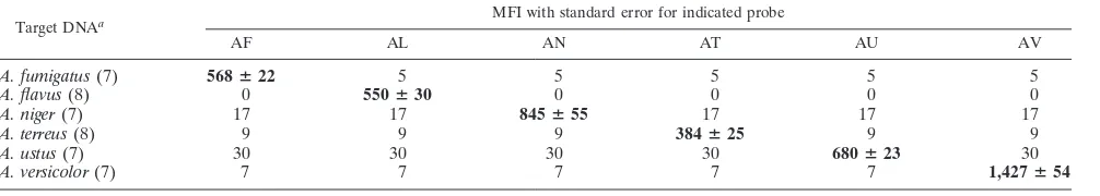
twice the background level with nontarget DNA. As can be
seen from Table 3, the Luminex probes AF, AL AN, AT, AU,
and AV hybridized with their respective targets,
A. fumigatus,
A. flavus,
A. niger,
A. terreus,
A. ustus, and
A. versicolor, yielding
MFIs that ranged from a mean MFI of 384 (A. terreus) to 1,427
(A. versicolor). In spite of this species-specific variability in MFI
values, each of the probes produced MFIs that were more than
twice the background level, thus generating an MFI that was,
on average, 167% higher than the background.
When the assay was tested for reproducibility, it was found
that MFIs of the PCR products decreased after being
sub-jected to a freeze-thaw cycle compared to the MFIs of the fresh
PCR products. For instance, for all
A. fumigatus
isolates, the
mean MFIs on days 1, 2, and 3 were 895, 735, and 690,
respec-tively, after freezing and thawing, whereas the fresh PCR
prod-ucts for all
A. fumigatus
isolates (tested on day 4) generated a
mean MFI of 815. Thus, although the mean MFIs decreased
over the course of 3 days and varied between the two PCR
assays, the MFIs were always at least twice the background
level. This was a consistent trend observed for all
Aspergillus
reference isolates that included representative type isolates for
each species (data not shown).
Luminex assay validation.
Once the Luminex assay
param-eters were determined using the reference
Aspergillus
panel as
described above, the Luminex assay (a patent has been applied
for for this probe set) was tested with a set of 131
sequence-confirmed
Aspergillus
isolates. All the target PCR amplicons
hybridized to their species-specific Luminex probes, and the
species identifications generated by the Luminex assay
corre-lated 100% with the identities generated by a comparative
sequence identification of the ITS regions.
DISCUSSION
Over the last several years, molecular methods, including the
rolling-cycle amplification, repetitive sequence-based PCR,
PCR-restriction enzyme, reverse line blot assay, and DNA
microarray methods, have been evaluated for
Aspergillus
spe-cies identification (7, 14, 19, 22, 25). Although these methods
have been demonstrated to be useful for species identification,
most of these methods (except the reverse line blot assay) are
not amenable to multiplexing. In addition, some of the
meth-ods, such as DNA microarrays, are expensive to perform and
require sophisticated analyses to interpret the results. DNA
sequence-based methods are considered the gold standard for
fungal species identification and have been employed
increas-ingly for the identification of
Aspergillus
species. However,
comparative sequence-based methods can be labor-intensive
and time-consuming and cannot be multiplexed.
The microsphere-based Luminex xMAP technology builds
on the principles of flow cytometry and enzyme immunoassay,
resulting in a sensitive, specific genotyping method that is rapid
and has the additional flexibility of a multiplex format. To this
end, an
Aspergillus
Luminex assay was designed and validated
for the rapid identification of six medically important aspergilli,
A. fumigatus,
A. flavus,
A. niger,
A. terreus,
A. ustus, and
A.
versicolor, from culture. The results demonstrated that the
assay was specific to the target DNA, was easy to perform, and
had a rapid turnaround time of about 6 h (not including DNA
extraction).
[image:3.585.43.546.81.171.2]Previous studies have suggested that the probe GC content
and length and the length of the PCR amplicon can influence
hybridization profiles, thereby impacting the successful
out-come of the Luminex assay (13). A probe GC content of 30 to
50% was optimal for this study. In addition, all species-specific
probes were designed to be 21 to 25 mer in length, and this
yielded superior hybridization. One parameter that needed
optimization for the current assay was the PCR amplicon
length. Longer amplicons may inhibit hybridization due to
steric hindrance, but in some studies, larger amplicon targets
have been shown to be efficient for specific hybridization (13).
Diaz and Fell assessed the effect of amplicon length on
hybrid-ization efficiency by utilizing three sets of primers generating
amplicons 490 to 600 bp, 650 to 875 bp, and 950 to 1,200 bp
(11). For the most part, these investigators found a lower
TABLE 3. Specificity of probes used to detect clinically important
Aspergillus
species in the multiplex format
Target DNAa MFI with standard error for indicated probe
AF AL AN AT AU AV
A. fumigatus
(7)
568
ⴞ
22
5
5
5
5
5
A. flavus
(8)
0
550
ⴞ
30
0
0
0
0
A. niger
(7)
17
17
845
ⴞ
55
17
17
17
A. terreus
(8)
9
9
9
384
ⴞ
25
9
9
A. ustus
(7)
30
30
30
30
680
ⴞ
23
30
A. versicolor
(7)
7
7
7
7
7
1,427
ⴞ
54
a
The number of isolates included in the validation panel is indicated within parentheses.
on May 16, 2020 by guest
http://jcm.asm.org/
hybridization signal with the shortest amplicon target and a
higher hybridization signal with amplicon targets longer than
600 bp (11). In this study, an evaluation of three different
amplicon sizes demonstrated that a 250-bp amplicon length
provided optimal hybridization for all isolates within a given
Aspergillus
species. Additionally, our study also demonstrated
that the
Aspergillus
Luminex assay yielded lower but
reproduc-ible results with PCR amplicons that had been freeze-thawed
over time as well as between two independent assays.
After the conditions of the
Aspergillus
Luminex assay were
optimized with the reference panel of isolates (that included
type isolates), the assay was tested on an additional set of 131
Aspergillus
clinical isolates. There was 100% correlation
be-tween the results of the
Aspergillus
Luminex assay and the
identification derived by the comparative sequence analysis
method, thus yielding an assay specificity of 100%. Currently,
the
Aspergillus
Luminex assay includes only six probes and, as
designed, can identify the predominant
Aspergillus
species that
cause invasive aspergillosis (IA). Numerous studies have
dem-onstrated that IA is caused predominantly by these six
Aspergil-lus
species, with one large multicenter study showing that 56%
of IA was due to
A. fumigatus, 18.7% was caused by
A. flavus,
8% was caused by
A. niger, 16% was caused by
A. terreus, and
1.3% was caused by
A. versicolor.
Although multiple different
Aspergillus
species can exist in the environment and, in theory,
can cause IA, the Luminex assay was designed to identify the
relevant species that may be recovered from clinical specimens
and would serve as a first line of identification. For instance, if
the six-probe
Aspergillus
Luminex assay is used for the
identi-fication of an unknown
Aspergillus
isolate in a clinical
micro-biology laboratory and there is no hybridization with the target
DNA (because it is a species not included in the six-probe
panel), the target DNA can then be sequenced as a
second-step strategy for identification.
The
Aspergillus
species-specific probes were directed to the
ITS-1 locus, as this region has been demonstrated to be useful
for species complex-level identification within this genus (4).
However, the ITS locus is not suitable for the identification of
individual species within the species complex; for instance, the
ITS locus cannot discriminate between species within the
A.
fumigatus
complex that includes the newly described species
A.
lentulus
and other species such as
A. udagawae,
A.
thermomu-tatus, and
A. fumigatus
(3). Thus, with the current Luminex
panel, DNA from these isolates will hybridize to the
A.
fumiga-tus
probe and will therefore be identified as
A. fumigatus
com-plex. Recent studies have demonstrated that comparative
se-quence analyses of protein-coding regions such as that for

tubulin provide enough discrimination to differentiate taxa
within the
Aspergillus
species complexes (2, 23). For such levels
of identification, an additional set of probes directed to the

tubulin or any other suitable locus or loci can be designed and
added to the Luminex panel. Up to 100 different
Aspergillus
probes can be used on the Luminex platform; thus, in theory,
100 different genotypes can be distinguished using this assay.
As designed, the
Aspergillus
Luminex assay can be used for
the identification of isolates grown as pure culture. Other
stud-ies have employed Luminex assays for the direct detection of
pathogens from clinical specimens (10), and it remains to be
seen if the
Aspergillus
Luminex assays can be used as a
diag-nostic tool as well. In this center, the cost of sequencing
meth-ods is as low as $7 per sample, and though, at this time, the cost
of the Luminex assay is greater, time and labor can serve as a
definite trade-off. With continued and increased use of the
Luminex technology, the cost of the assay may decrease, thus
truly providing clinical microbiology laboratories with a
tech-nology that is high throughput as well as economical. In
sum-mary, a rapid, specific, and multiplex method, the
Aspergillus
Luminex assay, is described for the identification of various
clinically important aspergilli.
ACKNOWLEDGMENTS
K.A.E. was supported in part by an appointment to the Emerging
Infectious Disease (EID) fellowship program administered by the
As-sociation of Public Health Laboratories (APHL) and funded by the
CDC. R.K. was supported by a fellowship from the Academic Frontier
Project of the Ministry of Education, Culture, Sports, Science and
Technology (MEXT) and Nihon University.
We are grateful to Stephen Peterson, United States Department of
Agriculture (USDA), for providing the
Aspergillus
type isolates used in
this study.
The findings and conclusions in this article are those of the authors
and do not necessarily represent the views of the CDC.
REFERENCES
1.Araujo, R., C. Pina-Vaz, and A. G. Rodrigues.2007. Susceptibility of envi-ronmental versus clinical strains of pathogenic Aspergillus. Int. J. Antimi-crob. Agents29:108–111.
2.Balajee, S. A., J. Gribskov, M. Brandt, J. Ito, A. Fothergill, and K. A. Marr.
2005. Mistaken identity:Neosartorya pseudofischeriand its anamorph mas-querading asAspergillus fumigatus. J. Clin. Microbiol.43:5996–5999. 3.Balajee, S. A., J. L. Gribskov, E. Hanley, D. Nickle, and K. A. Marr.2005.
Aspergillus lentulussp. nov., a new sibling species ofA. fumigatus. Eukaryot. Cell4:625–632.
4.Balajee, S. A., J. Houbraken, P. E. Verweij, S. B. Hong, T. Yaghuchi, J. Varga, and R. A. Samson.2007. Aspergillus species identification in the clinical setting. Stud. Mycol.59:39–46.
5.Balajee, S. A., S. T. Tay, B. A. Lasker, S. F. Hurst, and A. P. Rooney.2007. Characterization of a novel gene for strain typing reveals substructuring of
Aspergillus fumigatusacross North America. Eukaryot. Cell6:1392–1399. 6.Bovers, M., M. R. Diaz, F. Hagen, L. Spanjaard, B. Duim, C. E. Visser, H. L.
Hoogveld, J. Scharringa, I. M. Hoepelman, J. W. Fell, and T. Boekhout.
2007. Identification of genotypically diverseCryptococcus neoformansand
Cryptococcus gattiiisolates by Luminex xMAP technology. J. Clin. Microbiol.
45:1874–1883.
7.Campa, D., A. Tavanti, F. Gemignani, C. S. Mogavero, I. Bellini, F. Bottari, R. Barale, S. Landi, and S. Senesi.2008. DNA microarray based on arrayed-primer extension technique for identification of pathogenic fungi responsible for invasive and superficial mycoses. J. Clin. Microbiol.46:909–915. 8.Dannaoui, E., D. Garcia-Hermoso, J. M. Naccache, I. Meneau, D. Sanglard,
C. Bouges-Michel, D. Valeyre, and O. Lortholary.2006. Use of voriconazole in a patient with aspergilloma caused by an itraconazole-resistant strain of
Aspergillus fumigatus. J. Med. Microbiol.55:1457–1459.
9.Das, S., T. M. Brown, K. L. Kellar, B. P. Holloway, and C. J. Morrison.2006. DNA probes for the rapid identification of medically importantCandida
species using a multianalyte profiling system. FEMS Immunol. Med. Micro-biol.46:244–250.
10.Diaz, M. R., T. Boekhout, B. Theelen, M. Bovers, F. J. Cabanes, and J. W. Fell.2006. Microcoding and flow cytometry as a high-throughput fungal identification system forMalasseziaspecies. J. Med. Microbiol.55:1197– 1209.
11.Diaz, M. R., and J. W. Fell.2004. High-throughput detection of pathogenic yeasts of the genusTrichosporon. J. Clin. Microbiol.42:3696–3706. 12.Diaz, M. R., and J. W. Fell. 2005. Use of a suspension array for rapid
identification of the varieties and genotypes of theCryptococcus neoformans
species complex. J. Clin. Microbiol.43:3662–3672.
13.Dunbar, S. A.2006. Applications of Luminex xMAP technology for rapid, high-throughput multiplexed nucleic acid detection. Clin. Chim. Acta363:
71–82.
14.Hansen, D., M. Healy, K. Reece, C. Smith, and G. L. Woods.2008. Repet-itive-sequence-based PCR using the DiversiLab system for identification of
Aspergillusspecies. J. Clin. Microbiol.46:1835–1839.
15.Howard, S. J., I. Webster, C. B. Moore, R. E. Gardiner, S. Park, D. S. Perlin, and D. W. Denning.2006. Multi-azole resistance inAspergillus fumigatus. Int. J. Antimicrob. Agents28:450–453.
16.Lasker, B. A.2002. Evaluation of performance of four genotypic methods for
on May 16, 2020 by guest
http://jcm.asm.org/
studying the genetic epidemiology ofAspergillus fumigatusisolates. J. Clin. Microbiol.40:2886–2892.
17.Lass-Florl, C., A. Rief, S. Leitner, C. Speth, R. Wurzner, and M. P. Dierich.
2005. In vitro activities of amphotericin B and voriconazole against aleurio-conidia fromAspergillus terreus. Antimicrob. Agents Chemother.49:2539– 2540.
18.Mellado, E., G. Garcia-Effron, L. Alcazar-Fuoli, W. J. Melchers, P. E. Ver-weij, M. Cuenca-Estrella, and J. L. Rodriguez-Tudela.2007. A new Aspergil-lus fumigatusresistance mechanism conferring in vitro cross-resistance to azole antifungals involves a combination of cyp51A alterations. Antimicrob. Agents Chemother.51:1897–1904.
19.Mirhendi, H., K. Diba, P. Kordbacheh, N. Jalalizand, and K. Makimura.
2007. Identification of pathogenicAspergillusspecies by a PCR-restriction enzyme method. J. Med. Microbiol.56:1568–1570.
20.O’Donnell, K., B. A. Sarver, M. Brandt, D. C. Chang, J. Noble-Wang, B. J. Park, D. A. Sutton, L. Benjamin, M. Lindsley, A. Padhye, D. M. Geiser, and T. J. Ward.2007. Phylogenetic diversity and microsphere array-based geno-typing of human pathogenic fusaria, including isolates from the multistate
contact lens-associated U.S. keratitis outbreaks of 2005 and 2006. J. Clin. Microbiol.45:2235–2248.
21.Panackal, A. A., A. Imhof, E. W. Hanley, and K. A. Marr.2006.Aspergillus ustusinfections among transplant recipients. Emerg. Infect. Dis.12:403–408. 22.Playford, E. G., F. Kong, Y. Sun, H. Wang, C. Halliday, and T. C. Sorrell.
2006. Simultaneous detection and identification ofCandida, Aspergillus, and
Cryptococcusspecies by reverse line blot hybridization. J. Clin. Microbiol.
44:876–880.
23.Varga, J., J. Houbraken, H. A. Van Der Lee, P. E. Verweij, and R. A. Samson.2008. Aspergillus calidoustus sp. nov., causative agent of human infections previously assigned to Aspergillus ustus. Eukaryot. Cell7:630– 638.
24.Yildiran, S. T., F. M. Mutlu, M. A. Saracli, Y. Uysal, A. Gonlum, G. Sobaci, and D. A. Sutton.2006. Fungal endophthalmitis caused byAspergillus ustus
in a patient following cataract surgery. Med. Mycol.44:665–669. 25.Zhou, X., F. Kong, T. C. Sorrell, H. Wang, Y. Duan, and S. C. Chen.2008.
Practical method for detection and identification ofCandida,Aspergillus, and
Scedosporiumspp. by use of rolling-circle amplification. J. Clin. Microbiol.
46:2423–2427.