0095-1137/05/$08.00⫹0 doi:10.1128/JCM.43.9.4880–4884.2005
Copyright © 2005, American Society for Microbiology. All Rights Reserved.
Direct Application of the INNO-LiPA Rif.TB Line-Probe Assay for
Rapid Identification of
Mycobacterium tuberculosis
Complex Strains
and Detection of Rifampin Resistance in 360 Smear-Positive
Respiratory Specimens from an Area of High Incidence of
Multidrug-Resistant Tuberculosis
Miguel Viveiros,
1Clara Leandro,
1Liliana Rodrigues,
1Josefina Almeida,
1Rosa
´rio Bettencourt,
1Isabel Couto,
1,2Lurdes Carrilho,
3Jose
´ Diogo,
4Ana Fonseca,
5Luı´s Lito,
6Joa
˜o Lopes,
7Teresa Pacheco,
8Mariana Pessanha,
9Judite Quirim,
10Luı´sa Sancho,
11Max Salfinger,
12and Leonard Amaral
1*
Unidade de Micobacterias, UPMM, Instituto de Higiene e Medicina Tropical, Universidade Nova de Lisboa, Lisboa, Portugal1;
Centro de Recursos Microbiolo´gicos (CREM), Faculdade de Cieˆncias e Tecnologia, Universidade Nova de Lisboa, Lisboa, Portugal2; Laboratorio de Micobacterias, Hospital Pulido Valente, Lisboa, Portugal3; Laboratorio de Microbiologia,
Hospital Garcia da Orta, Lisboa, Portugal4; Laboratorio de Microbiologia, Hospital Condes de Castro Guimara˜es,
Cascais, Portugal5; Laboratorio de Microbiologia, Hospital de Santa Maria, Lisboa, Portugal6; Laboratorio de
Microbiologia, Hospital de Nossa Senhora do Rosa´rio, Barreiro, Portugal7; Laboratorio de Microbiologia,
Hospital Egas Moniz, Lisboa, Portugal8; Laboratorio de Microbiologia, Hospital Sa˜o Francisco
Xavier, Lisboa, Portugal9; Laboratorio de Microbiologia, Hospital de Sa˜o Bernardo, Setu´bal,
Portugal10; Laboratorio de Microbiologia, Hospital Fernando da Fonseca, Amadora-Sintra,
Portugal11; and Clinical Mycobacteriology Laboratory, Wadsworth Center, New York State
Department of Health, Albany, New York 1220812
Received 11 June 2005/Accepted 15 June 2005
The INNO-LiPA Rif.TB assay for the identification ofMycobacterium tuberculosiscomplex strains and the
detection of rifampin (RIF) resistance has been evaluated with 360 smear-positive respiratory specimens from an area of high incidence of multidrug-resistant tuberculosis (MDR-TB). The sensitivity when compared to conventional identification/culture methods was 82.2%, and the specificity was 66.7%; the sensitivity and specificity were 100.0% and 96.9%, respectively, for the detection of RIF resistance. This assay has the potential to provide rapid information that is essential for the effective management of MDR-TB.
Portugal has a frequency of new cases of tuberculosis (TB) of 45.0 per 100,000 inhabitants (37), of which 21% are multi-drug-resistant TB (MDR-TB) cases, and of these, ⬎5% are resistant to four or five first-line anti-TB drugs (1, 23, 35). Despite control measures that have reduced the incidence of MDR-TB (6), in the major cities Lisbon and Oporto new cases exceed 50.0 per 100,000 inhabitants (19) and MDR-TB occurs in excess of 15% of cases (6, 36). The clinical laboratory has a major role in the control of TB (4, 20), inasmuch as effective management of TB patients from areas that have high rates of MDR-TB is dependent on the rapid identification of Mycobac-terium tuberculosiscomplex strains and their antibiotic suscep-tibility profiles (3, 35, 37). The role of the laboratory is even more critical for the management of AIDS patients who also have MDR-TB (3).
In Portugal, as is the case worldwide, the vast majority ofM. tuberculosiscomplex strains with resistance to rifampin (RIF) are also resistant to isoniazid (INH), and although monoresis-tance to INH is common (25), monoresismonoresis-tance to RIF is rare
(3, 6, 17, 24, 29). Thus, RIF resistance can be used for the identification of MDR-TB infections (8, 30). This makes it possible to treat MDR-TB patients aggressively (with four or five drugs) while sparing non-MDR-TB patients from areas with high MDR-TB frequencies from said therapy (3, 8, 21); a marked reduction in the frequency of noncompliance would consequently be expected (3, 8, 17, 21).
Ninety-five percent ofM. tuberculosisstrains with resistance to RIF contain distinct mutations located within an 81-bp (27-codon) region of the beta subunit of the RNA polymerase (rpoB) gene (29, 31). Several methods can rapidly detect these specific mutations and thereby identify RIF-resistantM. tuber-culosiscomplex strains (14, 22, 31, 32). One of these methods is the line probe assay INNO-LiPA Rif.TB (Innogenetics, Zwi-jndrecht, Belgium), a commercially available kit not yet ap-proved by the U.S. Food and Drug Administration which iden-tifiesM. tuberculosiscomplex strains and RIF resistance within a very short period of culture time (16, 26, 28). We have evaluated this assay for the identification of M. tuberculosis
complex strains and the detection of mutations in the rpoB
gene linked to RIF resistance directly from acid-fast smear-positive respiratory specimens obtained from patients who pre-sented with tuberculosis (clinical symptoms and radiological evidence). The assays were performed in parallel with
conven-* Corresponding author. Mailing address: Unidade de Micobacte-rias do Instituto de Higiene e Medicina Tropical, Rua Junqueira 96, 1349-008 Lisboa, Portugal. Phone: 351 21 3652653. Fax: 351 21 3632105. E-mail: [email protected].
4880
on May 15, 2020 by guest
http://jcm.asm.org/
tional isolation, identification, and susceptibility testing proce-dures routinely used in our mycobacteriology clinical labora-tory as part of the “TB Fast Track Program,” modeled after that of the New York State Department of Health (7, 8, 27). This program is under the supervision of the TB Task Force of Greater Lisbon, a cooperative joint venture involving the ma-jor hospitals of the Greater Lisbon area (33). From September 2002 to September 2003, a total of 360 acid-fast positive respi-ratory specimens consisting of sputa (n⫽318), bronchoalveo-lar lavage fluids (n ⫽23), and bronchial secretions (n⫽19) from patients presenting with presumptive active TB were re-ceived in our laboratory; each specimen was accompanied by a physician-completed questionnaire that included pertinent pa-tient demographics, clinical history, and MDR-TB risk factors. The patients, all from the Greater Lisbon area, ranged in age from 14 to 89 years (average, 42 years) and were mainly male (73.8%). The three major MDR-TB risk factors reported were, in order of importance, prior anti-TB treatment, contact with other MDR-TB patients, and origin from an area with a known high incidence of MDR-TB. The human immunodeficiency
virus status was determined for only 150 patients (41.7%), and of these, 82 patients were coinfected with human immunode-ficiency virus. Anti-TB treatment had already been initiated for 189 patients (52.5%) at the time of specimen collection.
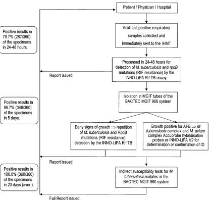
The TB Fast Track work algorithm, restricted to the work week of Monday to Friday, is summarized in Fig. 1. Briefly, all specimens received were processed by the conventional myco-bacteriological NaOH-NALC method (15), and aliquots were collected for acid-fast staining (Ziehl-Neelsen stain), for inoc-ulation of MGIT tubes employed by the BACTEC MGIT 960 system (Becton-Dickinson Diagnostic Instrument Systems, Towson, Md.) according to the manufacturer’s instructions, and for extraction of total DNA with a QIAamp DNA mini kit (QIAGEN, GmbH, Hilden, Germany). The identification of
M. tuberculosis complex and Mycobacterium avium complex strains present in full-grown cultures in the BACTEC MGIT 960 culture system was made with the aid of the Accuprobe system (Gen-Probe Inc., San Diego, California). A positive identification ofM. tuberculosiswas followed by susceptibility assays for RIF, INH, ethambutol, streptomycin, and
pyrazin-FIG. 1. TB Fast Track work algorithm. IHMT, Instituto de Higiene e Medicina Tropical; aver., average.
on May 15, 2020 by guest
http://jcm.asm.org/
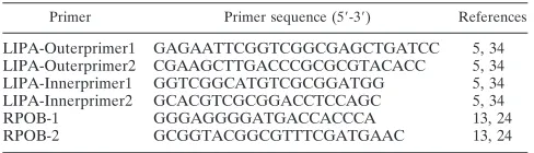
[image:2.585.86.500.68.464.2]amide afforded by the BACTEC MGIT 960 system. The iden-tification ofM. tuberculosisand detection ofrpoBgene muta-tions were made directly from acid-fast-bacillus (AFB)-positive respiratory specimens with the aid of the INNO-LiPA Rif.TB assay according to the following protocol, adapted for direct detection (5, 34). Five microliters of total DNA extract was used in a 50-l PCR mixture containing the outer LiPA prim-ers (Table 1), 1 microliter of the first-round product was trans-ferred to a 50-l second-round reaction mixture containing the inner primers biotinylated at the 5⬘ end (Table 1), and 10 microliters of the labeled PCR product was denatured and hybridized to membrane-bound capture-specific oligonucleo-tide probes, followed by a color detection step. One probe is specific forM.tuberculosiscomplex strains, five partially over-lapping probes hybridize exclusively to therpoBgene wild-type sequence, and four probes hybridize with amplicons carrying the following mutations: R2:D516V, R4a:H526Y, R4b:H526D, and R5:S531L (26). The absence of hybridization of one or more of the wild-type probes indicates a mutation that will be identified by hybridization with one of the mutation probes. The assay was used with appropriate controls, as described in previous evaluations (13, 30), namely, inclusion of a negative control containing no mycobacterial DNA (distilled sterile wa-ter) and a positive control containing DNA from anM. tuber-culosisH37Rv (ATCC 27294) MGIT culture. An initial screen-ing of the kit with nontuberculosis mycobacterial DNA samples (30) was also carried out. To control for potential inhibition of PCR amplification, a second separate aliquot of the extracted DNA (5l) served as the source for a parallel INNO-LiPA Rif.TB nested PCR supplemented with 5l ofM. tuberculosisH37Rv genomic DNA. Negative amplification re-sults in the presence of the spiked DNA were considered to be due to inhibition of amplification. For those specimens where no amplification was obtained by the direct application of the INNO-LiPA Rif.TB assay to the AFB-positive specimens, the amplification procedure was repeated as soon as a positive growth index was detected in the BACTEC MGIT 960 system tubes. If no amplification occurred, then the AFB present in the culture were subjected to the nucleic acid amplification (NAA) INNO-LiPA MYCOBACTERIA v2 assay (Innogenet-ics), which identifies 16 nontuberculosis mycobacterial strains (20). The reference BACTEC 460 TB system (Becton-Dickin-son) was used when samples presented a RIF susceptibility status that differed from that provided by the assay (12). Pre-liminary reports were sent to the physician at the completion of each phase, and a full report was issued after the completion of
the conventional isolation, identification, and susceptibility tests (Fig. 1). Each specimen identified as having a mutation in therpoBgene was subjected to sequence analysis of both DNA strands of a 350-bp fragment of therpoBgene (24).
Only the primary results, with no adjustment of false-nega-tive and false-posifalse-nega-tive results, were used for calculating sensi-tivity, specificity, and positive and negative predictive values for the INNO-LiPA Rif.TB assay. The isolation ofM. tubercu-losiscomplex strains in culture and identification by the Accu-probeM. tuberculosiscomplex DNA probe served as the “gold standard” for comparison to the results obtained with the INNO-LiPA Rif.TB assay. The susceptibility to RIF obtained with the BACTEC MGIT 960 system served as the “gold stan-dard” for comparison to that obtained with the INNO-LiPA Rif.TB assay.
A total of 287 (79.7%) of the 360 acid-fast positive respira-tory specimens assayed gave positive amplification results, of which 281 were identified as true-positive results by the culture isolation andM. tuberculosis Accuprobe identification proce-dures (Table 2). Retroactive reviews of the medical records of the six patients whose specimens yielded false-positive results revealed that all six patients were confirmed as positive for TB by clinical criteria and that at least three patients presented evidence of recent or current anti-TB therapy. Although these specimens could have been considered true-positive results for the final diagnosis of tuberculosis, for the above-defined assay performance evaluation compared to the “gold standard” af-forded by Accuprobe technology, they were considered false-positive results. Of the 73 specimens that were identified by the INNO-LiPA Rif.TB assay as negative for the presence ofM. tuberculosis, 12 were shown to be true-negative results by cul-ture isolation/M. tuberculosis Accuprobe identification. Of these 12 specimens, 2 were identified as containingM. avium
[image:3.585.42.286.80.150.2]complex isolates, 1 was identified as containingMycobacterium chelonae, and the remaining 9 were negative for any mycobac-teria. These last nine specimens came from patients whose radiological work-ups did not support a diagnosis of active TB. For the remaining 61 false-negative specimens, the nested PCR was repeated with the use of the DNA spike. Nineteen of these false-negative specimens yielded acid-fast smears with very small numbers of bacilli, and no DNA was detectable after extraction. For the remaining 42 specimens, extracted DNAs
TABLE 1. Primers and conditions used for this studya
Primer Primer sequence (5⬘-3⬘) References
LIPA-Outerprimer1 GAGAATTCGGTCGGCGAGCTGATCC 5, 34 LIPA-Outerprimer2 CGAAGCTTGACCCGCGCGTACACC 5, 34
LIPA-Innerprimer1 GGTCGGCATGTCGCGGATGG 5, 34
LIPA-Innerprimer2 GCACGTCGCGGACCTCCAGC 5, 34
RPOB-1 GGGAGGGGATGACCACCCA 13, 24
RPOB-2 GCGGTACGGCGTTTCGATGAAC 13, 24
aThe INNO-LiPA Rif.TB thermal cycling conditions were as follows. The
first- and second-round PCRs consisted of 40 cycles of denaturation (95°C for 30 s), annealing (62°C for 30 s), and extension (72°C for 30 s). The rpoB amplification thermal cycling conditions consisted of 30 cycles of denaturation (94°C for 60 s), annealing (60°C for 60 s), and extension (72°C for 60 s).
TABLE 2. Correlation of the INNO-LiPA Rif.TB method for direct identification ofM. tuberculosiscomplex in acid-fast-bacillus-positive respiratory specimens with conventional isolation in culture
(BACTEC 960 system) followed by AccuprobeM. tuberculosis
complex identification
INNO-Lipa Rif.TB test result for M. tuberculosisb
No. of specimens with correlation to conventional test resulta
Positive Negative Total
Positive 281 6 287
Negative 61 12 73
Total 342 18 360
a
Identification by AccuprobeM. tuberculosiscomplex probes and conven-tional biochemical tests with isolated cultures.
b
Sensitivity of INNO-LiPA Rif.TB direct identification compared toM. tu-berculosiscomplex identification after isolation in culture, 82.2%; specificity, 66.7%; positive predictive value, 97.9%; negative predictive value, 16.4%.
on May 15, 2020 by guest
http://jcm.asm.org/
were visualized by agarose gel electrophoresis, and the repeti-tion of the nested PCR revealed inhibirepeti-tion of amplificarepeti-tion for 32 of the 42 specimens. At this time, we are unable to explain the negative results for amplification of the remaining 10 spec-imens. Nevertheless, the 61 specimens were considered false-negative specimens for the identification of theM. tuberculosis
complex by the INNO-LiPA Rif.TB assay (Table 2). The assay yielded a sensitivity of 82.2% and a specificity of 66.7% for the direct identification of M. tuberculosis complex strains. The positive and negative predictive values for the identification of the M. tuberculosiscomplex were 97.9% and 16.4%, respec-tively (Table 2). This performance is compatible with that of other commercially available NAA assays certified for direct application to smear-positive respiratory samples (8, 9, 18).
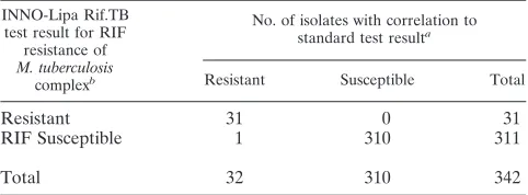
All M. tuberculosis complex isolates (n ⫽ 342) were sub-jected to susceptibility assays with the five first-line anti-TB drugs, and the overall resistance patterns are summarized in Table 3. Among the drug-resistant isolates, 8.8% were multi-drug resistant (INH plus RIF), and 5.6% were resistant to all five drugs (Table 3). The correlation of the RIF resistance/ susceptibility results obtained by standard indirect susceptibil-ity testing with those obtained with the INNO-LiPA Rif.TB kit
is summarized in Table 4. Thirty-oneM. tuberculosiscomplex isolates shown to be resistant to RIF by the standard suscep-tibility test were detected by the INNO-LiPA Rif.TB assay as carrying the following mutations: S531L (29 isolates), H526Y, and D516V (1 isolate each). Sequencing of the rpoB gene region confirmed the presence of the detected point mutations. The INNO-LiPA Rif.TB assay failed to detect any mutation in one sample that was repeatedly identified as RIF resistant by both the BACTEC MGIT 960 and BACTEC 460TB systems. No point mutation in therpoBgene was detected by sequenc-ing, and thus this phenotype must be due to another, as yet undescribed cause (10, 29, 31). The predominant mutation detected in therpoBgene is S531L, the one most frequently detected in European and Asian countries (2, 11, 13, 16, 26). All 310 RIF-susceptible isolates were correctly identified by the INNO-LiPA Rif.TB assay as being negative for mutations in therpoBgene. The specificity and sensitivity of the INNO-LiPA Rif.TB assay for the detection of the RIF resistance profile were 100.0% and 96.9%, respectively. The positive and negative predictive values obtained were 100.0% and 99.7%, respectively (Table 4), in agreement with those obtained by others (13, 30). The INNO-LiPA Rif.TB PCR-based hybrid-ization assay, adjusted for the use of a nested PCR to increase its sensitivity, rapidly identifies M. tuberculosis and its resis-tance to RIF. The assay is simple, convenient, cost-effective, and highly reliable when run in parallel with a conventional TB laboratory diagnostic algorithm. It directly identified 79.7% of the samples within 24 to 48 h of the arrival of the specimens. This percentage was raised to 96.7% with the application of the same amplification and detection protocol to growing cultures of specimens with initial negative direct amplification results. The major limitations found were (i) false-negative results due to inhibition of amplification, (ii) the absence of an internal amplification control, and (iii) the fact that this assay, as well as other NAA assays, identifies DNA from dead mycobacteria from patients undergoing therapy. Regarding the RIF resis-tance profile, the assay yielded results that were 96.9% in agreement with those obtained much later by the use of a culture-based susceptibility method. The ability to rapidly identify an MDR-TB-type infection affords the effective man-agement of MDR-TB, reduces the frequency of noncompli-ance since patients that do not have MDR-TB can be treated less aggressively, and therefore contributes to the control of TB.
M.V. and C.L. contributed equally to this study.
We are grateful for the participation of the physicians and labora-tory personnel from hospitals involved in the TB Task Force Program for Greater Lisbon, especially to our colleagues Gabriela Abreu (Hos-pital do Barreiro), Isabel Oliveira (Hos(Hos-pital Cascais), Judite Batista (Hospital Egas Moniz), Clara Portugal (Hospital Amadora-Sintra), Ana Rodrigues (Hospital de Almada), Ca¯ndida Canha (Hospital Pu-lido Valente), Maria Jose´ Salgado and Carvalho de Sousa (Hospital Santa Maria), Jesuina Duarte (Hospital de Setu´bal), and Filomena Martins (Hospital Sa˜o Francisco Xavier). We give special thanks to Francisco Antunes, Jaime Pina, Jose´ Moniz-Pereira, Fernando Ven-tura, and Emı´lia Valadas for their support, encouragement, and par-ticipation in the local and nationwide meetings held by the TB Task Force of Greater Lisbon.
[image:4.585.43.282.86.228.2]This study was supported by grant SDH.IC.I.01.17-TB from the Task Force for Greater Lisbon and by the TB-Fast-Track Program from the Calouste Gulbenkian Foundation. We thank the Quilaban Corpora-tion for generous technical support.
TABLE 3. Resistance patterns of the 342M. tuberculosiscomplex isolates obtained in this study
Resistance patterna
No. of isolates
HRSEZ ... 19
HRSE... 1
HRSZ... 2
HREZ ... 5
HRS... 2
HR... 1
SE ... 1
HS... 10
H... 6
R ... 2
S... 33
Susceptible to all five drugs ... 260
Total ... 342
aH, isoniazid; R, rifampin; S, streptomycin; E, ethambutol; and Z,
[image:4.585.43.284.570.659.2]pyrazin-amide. Resistance was determined with the indirect drug susceptibility assay of the BACTEC MGIT 960 system.
TABLE 4. Correlation of INNO-LiPA Rif.TB and standard susceptibility tests in the BACTEC 960 system
INNO-Lipa Rif.TB test result for RIF
resistance of M. tuberculosis
complexb
No. of isolates with correlation to standard test resulta
Resistant Susceptible Total
Resistant 31 0 31
RIF Susceptible 1 310 311
Total 32 310 342
aSusceptibility was determined by the BACTEC MGIT 960 system’s indirect
susceptibility assay and confirmed by the BACTEC 460TB system’s indirect susceptibility assay when necessary.
bSensitivity of INNO-LiPA Rif.TBM. tuberculosiscomplex direct detection of
rpoBgene mutations that confer RIF resistance in acid-fast-bacillus-positive respiratory specimens compared to RIF resistance determined by standard in-direct susceptibility assays, 96.9%; specificity, 100.0%; positive predictive value, 100.0%; negative predictive value, 99.7%.
on May 15, 2020 by guest
http://jcm.asm.org/
REFERENCES
1.Antunes, M. L., J. Aleixo-Dias, A. F. Antunes, M. F. Pereira, F. Raymundo, and M. F. Rodrigues.2000. Anti-tuberculosis drug resistance in Portugal. Int. J. Tuberc. Lung Dis.4:223–231.
2.Bartfai, Z., A. Somoskovi, C. Kodmon, N. Szabo, E. Puskas, L. Kosztolanyi, E. Farago, J. Mester, L. M. Parsons, and M. Salfinger.2001. Molecular characterization of rifampin-resistant isolates ofMycobacterium tuberculosis from Hungary by DNA sequencing and the line probe assay. J. Clin. Micro-biol.39:3736–3739.
3.Centers for Disease Control and Prevention.1993. Initial therapy for tuber-culosis in the era of multidrug resistance: recommendations of the Advisory Council for the Elimination of Tuberculosis. Morb. Mortal. Wkly. Rep. 42:1–8.
4.Centers for Disease Control and Prevention.2002. TB elimination cooper-ative agreement. Centers for Disease Control and Prevention, Atlanta, Ga. 5.De Beenhouwer, H., Z. Lhiang, G. Jannes, W. Mijs, L. Machtelinckx, R. Rossau, H. Traore, and F. Portaels.1995. Rapid detection of rifampin resistance in sputum and biopsy specimens from tuberculosis patients by PCR and line probe assay. Tuber. Lung Dis.76:425–430.
6.Direcc¸a˜o Geral de Sau´de.2004. Programa Nacional de Luta contra a Tu-berculose (PNT). Ponto da situac¸a˜o epidemiolo´gica e de desempenho ano 2003, p. 1–12. DGS, Lisboa, Portugal.
7.Frieden, T. R., T. Sterling, A. Pablos-Mendez, J. O. Kilburn, G. M. Cauthen, and S. W. Dooley.1993. The emergence of drug-resistant tuberculosis in New York City. N. Engl. J. Med.328:521–526.
8.Hale, Y. M., E. P. Desmond, K. C. Jost, Jr., and M. Salfinger.2000. Access to newer laboratory procedures: a call for action. Int. J. Tuberc. Lung Dis. 4:171–175.
9.Hale, Y. M., G. E. Pfyffer, and M. Salfinger.2001. Laboratory diagnosis of mycobacterial infections: new tools and lessons learned. Clin. Infect. Dis. 33:834–846.
10.Heep, M., B. Brandstatter, U. Rieger, N. Lehn, E. Richter, S. Rusch-Gerdes, and S. Niemann.2001. Frequency ofrpoBmutations inside and outside the cluster I region in rifampin-resistant clinicalMycobacterium tuberculosis iso-lates. J. Clin. Microbiol.39:107–110.
11.Hirano, K., C. Abe, and M. Takahashi.1999. Mutations in therpoBgene of rifampin-resistant Mycobacterium tuberculosis strains isolated mostly in Asian countries and their rapid detection by line probe assay. J. Clin. Mi-crobiol.37:2663–2666.
12.Inderlied, C. B., and K. A. Nash.1996. Antimycobacterial agents: in vitro susceptibility testing, spectra of activity, mechanisms of action and resistance, and assays for activity in biologic fluids, p. 127–175.InV. Lorian (ed.), Antibiotics in laboratory medicine, 4th ed. Williams and Wilkins, Baltimore, Md.
13.Johansen, I. S., B. Lundgren, A. Sosnovskaja, and V. O. Thomsen.2003. Direct detection of multidrug-resistantMycobacterium tuberculosisin clinical specimens in low- and high-incidence countries by line probe assay. J. Clin. Microbiol.41:4454–4456.
14.Kapur, V., L. L. Li, S. Iordanescu, M. R. Hamrick, A. Wanger, B. N. Kreiswirth, and J. M. Musser.1994. Characterization by automated DNA sequencing of mutations in the gene (rpoB) encoding the RNA polymerase beta subunit in rifampin-resistantMycobacterium tuberculosisstrains from New York City and Texas. J. Clin. Microbiol.32:1095–1098.
15.Kent, P. T., and G. P. Kubica.1985. Public health mycobacteriology: a guide for the level III laboratory. Centers for Disease Control and Prevention, U.S. Department of Health and Human Services, Atlanta, Ga.
16.Matsiota-Bernard, P., G. Vrioni, and E. Marinis.1998. Characterization of rpoBmutations in rifampin-resistant clinicalMycobacterium tuberculosis iso-lates from Greece. J. Clin. Microbiol.36:20–23.
17.Mitchison, D. A.1998. How drug resistance emerges as a result of poor compliance during short course chemotherapy for tuberculosis. Int. J. Tu-berc. Lung Dis.2:10–15.
18.Noordhoek, G. T., J. D. A. Van Embden, and A. H. J. Kolk.1996. Reliability of nucleic acid amplification for detection ofMycobacterium tuberculosis: an international collaborative quality control study among 30 laboratories. J. Clin. Microbiol.34:2522–2525.
19.Ordway, D. J., L. Costa, M. Martins, H. Silveira, L. Amaral, M. J. Arroz, F. Ventura, and H. M. Dockrell.2004. Increased interleukin-4 production by CD8 and gammadelta T cells in health-care workers is associated with the subsequent development of active tuberculosis. J. Infect. Dis.190:756–766. 20.Padilla, E., V. Gonza´lez, J. M. Manterola, A. Pe´rez, M. D. Quesada, S. Gordillo, C. Vilaplana, M. A. Pallare´s, S. Molinos, M. D. Sa´nchez, and V. Ausina.2004. Comparative evaluation of the new version of the INNO-LiPA Mycobacteria and GenoType Mycobacterium assays for identification of
Mycobacteriumspecies from MB/BacT liquid cultures artificially inoculated
with mycobacterial strains. J. Clin. Microbiol.42:3083–3088.
21.Parsons, L. M., A. Somoskovi, R. Urbanczik, and M. Salfinger.2004. Lab-oratory diagnostic aspects of drug resistant tuberculosis. Front. Biosci. 9:2086–2105.
22.Piatek, A. S., S. Tyagi, A. C. Pol, A. Telenti, L. P. Miller, F. R. Kramer, and D. Alland.1998. Molecular beacon sequence analysis for detecting drug resistance inMycobacterium tuberculosis. Nat. Biotechnol.16:359–363. 23.Portugal, I., M. J. Covas, L. Brum, M. Viveiros, P. Ferrinho, J.
Moniz-Pereira, and H. David.1999. Outbreak of multiple drug-resistant tubercu-losis in Lisbon: detection by restriction fragment length polymorphism anal-ysis. Int. J. Tuberc. Lung Dis.3:207–213.
24.Portugal, I., S. Maia, and J. Moniz-Pereira.1999. Discrimination of multi-drug-resistantMycobacterium tuberculosisIS6110fingerprint subclusters by rpoBgene mutation analysis. J. Clin. Microbiol.37:3022–3024.
25.Ridzon, R., C. G. Whitney, M. T. McKenna, J. P. Taylor, S. H. Ashkar, A. T. Nitta, S. M. Harvey, S. Valway, C. Woodley, R. Cooksey, and I. M. Onorato. 1998. Risk factors for rifampin mono-resistant tuberculosis. Am. J. Respir. Crit. Care Med.157:1881–1884.
26.Rossau, R., H. Traore, H. De Beenhouwer, W. Mijs, G. Jannes, P. De Rijk, and F. Portaels.1997. Evaluation of the INNO-LiPA Rif. TB assay, a reverse hybridization assay for the simultaneous detection ofMycobacterium
tuber-culosiscomplex and its resistance to rifampin. Antimicrob. Agents
Che-mother.41:2093–2098.
27.Salfinger, M.1977. Diagnosis of tuberculosis and other diseases caused by mycobacteria. Infection25:60–62.
28.Sharma, M., S. Sethi, B. Mishra, C. Sengupta, and S. K. Sharma.2003. Rapid detection of mutations in rpoB gene of rifampin resistant Mycobac-terium tuberculosisstrains by line probe assay. Ind. J. Med. Res.117:76–80. 29.Somoskovi, A., L. M. Parsons, and M. Salfinger.2001. The molecular basis of resistance to isoniazid, rifampin, and pyrazinamide in Mycobacterium tuberculosis. Respir. Res.2:164–168.
30.Somoskovi, A., Q. Song, J. Mester, C. Tanner, Y. M. Hale, L. M. Parsons, and M. Salfinger.2003. Use of molecular methods to identify the
Mycobac-terium tuberculosiscomplex (MTBC) and other mycobacterial species and to
detect rifampin resistance in MTBC isolates following growth detection with the BACTEC MGIT 960 system. J. Clin. Microbiol.41:2822–2826. 31.Telenti, A., P. Imboden, F. Marchesi, D. Lowrie, S. Cole, M. J. Colston, L.
Matter, K. Schopfer, and T. Bodmer.1993. Detection of rifampin-resistance mutations inMycobacterium tuberculosis. Lancet341:647–650.
32.Victor, T. C., A. M. Jordaan, A. van Rie, G. D. van der Spuy, M. Richardson, P. D. van Helden, and R. Warren.1999. Detection of mutations in drug resistance genes ofMycobacterium tuberculosisby a dot-blot hybridization strategy. Tuber. Lung Dis.79:343–348.
33.Viveiros, M., and the TB Task Force for Greater Lisbon.2003. The Fast-Track TB Programme—preliminary results. Rev. Port. Dis. Infect.2:28. 34.Watterson, S. A., S. M. Wilson, M. D. Yates, and F. A. Drobniewski.1998.
Comparison of three molecular assays for rapid detection of rifampin resis-tance inMycobacterium tuberculosis. J. Clin. Microbiol.36:1969–1973. 35.World Health Organization.1998. The W.H.O./IUTALD Global Project on
Anti-Tuberculosis Drug Resistance Surveillance 1994–1997, p. 1–227. World Health Organization, Geneva, Switzerland.
36.World Health Organization.2003. Surveillance of tuberculosis in Europe. EuroTB (InVS/KNCV). Draft report on tuberculosis cases notified in 2002, p. 1–18. World Health Organization, Geneva, Switzerland.
37.World Health Organization. 2005. Global tuberculosis control—surveil-lance, planning, financing. W.H.O. report 2005, p. 1–247. World Health Organization, Geneva, Switzerland.