Copyright © 2004, American Society for Microbiology. All Rights Reserved.
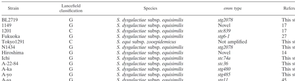
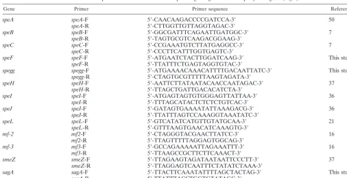
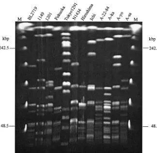
Characterization of Group C and G Streptococcal Strains That Cause
Streptococcal Toxic Shock Syndrome
Shinnosuke Hashikawa,
1Yoshitsugu Iinuma,
2Manabu Furushita,
1,3Teruko Ohkura,
1,4Toshi Nada,
4Keizo Torii,
1Tadao Hasegawa,
1and Michio Ohta
1*
Departments of Bacteriology
1and Clinical Laboratory Medicine,
4Nagoya University Graduate School of Medicine, Showa-ku,
Nagoya 466-8550, Department of Central Clinical Laboratory, Kyoto University Hospital, Konoe-cho, Yoshida, Sakyo-ku,
Kyoto 606-8501,
2and Department of Food Science and Technology, National Fisheries University, Shimonoseki,
Yamaguchi 759-6595,
3Japan
Received 29 March 2003/Returned for modification 2 June 2003/Accepted 7 October 2003
Twelve strains (the largest number ever reported) of group C and G
1streptococci (GCS and GGS,
respec-tively) that caused streptococcal toxic shock syndrome (STSS) were collected and characterized. Eleven strains
were identified as
Streptococcus dysgalactiae
subsp.
equisimilis, and one strain was identified as
Streptococcus equi
subsp.
zooepidemicus. We found that it was the first reported case of STSS caused by
S. equi
subsp.
zooepi-demicus. Cluster analysis according to the 16S rRNA gene (rDNA) sequences revealed that the
S. dysgalactiae
strains belonged to clusters I and II, both of which were closely related. The
emm
types and the restriction
patterns of chromosomal DNA measured by pulsed-field gel electrophoresis were highly variable in these
strains except BL2719 and N1434. The 16S rDNA sequences and other characteristics of these two strains were
indistinguishable, suggesting the clonal dissemination of this particular
S. dysgalactiae
strain in Japan. As the
involvement of superantigens in the pathogenesis of group A streptococcus-related STSS has been suggested,
we tried to detect known streptococcal superantigens in GCS and GGS strains. However, only the
spegg
gene
was detected in seven
S. dysgalactiae
strains, with none of the other superantigen genes being detected in any
of the strains. However, the
sagA
gene was detected in all of the strains except Tokyo1291. In the present study
no apparent factor(s) responsible for the pathogenesis of STSS was identified, although close genetic
rela-tionships of GCS and GGS strains involved in this disease were suggested.
A toxic shock-like syndrome due to group A streptococcus
(GAS) (
Streptococcus pyogenes
) was reported in the late 1980s
(8). Since then the number of patients of this serious infectious
disease has been increasing in North America, Europe, and
other parts of the world (2, 10, 18, 21, 34). The Working Group
on Severe Streptococcal Infections suggested a case definition
of streptococcal toxic shock syndrome (STSS) and necrotizing
fasciitis (52). STSS caused by GAS was characterized as a
severe infectious disease which progresses to septic shock and
multiple organ failure with a frequent occurrence of
necrotiz-ing fasciitis.
Recently, case reports referring to STSS caused by the
Lancefield group C streptococci (GCS and G
iand GGS,
re-spectively) have also accumulated (1, 2, 11, 14, 17, 24, 27, 30,
35, 38, 45, 49; H. Watanabe (ed.), Rep. 24th Hyg. Microbe
Meet., p. 23–27, 2002 [In Japanese]). However,
epidemiologi-cal analysis and study for toxin production profiles of the
caus-ative organisms have not been fully conducted yet, whereas
such analysis and study have been carried out for those
organ-isms associated with GAS-induced STSS. Most clinical isolates
of GCS and GGS show beta-hemolysis on sheep blood agar,
and some isolates are susceptible to bacitracin. Therefore,
these beta-hemolytic GCS and GGS may be misidentified as
GAS unless Lancefield serologic testing is performed.
Beta-hemolytic GCS include
S. equi
(subspecies
equi
and
zooepi-demicus
),
S. dysgalactiae
(subspecies
dysgalactiae
and
equisi-milis
),
S. anginosus
, and
S. constellatus
, and beta-hemolytic
GGS include
S. dysgalactiae
,
S. canis
,
S. uberis
,
S. intestinalis
,
and
S. anginosus
, respectively. Some of these bacteria cause
various infections in domestic animals and, sometimes, in
hu-mans. Previous studies have demonstrated that none of these
beta-hemolytic GCS and GGS secrete any major exotoxins (17,
27, 35, 38, 45, 49). Unlike GAS-induced STSS, underlying
conditions have been noted in most patients with STSS related
to GCS and GGS. These patients had cardiopulmonary
dis-ease, diabetes mellitus, malignancy, and renal or hepatic
fail-ure, etc.
In the present study we collected and characterized GCS
and GGS strains that caused STSS. We determined the 16S
rRNA gene (rDNA) sequences of these strains for
identifica-tion of the species, since the 16S rDNA is highly conserved
within a streptococcal species and can be used as the “gold
standard” for identification of the species of bacteria (51). We
performed epidemiological analyses with chromosomal
pulsed-field gel electrophoresis (PFGE) and
emm
typing of these GCS
and GGS strains. We further searched for the genes encoding
superantigenic or mitogenic toxins, including a number of
novel streptococcal exotoxins that are believed to express
su-perantigenic or mitogenic activities (1, 39, 49), for more
un-derstanding of the pathogenesis of this rare, severe infection.
MATERIALS AND METHODS
Bacterial strains.GGS and GCS strains collected in this study are shown in Table 1. All these strains were recently isolated as causative organisms from STSS patients in Japan.S. pyogenesSF370 was provided courtesy of J. J. Ferretti
* Corresponding author. Mailing address: Department of
Bacteriol-ogy, Nagoya University Graduate School of Medicine, 65
Tsurumai-cho, Showa-ku, Nagoya 466-8550, Japan. Phone: 81-52-744-2106. Fax:
81-52-744-2107. E-mail: mohta@med.nagoya-u.ac.jp.
186
on May 15, 2020 by guest
http://jcm.asm.org/
(13, 44). This strain carried superantigenic genes ofspeB,speC,speF,mf-2,mf-3,
speG,speH,speI,speJ,smeZ, andsagAand was used as a positive control.S. pyogenes1246 (29) andS. pyogenesJi are clinical isolates harboringspeA,speC, andspeLand were also used as positive controls to detect these superantigenic genes.
Identification of bacteria.The GGS and GCS strains that caused STSS were identified on the basis of phenotypic characteristics as determined by a rapid ID 32 STREP kit (bioMe´rieux, Marcy-l’Etoile, France). Phylogenetic analysis of these strains was performed with 16S rDNA sequence. The 16S rDNA was amplified by PCR using a thermal cycler (GeneAmp PCR 9700; Applied Bio-systems, Foster City, Calif.) and degenerate primers 27F [AGAGTTTGATC(A /C)TGGCTCAG] and 1492R (GGCTACCTTGTTACGACTT). PCR template DNA was prepared according to theemmtyping protocol (http://www.cdc.gov /ncidod/biotech/strep/strepindex.html). The PCR for 16S rDNA was carried out by denaturation over 2 min at 94°C, followed by 30 cycles of 60 s at 94°C, 45 s at 55°C, and 90 s at 72°C with a final extension at 72°C for 5 min. PCR products were purified with a Qiaquick PCR purification kit (Qiagen, Hilden, Germany) and were sequenced on a CEQ 2000 DNA analysis system (Beckman Coulter, Fullerton, Calif.) using a Beckman Dye terminator cycle sequencing kit (CEQ DTCS kit) with the primers 27F, 323F (AGACACGGCCCAGACTCCTA), 702F (GTAGCGGTGAAATGCGTAGA), 683R (CTACGCATTTCACCGCT AC), 1238R (TTGTAGCACGTGTGTAGCCC), and 1492R. These kits were used according to the manufacturer’s instructions. The search for 16S rDNA sequences of streptococci was performed using Similar Rank in the RDP data-base (28). The 16S rDNA sequences were aligned using CLUSTAL W software (46). Kimura’s two-parameter model was applied for the calculation of evolu-tionary distance (25), and a phylogenetic tree was constructed by using the neighbor-joining method. Bootstrap analyses of 1,000 replicates were carried out using CLUSTAL W. The classification ofS. dysgalactiaeinto two subgroups was based on the recent approval of this classification (47, 48). The 16S rDNA sequences used for depicting a phylogenetic tree include the sequences ofS. dysgalactiaesubsp.dysgalactiaeATCC 43078 (GenBank accession no. AB002485) and ATCC 27957 (AB002484);S. dysgalactiaeV26 (AB002512), A5 (AB002490), L7 (AB002507), A24 (AB002488), A7 (AB002492), L1 (AB002494), V24 (AB002510), L33 (AB002501), and A20 (AB002487);S. dysgalactiaesubsp. eq-uisimilisAC-2074 (AJ314611), AC-2832 (AJ314611), AC-2713 (AJ314609), and NCFB1356 (AB008926);S. uberisJCM 5709 (AB023573) and HN1 (AB023576);
S. equisubsp.zooepidemicusATCC 19615 (AB002516);S. equisubsp.equiATCC 33398 (AB002515); S. pyogenes ATCC 19613 (Y12924); andS. pneumoniae
MFF911410 (AB002522).
Typing of M protein gene (emm).The M protein types of the GCS and GGS strains were determined according to the M protein gene (emm) typing protocol by the sequencing of theemmgene (http://www.cdc.gov/ncidod/biotech/strep /strepindex.html).
Susceptibility testing and detection of resistance genes.Antimicrobial suscep-tibilities were determined by a MicroScan (Dade Behring, Deerfield, Ill.) mi-crodilution method and Etest (AB Biodisk, Solna, Sweden) and were confirmed by an agar dilution method.
Erythromycin resistance genes (ermB,ermTR, andmefA) were searched by PCR (12, 42, 43). For amplification of erythromycin resistance genes, the fol-lowing primers were used: 5⬘-GAAAARGTACTCAACCAAATA-3⬘ (R indi-cates A or G) and 5⬘-AGTAACGGTACTTAAATTGTTTAC-3⬘forermB(43), 5⬘-TCAGGAAAAGGACATTTTAC-3⬘ and 5⬘-ATACTTTTTGTAGTCCTTC
TTT-3⬘forermTR(designed on the basis of the homologous region ofermTR
andermA), and 5⬘-AGTATCATTAATCACTAGTGC-3⬘and 5⬘-TTCTTCTGG TACTAAAAGTGG-3⬘formefA(43).
The tetracycline resistance gene (tetM) was detected by PCR (4, 23). The sequences of the primers for amplification oftetMwere 5⬘-TTATCAACGGTT TATCAGG-3⬘and 5⬘-CGTATATATGCAAGACG-3⬘.
PCR for erythromycin resistance genes (ermB,ermTR, andmefA) was carried out by denaturation over 2 min at 94°C, followed by 30 cycles of 30 s at 94°C, 30 s at 49°C, and 90 s at 72°C with a final extension at 72°C for 5 min. FortetM, the annealing temperature was changed to 50°C.
Detection of superantigenic genes and streptolysin S gene (sagA) by PCR.
PCR primers were designed to detect superantigenic genesspeA,speB,speC,
speF,speH,speI,speJ,speL,mf-2,mf-3, andsmeZfound inS. pyogenesand a superantigenic gene,spegg(GenBank accession no. AJ294849).speggis a homo-logue of theS. pyogenessuperantigenic genespeGand was found inS. dysgalac-tiaesubsp.equisimilis(39). PCR template DNA was the same as that foremm
typing. For amplification of superantigenic genes as well as thesagA gene, primers listed in Table 2 were designed according to the indicated references.
PCR forspeA,speC,spegg, andspeIwas carried out by denaturation over 2 min at 94°C, followed by 30 cycles of 45 s at 94°C, 45 s at 51°C, and 90 s at 72°C with a final extension at 72°C for 5 min. ForspeB,speF,mf-2, andmf-3, the annealing temperature was changed to 55°C. ForspeH,speJ,speL, andsmeZ, the annealing temperature was changed to 49°C. ForsagA, the annealing temperature was changed to 50°C.
PFGE.PFGE of restriction enzyme-digested chromosomal DNA was carried out according to the previously described method (20). Briefly, chromosomal DNA was digested overnight withSmaI (Takara, Kyoto, Japan), and the digested sample was electrophoresed through 1% agarose gel in 0.5⫻TBE buffer (0.05 M Tris, 0.05 M boric acid, 1 mM EDTA [pH 8]) by using a Gene Navigator pulsed-field system (Amersham Pharmacia Biotech, Uppsala, Sweden). Electro-phoresis was carried out at 200 V for 18 h, with the pulse time ranging from 1 to 20 s. Thereafter, the gel was stained with ethidium bromide, washed with distilled water, and photographed. Lambda DNA PFGE markers (Amersham Pharmacia Biotech) were used as size standards.
Nucleotide sequence accession numbers. The nucleotide sequences deter-mined in this study were submitted to the DDBJ nucleotide sequence database, and the accession numbers for each strain were given as follows: BL2719 (AB10483), 1149 (AB104840), 1201 (AB104841), Fukuoka (AB104842), To-kyo1291 (AB104843), N1434 (AB104844), Hiroshima (AB104845), Ichi (AB104846), A-22-84 (AB104847), A-ka (AB104848), A-yo (AB104849), and A-sa (AB104850).
RESULTS
Differentiation of GCS and GGS strains and typing of the M
protein gene (emm).
The species of the GCS and GGS strains
[image:2.603.42.543.81.219.2]isolated from STSS patients were determined with a rapid ID
32 STREP kit (bioMe´rieux). The PCR-amplified 16S rDNA
fragments ranging from positions 27 to 1511 of the 16S rDNA
sequence of
Escherichia coli
were directly sequenced. These
sequences corresponded to an estimated 93% of the total 16S
TABLE 1. Species and
emm
type of GCS and GGS strains used in this study
Strain classificationLancefield Species emmtype Reference
BL2719
G
S. dysgalactiae
subsp.
equisimilis
stg2078
This study
1149
G
S. dysgalactiae
subsp.
equisimilis
Novel
17
1201
C
S. dysgalactiae
subsp.
equisimilis
stc839
17
Fukuoka
G
S. dysgalactiae
subsp.
equisimilis
stg6-1
27
Tokyo1291
C
S. equi
subsp.
zooepidemicus
Not amplified
This study
N1434
G
S. dysgalactiae
subsp.
equisimilis
stg2078
This study
Hiroshima
G
S. dysgalactiae
subsp.
equisimilis
Novel
14
Ichi
G
S. dysgalactiae
subsp.
equisimilis
stc74a
This study
A-22-84
G
S. dysgalactiae
subsp.
equisimilis
stc36
This study
A-ka
G
S. dysgalactiae
subsp.
equisimilis
stg480
This study
A-yo
G
S. dysgalactiae
subsp.
equisimilis
stg485
This study
A-sa
G
S. dysgalactiae
subsp.
equisimilis
stg11
45
on May 15, 2020 by guest
http://jcm.asm.org/
rDNA primary sequence. These sequences were compared
with the published 16S rDNA sequences of the streptococcal
reference strains as described in Materials and Methods.
Ac-cording to the 16S rDNA sequences a dendrogram depicting
the estimated phylogenetic relationships was constructed by
the neighbor-joining clustering method (Fig. 1). Three
se-quence clusters could be defined in
S. dysgalactiae
subsp.
eq-uisimilis
: cluster I contained strains Ichi, BL2719, N1434, 1149,
1201, and A-yo and had a mean 16S rDNA sequence similarity
of 99.84 to 100%; cluster II contained Hiroshima, Fukuoka,
A-22-84, A-ka, and A-sa and had a mean 16S rDNA sequence
similarity of 99.77 to 100%; and cluster III had a mean 16S
rDNA sequence similarity of 99.77 to 100%. The 16S rDNA
sequences of strains belonging to clusters I and II were close to
each other (99.62 to 99.92%), with 99.39 to 99.84% differences
from cluster III. The reference strains belonging to cluster III
have not been divided into subspecies based on the new
clas-sification (47, 48). We identified strains BL2719, 1149, 1201,
Fukuoka, N1434, Hiroshima, Ichi, A-22-84, A-ka, A-yo, and
A-sa as
S. dysgalactiae
subsp.
equisimilis
and identified strain
Tokyo1291 as
S. equi
subsp.
zooepidemicus
based on the results
of the rapid ID 32 STREP assay and of 16S rDNA analysis
(Table 1).
The M protein type was determined according to the
emm
typing protocol. As Table 1 shows, the
emm
types of BL2719,
1201, Fukuoka, N1434, Ichi, A-22-84, A-ka, A-yo, and A-sa
were
stg2078
,
stc839
,
stg6
-
1
,
stg2078
,
stc74a
,
stc36
,
stg480
,
stg485
, and
stg11
, respectively. The
emm
types of strains 1149
and Hiroshima were novel types. The
emm
gene of Tokyo1291
was not amplified. These results indicate that no specific M
proteins are related to the pathogenesis of STSS caused by
GCS and GGS.
Detection of superantigenic genes and the
sagA
gene.
In
previous reports the involvement of superantigens in the
pathogenesis of GAS-related STSS has been strongly
sus-pected (26, 32, 33). We therefore tried to detect known
strep-tococcal superantigens and putative superantigens by PCR
with specific primers in the GCS and GGS strains that caused
STSS. As shown in Table 3,
spegg
was detected in 1149, 1201,
Ichi, A-22-84, A-ka, A-yo, and A-sa, but none of the other
superantigenic genes were detected in any strains. The
sagA
gene was detected in all strains except Tokyo1291 (Table 3). It
should be noted that a negative reaction in PCR implies simply
loss or alteration of the target sequences for the primers and
the presence of the entire superantigenic gene may not be
excluded, although such a possibility is quite low.
Antimicrobial susceptibility and detection of resistance
genes.
All strains were sensitive to

-lactam antibiotics such as
penicillin G (MIC
ⱕ
0.03
g/ml). These strains were also
sensitive to vancomycin (MIC
ⱕ
0.5
g/ml),
trimethoprim-sulfamethoxazole (MIC
ⱕ
0.5
g/ml), levofloxacin (MIC
ⱕ
1.0
g/ml), chloramphenicol (MIC
ⱕ
0.5
g/ml), and rifampin
(MIC
ⱕ
1.0
g/ml). However, for 6 out of 12 strains resistance
to tetracycline was high (MIC
⫽
32
g/ml) or moderate (MIC
⫽
2 to 8
g/ml). The tetracycline resistance gene
tetM
was
detected in both 1149 and Ichi, for which resistance to
tetra-cycline was high. The strain A-sa was resistant to erythromycin,
clarithromycin, and clindamycin (MICs
ⱖ
256
g/ml) and
other strains were susceptible to these antibiotics. We detected
the erythromycin resistance gene
ermB
in A-sa.
PFGE.
PFGE patterns of all 12 GCS and GGS strains are
[image:3.603.41.547.78.338.2]shown in Fig. 2. The restriction patterns of BL2719 and N1434
are indistinguishable from each other. Note that these two
TABLE 2. PCR primer sets for detection of superantigenic genes and streptolysin S gene (sagA)
Gene Primer Primer sequence Reference
speA
speA-F
5⬘-CAACAAGACCCCGATCCA-3⬘
50
speA-R
5⬘-CTTGGTTGTTAGGTAGAC-3⬘
speB
speB-F
5⬘-GGCGATTTCAGAATTGATGGC-3⬘
7
speB-R
5⬘-TAGTGCGTCAAGACGGAAG-3⬘
speC
speC-F
5⬘-CCGAAATGTCTTATGAGGCC-3⬘
7
speC-R
5⬘-CCCTTCATTTGGTGAGTC-3⬘
speF
speF-F
5⬘-ATGAATCTACTTGGATCAAG-3⬘
This study
speF-R
5⬘-TTATTTCTGAGTAGGTGTAC-3⬘
spegg
spegg-F
5⬘-ATGAAAACAAACATTTTGACAATTATC-3⬘
This study
spegg-R
5⬘-CTAGTGCGTTTTTAAGTAGATA-3⬘
speH
speH-F
5⬘-AATTCTTATAATACAACCAATAGAC-3⬘
37
speH-R
5⬘-TTAGCTGATTGACACATCTA-3⬘
speI
speI-F
5⬘-ATGAGTAGTGTGGGAGTTATTAA-3⬘
36
speI-R
5⬘-TTTAGCATACTCTCTCTGTCAC-3⬘
speJ
speJ-F
5⬘-GATAGTGAAAATATTAAAGACG-3⬘
36
speJ-R
5⬘-TTATTTAGTCCAAAGGTAAATATC-3⬘
speL
speL-F
5⬘-GTCATATCATGTTGTATGCAA-3⬘
21
speL-R
5⬘-GTTTAAGTGAACATCAAAGTG-3⬘
mf-2
mf2-F
5⬘-CTAGGGTACGAACTTATCC-3⬘
16
mf2-R
5⬘-TTAGTTTTTAGGAGTGGCAG-3⬘
mf-3
mf3-F
5⬘-GCCAGAAAAATTAGAAATTT-3⬘
16
mf3-R
5⬘-TTAAGCCGCTTCTTCAAACT-3⬘
smeZ
smeZ-F
5⬘-TTAGAAGTAGATAATAATTCCCTT-3⬘
37
smeZ-R
5⬘-TTAGGAGTCAATTTCTATATCTAAA-3⬘
sagA
sagA-F
5⬘-TTACTTCAAATATTTTAGCTACTAG-3⬘
This study
sagA-R
5⬘-TTATTTACCTGGTGTATAGC-3⬘
on May 15, 2020 by guest
http://jcm.asm.org/
strains were isolated in different places and years. Other strains
were genetically diverse.
DISCUSSION
In the past case reports of STSS caused by GCS and GGS (1,
2, 11, 14, 17, 24, 27, 30, 35, 38, 45, 49; H. Watanabe, Rep. 24th
Hyg. Microbe Meet.), the causative bacteria in six cases were
identified as
S. dysgalactiae
subsp.
equisimilis
(2, 27, 38) and
S.
equisimilis
(24, 45), which was renamed as
S. dysgalactiae
subsp.
equisimilis
according to the latest classification (47, 48), while
other reports did not identify the species of the causative
bacteria. To the best of our knowledge, in the present study we
report the first case of STSS caused by
S. equi
subsp.
zooepi-demicus
. Clinical information is as follows. A 64-year-old male
patient visited an emergency room on foot with lumbago, and
2 h later he lost consciousness and died. Autopsy of the patient
indicated necrotic fasciitis of the left arm and underlying
cir-rhosis. The organism was isolated from the blood culture and
the left arm. Strains of
S. dysgalactiae
subsp.
equisimilis
belong
to Lancefield group C or G, whereas strains of
S. equi
subsp.
zooepidemicus
belongs to only group C. While
S. dysgalactiae
subsp.
equisimilis
colonizes and causes various infections in
humans (51, 53),
S. equi
subsp.
zooepidemicus
is a common
pathogen in domestic animals and causes various infections in
animals. Therefore, most cases of human infection can be
traced to an animal source (5). As our present study and earlier
reports have indicated, these organisms secrete only a few
superantigenic exotoxins or unknown exotoxins (1, 17, 27, 35,
38, 45, 49). Although clinical manifestations of STSS related to
GCS and GGS are the same as those caused by GAS, the
involvement of major superantigens such as SpeA, SpeB, and
SpeC secreted from GAS are excluded. It has been proposed
that streptococcal superantigens, especially pyrogenic
exotox-FIG. 1. Phylogenetic tree of GCS and GGS strains isolated from patients with STSS. A phylogenetic tree based on the 16S rDNA sequences
was constructed by the neighbor-joining method. Bootstrap analyses of 1,000 replicates were carried out using CLUSTAL W software. Each of the
STSS-related strains analyzed in this study is indicated as a square. Sequences of other strains were derived from the GenBank database. Species
names of reference strains
S. dysgalactiae
V26, A5, L7, A24, A7, L1, V24, L33, and A20 appeared in the database under the old classification and
were not identified into subspecies.
on May 15, 2020 by guest
http://jcm.asm.org/
[image:4.603.50.525.74.467.2]ins, are potentially responsible for at least some of the
mani-festations of STSS caused by GAS through the activation of
Th1 cells and the liberation of large amounts of interleukins as
well as inflammatory cytokines, such as tumor necrosis factor
(9). Therefore, unidentified superantigenic exotoxins or
exoen-zymes may play a role in the development of STSS caused by
GCS and GGS. Indeed, it has been reported that some GAS
strains that caused STSS lacked one or more major
superan-tigens (2, 18, 21).
[image:5.603.43.545.81.272.2]Streptolysin S encoded by
sagA
was considered to be a
con-tributing factor in the pathogenesis of streptococcal
necrotiz-ing soft tissue infection (19). The
sagA
gene was detected in all
[image:5.603.134.458.393.709.2]FIG. 2. PFGE separation of
SmaI-restricted fragments of the chromosomal DNA of the GCS and GGS strains.
TABLE 3. Detection of superantigenic genes and streptolysin S gene (sagA) by PCR
Strain Detection of:
speA speB speC speF spegg speH speI speJ speL mf-2 mf-3 smeZ sagA
BL2719
ND
aND
ND
ND
ND
ND
ND
ND
ND
ND
ND
ND
Positive
1149
ND
ND
ND
ND
Positive ND
ND
ND
ND
ND
ND
ND
Positive
1201
ND
ND
ND
ND
Positive ND
ND
ND
ND
ND
ND
ND
Positive
Fukuoka
ND
ND
ND
ND
ND
ND
ND
ND
ND
ND
ND
ND
Positive
Tokyo1291
ND
ND
ND
ND
ND
ND
ND
ND
ND
ND
ND
ND
ND
N1434
ND
ND
ND
ND
ND
ND
ND
ND
ND
ND
ND
ND
Positive
Hiroshima
ND
ND
ND
ND
ND
ND
ND
ND
ND
ND
ND
ND
Positive
Ichi
ND
ND
ND
ND
Positive ND
ND
ND
ND
ND
ND
ND
Positive
A-22-84
ND
ND
ND
ND
Positive ND
ND
ND
ND
ND
ND
ND
Positive
A-ka
ND
ND
ND
ND
Positive ND
ND
ND
ND
ND
ND
ND
Positive
A-yo
ND
ND
ND
ND
Positive ND
ND
ND
ND
ND
ND
ND
Positive
A-sa
ND
ND
ND
ND
Positive ND
ND
ND
ND
ND
ND
ND
Positive
Positive controls
S. pyogenes
SF370 ND
Positive Positive Positive Positive Positive Positive Positive ND
Positive Positive Positive Positive
S. pyogenes
1246
Positive Positive Positive Positive Positive ND
ND
Positive ND
Positive Positive Positive Positive
S. pyogenes
Ji
Positive Positive ND
ND
ND
Positive ND
ND
Positive Positive
aND, not detected.
on May 15, 2020 by guest
http://jcm.asm.org/
of the investigated GCS and GGS strains except Tokyo1291.
However, the patient infected with Tokyo1291 showed
necro-tizing fasciitis, suggesting that streptolysin S may not be
essen-tial for the development of streptococcal necrotizing soft tissue
infection, or in this case, underlying host factors might have an
important role in the pathogenesis.
M protein is known to be the major virulence factor of GAS
that inhibits the activation of the alternative complement
path-way (9) and impedes phagocytosis by polymorphonuclear
leu-kocytes (9). In epidemiological studies of STSS caused by
GAS, M1 and M3 strains were most commonly isolated (2, 22,
34; H. Watanabe, Rep. 23rd Hyg. Microbe Meet.). However,
other M serotypes of GAS have infrequently also been isolated
from STSS patients. Therefore, the role of specific M proteins
in the pathogenesis of STSS is still unknown. Since GCS and
GGS have been shown to express M proteins that show high
sequence homology to those of GAS (3, 15, 41), M proteins of
GCS and GGS may also have antiphagocytic activity. However,
whether or not GCS and GGS strains of specific
emm
(M)
types are involved in the pathogenesis of STSS remains
un-known, since only a few epidemiological studies on
emm
types
of GCS and GGS that caused STSS have been performed (H.
Watanabe, Rep. 23rd Hyg. Microbe Meet.). We found that the
M types of GCS and GGS strains isolated from STSS patients
were highly variable, suggesting that no specific types of M
proteins were responsible for the pathogenesis of STSS caused
by GCS and GGS. Interestingly, M type
stg2078
was identified
in two strains that were isolated in different places and on
different occasions. Moreover, the PFGE patterns and the
sequences of 16S rDNA of these strains were indistinguishable
from each other. We therefore suspect the clonal
dissemina-tion of this particular
S. dysgalactiae
subsp.
equisimilis
strain in
Japan. Additionally, we add two new
emm
types found in this
study to the M protein group of
S. dysgalactiae
.
Unlike those with GAS-induced STSS, most patients with
STSS caused by GCS and GGS have been noted to have
serious underlying diseases. Patients with STSS in this study
also had various underlying diseases such as malignancy,
chronic renal or hepatic failure, diabetes mellitus, and collagen
diseases. Most of these patients were given antibiotic therapy
with penicillins and/or other antibiotics. As the present study
indicated, all STSS strains were penicillin-sensitive. However,
six strains were resistant to tetracycline and one strain was
resistant to erythromycin. Among six strains that were resistant
to tetracycline, strains 1149 and Ichi harbored
tetM
. Strain
A-sa, which was resistant to erythromycin, harbored
ermB
.
These sensitivity patterns were similar to those of other reports
(6, 31) and GAS strains (6, 31, 40).
A nationwide surveillance for GAS-induced STSS started in
Japan in 1999 by the enforcement of a new infection control
law. However, cases of GCS and GGS-induced STSS were
excluded in that surveillance. Because it seems likely that the
frequency of STSS caused by GCS and GGS has increased
recently, a surveillance study must be required for more
un-derstanding of the disease. In the present study we analyzed a
larger number of STSS-causing GCS and GGS strains than had
been analyzed in previous reports. Although we found no
ap-parent STSS-related pathogenic factors in these strains, most
of the strains belonged to the specific groups of
S. dysgalactiae
subsp.
equisimilis
. Epidemiological studies for more GCS and
GGS strains related to STSS are expected to be helpful for the
identification of the common pathogenic factors in this disease.
ACKNOWLEDGMENTS
We warmly thank J. J. Ferretti, University of Oklahoma Health
Sciences Center, for providing the
S. pyogenes
strain SF370. We also
thank Nihon University School of Medicine, Tokyo Metropolitan
Re-search Laboratory of Public Health, Fukuoka University Hospital,
Tokyo Medical University Department Anesthesia, Asahi General
Hospital, Ichinomiya City Hospital, and Hiroshima City Hospital for
providing us patient information with GCS or GGS clinical isolates
that caused STSS.
This study was supported by Grants-in-Aid for Scientific Research
(12557027 and 14370090) from the Ministry of Education and a grant
for research on emerging and reemerging infectious disease (H12
Shinkou-27) from the Ministry of Health and Welfare, Japan.
REFERENCES
1. Assimacopoulos, A. P., J. A. Stoehr, and P. M. Schlievert.1997. Mitogenic factors from group G streptococci associated with scarlet fever and strepto-coccal toxic shock syndrome. Adv. Exp. Med. Biol.418:109–114. 2. Barnham, M. R., N. C. Weightman, A. W. Anderson, and A. Tanna.2002.
Streptococcal toxic shock syndrome: a description of 14 cases from North Yorkshire, UK. Clin. Microbiol. Infect.8:174–181.
3. Ben Nasr, A., A. Wistedt, U. Ringdahl, and U. Sjobring.1994. Streptokinase activates plasminogen bound to human group C and G streptococci through M-like proteins. Eur. J. Biochem.222:267–276.
4. Blanchard, A., D. M. Crabb, K. Dybvig, L. B. Duffy, and G. H. Cassell.1992. Rapid detection of tetM in Mycoplasma hominis and Ureaplasma urealyti-cum by PCR: tetM confers resistance to tetracycline but not necessarily to doxycycline. FEMS Microbiol. Lett.74:277–281.
5. Bradley, S. F., J. J. Gordon, D. D. Baumgartner, W. A. Marasco, and C. A. Kauffman.1991. Group C streptococcal bacteremia: analysis of 88 cases. Rev. Infect. Dis.13:270–280.
6. Carroll, K. C., P. Monroe, S. Cohen, M. Hoffman, L. Hamilton, K. Korgen-ski, L. Reimer, D. Classen, and J. Daly.1997. Susceptibility of beta-hemo-lytic streptococci to nine antimicrobial agents among four medical centers in Salt Lake City, Utah, USA. Diagn. Microbiol. Infect. Dis.27:123–128. 7. Chaussee, M. S., J. Liu, D. L. Stevens, and J. J. Ferretti.1996. Genetic and
phenotypic diversity among isolates of Streptococcus pyogenes from invasive infections. J. Infect. Dis.173:901–908.
8. Cone, L. A., D. R. Woodard, P. M. Schlievert, and G. S. Tomory.1987. Clinical and bacteriologic observations of a toxic shock-like syndrome due to Streptococcus pyogenes. N. Engl. J. Med.317:146–149.
9. Cunningham, M. W.2000. Pathogenesis of group A streptococcal infections. Clin. Microbiol. Rev.13:470–511.
10. Davies, H. D., A. McGeer, B. Schwartz, K. Green, D. Cann, A. E. Simor, D. E. Low, et al.1996. Invasive group A streptococcal infections in Ontario, Can-ada. N. Engl. J. Med.335:547–554.
11. Eskens, F. A., P. E. Verweij, J. F. Meis, and A. Soomers.1995. Septic shock caused by group G beta-haemolytic streptococci as presenting symptom of acute myeloid leukaemia. Neth. J. Med.46:153–155.
12. Facinelli, B., C. Spinaci, G. Magi, E. Giovanetti, and E. V. P.2001. Associ-ation between erythromycin resistance and ability to enter human respiratory cells in group A streptococci. Lancet358:30–33.
13. Ferretti, J. J., W. M. McShan, D. Ajdic, D. J. Savic, G. Savic, K. Lyon, C. Primeaux, S. Sezate, A. N. Suvorov, S. Kenton, H. S. Lai, S. P. Lin, Y. Qian, H. G. Jia, F. Z. Najar, Q. Ren, H. Zhu, L. Song, J. White, X. Yuan, S. W. Clifton, B. A. Roe, and R. McLaughlin.2001. Complete genome sequence of an M1 strain of Streptococcus pyogenes. Proc. Natl. Acad. Sci. USA98:
4658–4663.
14. Fujiwara, Y., et al.1999. Two case reports of toxic shock-like syndrome (TSLS). Cent. Jpn. J. Orthop. Traumat.42:1187–1188. (In Japanese.) 15. Geyer, A., A. Roth, S. Vettermann, E. Gunther, A. Groh, E. Straube, and K.
Schmidt.1999. M protein of a Streptococcus dysgalactiae human wound isolate shows multiple binding to different plasma proteins and shares epitopes with keratin and human cartilage. FEMS Immunol. Med. Micro-biol.26:11–24.
16. Hasegawa, T., K. Torii, S. Hashikawa, Y. Iinuma, and M. Ohta.2002. Cloning and characterization of two novel DNases from Streptococcus pyo-genes. Arch. Microbiol.177:451–456.
17. Hirose, Y., K. Yagi, H. Honda, H. Shibuya, and E. Okazaki.1997. Toxic shock-like syndrome caused by non-group A beta-hemolytic streptococci. Arch. Intern. Med.157:1891–1894.
18. Hsueh, P. R., J. J. Wu, P. J. Tsai, J. W. Liu, Y. C. Chuang, and K. T. Luh.
1998. Invasive group A streptococcal disease in Taiwan is not associated with the presence of streptococcal pyrogenic exotoxin genes. Clin. Infect. Dis.
26:584–589.
on May 15, 2020 by guest
http://jcm.asm.org/
19. Humar, D., V. Datta, D. J. Bast, B. Beall, J. C. De Azavedo, and V. Nizet.
2002. Streptolysin S and necrotising infections produced by group G strep-tococcus. Lancet359:124–129.
20. Ichiyama, S., M. Ohta, K. Shimokata, N. Kato, and J. Takeuchi.1991. Genomic DNA fingerprinting by pulsed-field gel electrophoresis as an epi-demiological marker for study of nosocomial infections caused by methicil-lin-resistantStaphylococcus aureus. J. Clin. Microbiol.29:2690–2695. 21. Ikebe, T., A. Wada, Y. Inagaki, K. Sugama, R. Suzuki, D. Tanaka, A.
Tamaru, Y. Fujinaga, Y. Abe, Y. Shimizu, and H. Watanabe.2002. Dissem-ination of the phage-associated novel superantigen genespeLin recent invasive and noninvasiveStreptococcus pyogenesM3/T3 isolates in Japan. Infect. Immun.70:3227–3233.
22. Inagaki, Y., T. Konda, S. Murayama, S. Yamai, A. Matsushima, Y. Gyobu, D. Tanaka, A. Tamaru, C. Katsukawa, A. Katayama, M. Tomita, Y. Fuchi, K. Hoashi, H. Watanabe, et al.1997. Serotyping of Streptococcus pyogenes isolated from common and severe invasive infections in Japan, 1990–5: implication of the T3 serotype strain-expansion in TSLS. Epidemiol. Infect.
119:41–48.
23. Jeric, P. E., H. Lopardo, P. Vidal, S. Arduino, A. Fernandez, B. E. Orman, D. O. Sordelli, and D. Centron.2002. Multicenter study on spreading of the
tet(M) gene in tetracycline-resistantStreptococcusgroup G and C isolates in Argentina. Antimicrob. Agents Chemother.46:239–241.
24. Keiser, P., and W. Campbell.1992.⬘Toxic strep syndrome’ associated with group C Streptococcus. Arch. Intern. Med.152:882–884.
25. Kimura, M.1980. A simple method for estimating evolutionary rates of base substitutions through comparative studies of nucleotide sequences. J. Mol. Evol.16:111–120.
26. Kotb, M.1995. Bacterial pyrogenic exotoxins as superantigens. Clin. Micro-biol. Rev.8:411–426.
27. Kugi, M., H. Tojo, I. Haraga, T. Takata, K. Handa, and K. Tanaka.1998. Toxic shock-like syndrome caused by group G Streptococcus. J. Infect.37:
308–309.
28. Maidak, B. L., J. R. Cole, T. G. Lilburn, C. T. Parker, Jr., P. R. Saxman, R. J. Farris, G. M. Garrity, G. J. Olsen, T. M. Schmidt, and J. M. Tiedje.2001. The RDP-II (Ribosomal Database Project). Nucleic Acids Res.29:173–174. 29. Matsumoto, M., T. Murai, S. Ichiyama, M. Saito, Y. Arakawa, and M. Ohta.
1997. Prevalence of the speA2 and speA3 alleles in Streptococcus pyogenes isolated from TSLS patients in Japan. FEMS Microbiol. Lett.150:233–237. 30. Natoli, S., C. Fimiani, N. Faglieri, L. Laurenzi, A. Calamaro, A. M. Frasca, and E. Arcuri.1996. Toxic shock syndrome due to group C streptococci. A case report. Intensive Care Med.22:985–989.
31. Navaneeth, B. V., N. Ray, S. Chawda, P. Selvarani, M. Bhaskar, and N. Suganthi.2001. Prevalence of beta hemolytic streptococci carrier rate among schoolchildren in Salem. Indian J. Pediatr.68:985–986.
32. Norrby-Teglund, A., S. Chatellier, D. E. Low, A. McGeer, K. Green, and M. Kotb.2000. Host variation in cytokine responses to superantigens determine the severity of invasive group A streptococcal infection. Eur. J. Immunol.
30:3247–3255.
33. Norrby-Teglund, A., R. Lustig, and M. Kotb.1997. Differential induction of Th1 versus Th2 cytokines by group A streptococcal toxic shock syndrome isolates. Infect. Immun.65:5209–5215.
34. O’Brien, K. L., B. Beall, N. L. Barrett, P. R. Cieslak, A. Reingold, M. M. Farley, R. Danila, E. R. Zell, R. Facklam, B. Schwartz, and A. Schuchat.
2002. Epidemiology of invasive group a streptococcus disease in the United States, 1995–1999. Clin. Infect. Dis.35:268–276.
35. Ojukwu, I. C., D. W. Newton, A. E. Luque, M. Y. Kotb, and M. Menegus.
2001. Invasive Group C Streptococcus infection associated with rhabdomy-olysis and disseminated intravascular coagulation in a previously healthy adult. Scand. J. Infect. Dis.33:227–229.
36. Proft, T., V. L. Arcus, V. Handley, E. N. Baker, and J. D. Fraser.2001. Immunological and biochemical characterization of streptococcal pyrogenic
exotoxins I and J (SPE-I and SPE-J) from Streptococcus pyogenes. J. Im-munol.166:6711–6719.
37. Proft, T., S. L. Moffatt, C. J. Berkahn, and J. D. Fraser.1999. Identification and characterization of novel superantigens from Streptococcus pyogenes. J. Exp. Med.189:89–102.
38. Roth, S., K. Andrassy, K. H. Schmidt, E. Gunther, and E. Ritz.1999. Febrile lady with acute renal failure and desquamating erythema. Am. J. Kidney Dis.
34:150–154.
39. Sachse, S., P. Seidel, D. Gerlach, E. Gunther, J. Rodel, E. Straube, and K. H. Schmidt.2002. Superantigen-like gene(s) in human pathogenic Streptococ-cus dysgalactiae, subsp equisimilis: genomic localisation of the gene encod-ing streptococcal pyrogenic exotoxin G (speG(dys)). FEMS Immunol. Med. Microbiol.34:159–167.
40. Sauermann, R., R. Gattringer, W. Graninger, A. Buxbaum, and A. Georgo-poulos.2003. Phenotypes of macrolide resistance of group A streptococci isolated from outpatients in Bavaria and susceptibility to 16 antibiotics. J. Antimicrob. Chemother.51:53–57.
41. Schnitzler, N., A. Podbielski, G. Baumgarten, M. Mignon, and A. Kaufhold.
1995. M or M-like protein gene polymorphisms in human group G strepto-cocci. J. Clin. Microbiol.33:356–363.
42. Seppa¨la¨, H., M. Skurnik, H. Soini, M. C. Roberts, and P. Huovinen.1998. A novel erythromycin resistance methylase gene (ermTR) inStreptococcus pyo-genes. Antimicrob. Agents Chemother.42:257–262.
43. Sutcliffe, J., T. Grebe, A. Tait-Kamradt, and L. Wondrack.1996. Detection of erythromycin-resistant determinants by PCR. Antimicrob. Agents Che-mother.40:2562–2566.
44. Suvorov, A. N., and J. J. Ferretti.1996. Physical and genetic chromosomal map of an M type 1 strain ofStreptococcus pyogenes. J. Bacteriol.178:5546– 5549.
45. Tanno, M., K. Oe, Y. Shimizu, K. Murata, T. Kubota, and J. Suzuki.2000. A case of acute group G streptococcal myositis associated with toxic shock like syndrome. J. Jpn. Soc. Intensive Care Med.7:115–120. (In Japanese.) 46. Thompson, J. D., D. G. Higgins, and T. J. Gibson.1994. CLUSTAL W:
improving the sensitivity of progressive multiple sequence alignment through sequence weighting, position-specific gap penalties and weight matrix choice. Nucleic Acids Res.22:4673–4680.
47. Vandamme, P., B. Pot, E. Falsen, K. Kersters, and L. A. Devriese.1996. Taxonomic study of Lancefield streptococcal groups C, G, and L ( Strepto-coccus dysgalactiae) and proposal ofS. dysgalactiaesubsp.equisimilissubsp. nov. Int. J. Syst. Bacteriol.46:774–781.
48. Vieira, V. V., L. M. Teixeira, V. Zahner, H. Momen, R. R. Facklam, A. G. Steigerwalt, D. J. Brenner, and A. C. Castro.1998. Genetic relationships among the different phenotypes ofStreptococcus dysgalactiaestrains. Int. J. Syst. Bacteriol.48:1231–1243.
49. Wagner, J. G., P. M. Schlievert, A. P. Assimacopoulos, J. A. Stoehr, P. J. Carson, and K. Komadina.1996. Acute group G streptococcal myositis associated with streptococcal toxic shock syndrome: case report and review. Clin. Infect. Dis.23:1159–1161.
50. Weeks, C. R., and J. J. Ferretti.1986. Nucleotide sequence of the type A streptococcal exotoxin (erythrogenic toxin) gene fromStreptococcus pyogenes
bacteriophage T12. Infect. Immun.52:144–150.
51. Woo, P. C., A. M. Fung, S. K. Lau, S. S. Wong, and K. Y. Yuen.2001. Group G beta-hemolytic streptococcal bacteremia characterized by 16S ribosomal RNA gene sequencing. J. Clin. Microbiol.39:3147–3155.
52. The Working Group on Severe Streptococcal Infections.1993. Defining the group A streptococcal toxic shock syndrome. Rationale and consensus def-inition. JAMA269:390–391.
53. Zaoutis, T., B. Schneider, L. Steele Moore, and J. D. Klein.1999. Antibiotic susceptibilities of group C and group G streptococci isolated from patients with invasive infections: evidence of vancomycin tolerance among group G serotypes. J. Clin. Microbiol.37:3380–3383.