Copyright © 2003, American Society for Microbiology. All Rights Reserved.
Development and Evaluation of a Seminested PCR for Detection and
Differentiation of
Babesia gibsoni
(Asian Genotype) and
B. canis
DNA in Canine Blood Samples
Adam J. Birkenheuer,
1Michael G. Levy,
2and Edward B. Breitschwerdt
1*
Departments of Clinical Sciences1and Food Animal and Health Resource Management,2College of Veterinary
Medicine, North Carolina State University, Raleigh, North Carolina 27606-1428 Received 6 November 2002/Returned for modification 18 March 2003/Accepted 8 May 2003
Canine babesiosis has recently been recognized as an emerging infectious disease of dogs in North America. We sought to develop a seminested PCR to detect and differentiateBabesia gibsoni(Asian genotype),B. canis
subsp.vogeli,B. canissubsp.canis, andB. canissubsp.rossiDNA in canine blood samples. An outer primer pair was designed to amplify an⬃340-bp fragment of the 18S rRNA genes fromB. gibsoni(Asian genotype),B. canis
subsp.vogeli,B. canissubsp.rossi, andB. canissubsp.canisbut not mammalian DNA. Forward primers were designed that would specifically amplify a smaller fragment from each organism in a seminested PCR. The practical limit of detection was 50 organisms/ml of mock-infected EDTA anticoagulated whole blood. The primer pair also amplified an⬃370-bp fragment of theB. gibsoni(USA/California genotype) 18S rRNA gene from the blood of an experimentally infected dog with a high percentage of parasitemia. Amplicons were not detected when DNA extracted from the blood of a dog that was naturally infected withTheileria annaeat a low percentage of parasitemia was amplified. Due to limited sensitivity, this test is not recommended for the routine diagnosis ofB. gibsoni(USA/California genotype) orT. annae. The PCR test did not amplifyToxoplasma gondii, Neospora caninum, Leishmania infantum, Cryptosporidium parvum, or canine DNA under any of the conditions tested. The seminested PCR test was able to detect and discriminateB. gibsoni(Asian genotype),
B. canissubsp.vogeli,B. canissubsp.canis, andB. canissubsp.rossiDNA in blood samples from infected dogs.
Babesiosis is an important disease of domestic dogs in the United States caused by intraerythrocytic protozoan parasites of the genus Babesia. These hemoprotozoan parasites, along withTheileriaspp., are often referred to as piroplasms. Babe-siosis is typically characterized by hemolytic anemia, thrombo-cytopenia, fever, and splenomegaly. Historically, canine babe-siosis has been attributed to infection with eitherBabesia canis
orB. gibsoni, based on parasite size and the geographic loca-tion in which the infecloca-tion was acquired. Recently, an in-creased number of genetically unique piroplasms have been identified, and the recognized geographic ranges of various canine piroplasms appear to be expanding. Since Conrad et al. reported the first outbreak of canine babesiosis caused by a smallBabesiasp. in the United States in 1991, there have been an increasing number of reports of dogs infected with small
Babesiasp. (4, 9, 14–17, 24). During the California outbreak in 1991,B. gibsoniwas presumed to be the only smallBabesiasp. to infect dogs. However, recent studies demonstrate that there are at least three genetically distinct smallBabesia-like organ-isms or piroplasms that can infect dogs (15, 24, 25). Based on evolving data, the nomenclature of these small piroplasms is likely to undergo revision.
It is diagnostically important to determine the species, sub-species, and genotype that causes canine babesiosis, since the virulence, prognosis, and response to antibabesial drugs may be different for each organism. We refer here to the Asian
genotype ofB. gibsoni(GenBank accession numbers AF271081, AF271082, AF205636, AF175300, and AF175301) asB. gibsoni
(Asian genotype) (15, 16, 25), the North American genotype of
B. gibsoni (AF158702, AF231350, and L13729) as B. gibsoni
(USA/California genotype) (11, 15, 25), and the European small canine piroplasm (AF188001) asTheileria annae, as has been proposed by Zahler et al. (24). BothB. gibsoni (Asian genotype) andB. gibsoni(USA/California genotype) have been identified in dogs from North America, whereasT. annaehas only been reported in Europe (9, 24).B. gibsoni(Asian geno-type) is considered to be virulent in dogs and, to date, no antibabesial treatment has been able to eliminate the infection (22).B. gibsoni(USA/California genotype) is also virulent, but its susceptibility to antibabesial therapy has not been well char-acterized (21, 23). To our knowledge, comparative pathoge-nicities or responsiveness to antibabesial therapy forT. annae
has not been studied.
There is support for the existence of three subspecies of large canine piroplasms—B. canissubsp.vogeli,B. canissubsp.
canis, andB. canissubsp.rossi—based on genetic data, vector specificity, and variations in pathogenicity (10, 20, 26).B. canis
subsp.vogeli, which is found in North America, Europe, and Asia, is considered to be a moderately virulent species, and it is presumed that antibabesial therapy will eliminate the infec-tion.B. canissubsp.canis, which is mostly found in Europe, has somewhat variable virulence, and the organism is generally considered to respond to antibabesial drugs (10, 20, 26).B. ca-nis subsp. rossi, which has only been identified in Africa, is considered to be a highly virulent piroplasm that may not be susceptible to the currently available drugs (10, 20, 26).
The definitive diagnosis of canine babesiosis, as well as the
* Corresponding author. Mailing address: North Carolina State University, College of Veterinary Medicine, 4700 Hillsborough St., Raleigh, NC 27606-1428. Phone: (919) 513-6357. Fax: (919) 513-6336. E-mail: [email protected].
4172
on May 15, 2020 by guest
http://jcm.asm.org/
visual differentiation of the species of piroplasms, can be dif-ficult for the clinician. The generally accepted “gold stan-dard(s)” for ruling out babesiosis are splenectomy with or without immune suppression or blood transfusion from the suspect dog into a splenectomized dog (5). For obvious rea-sons, these procedures are rarely, if ever, performed in the clinical setting to achieve a diagnosis. Historically, light micro-scopic examination of stained blood smears and serology have provided the primary means of diagnosing babesiosis in dogs. Light microscopic examination cannot be used to establish the genotype of any piroplasm. Antibodies to Babesia spp. are often cross-reactive; therefore, serology may not definitively discriminate species or subspecies. In addition, there are re-ports of canineBabesiainfections in which piroplasms were not identified by light microscopic examination and/or in which serologic testing yielded false-negative results in dogs that were infected withBabesia(4, 6, 17). Since the geographic range of specific piroplasms appears to be expanding, location should not be used as the sole criterion for species or subspecies identification. Although not without limitations, the PCR of-fers a practical and noninvasive means to detect and differen-tiate infections with variousBabesiaspp. and also provides a sensitive tool for assessing treatment outcomes. PCR is likely to be more sensitive than light microscopic examination of stained blood smears based on the reported limits of detection for each test (5). Since infected dogs may have antibodies that are unpredictably cross-reactive against otherBabesiaspecies or subspecies, PCR is more specific than serology. To our knowledge, there are no studies directly comparing all of the available diagnostic tests for babesiosis in a canine population in which the true disease prevalence is known. In the present study we sought to develop a PCR test for canine babesiosis that can detect and differentiateB. gibsoni(Asian genotype),
B. canissubsp.vogeli,B. canissubsp.canis, andB. canissubsp.
rossiand to define the test’s limits of detection in canine blood samples.
MATERIALS AND METHODS
Samples.B. gibsoni(Asian genotype)-infected whole-blood samples were ei-ther obtained from a specific-pathogen-free splenectomized dog that was in-fected intravenously with blood from a dog that was confirmed to be inin-fected withB. gibsoni(Asian genotype) or from dogs (n⫽5) from North America that were confirmed to be infected with a small piroplasm.B. canissubsp.vogeli -infected canine whole-blood samples were obtained from dogs (n⫽3) from North America that were confirmed via light microscopy to be infected with large piroplasms. AB. canissubsp.canis-infected canine whole-blood sample was obtained from a specific-pathogen-free splenectomized dog that was infected intravenously with blood from a dog that was confirmed to be infected with
B. canissubsp.canis.B. canissubsp.rossi-infected canine whole-blood samples, kindly provided by George Moore (U.S. Army Medical Department Center and School, Fort Sam Houston, Tex.), were obtained from dogs (n⫽2) from South Africa that were confirmed via light microscopy to be infected with large piro-plasms.B. gibsoni(California/USA genotype)-infected canine whole-blood sam-ples were obtained from a specific-pathogen-free splenectomized dog that was infected intravenously with blood from a dog that was confirmed to be infected withB. gibsoni(California/USA genotype). The originalB. gibsoni(California/ USA genotype) isolate was kindly provided by Patricia Conrad (University of California, Davis). AT. annae-infected canine whole-blood sample from Spain, kindly provided by Sam Telford III (Harvard University, Boston, Mass.), was obtained from a dog confirmed via light microscopy to be infected with small piroplasms.Toxoplasma gondiiandNeospora caninumDNAs were kindly pro-vided by Nick Sharp (Animal Critical Care Group of Vancouver, Burnaby, British Columbia, Canada). DNA was also extracted from the anticoagulated whole blood of a dog that was naturally infected withLeishmania infantum(13).
Cryptosporidium parvumDNA was kindly provided by Lance Perryman (Colo-rado State University, Fort Collins).
Primer design.Oligonucleotide primers were designed based on the canine
Babesia18S rRNA genes reported in GenBank (3). For the amplification of the nearly full-lengthBabesia18S rRNA genes, primers (5-22F and 1661R) were designed to amplifyBabesia18S rRNA genes but not mammalian 18S rRNA genes.
For the seminested PCR, an outer primer pair (455-479F and 793-772R) was designed that would amplify an approximately⬃340-bp fragment fromB. gibsoni
(Asian genotype) (AF271081, AF271082, AF205636, AF175300, and AF175301),
B. canissubsp.vogeli(AJ009796 and AY072925),B. canissubsp.canis(AJ009795 and AY072926), andB. canissubsp.rossi(L19079) that spanned a hypervariable region of the 18S rRNA gene. Then, specific internal primers were designed for
B. gibsoni(Asian genotype) (BgibAsia-F),B. canissubsp.vogeli(BCV-F),B. ca-nissubsp.canis(BCC-F), andB. canissubsp.rossi(BCR-F) that were paired with the outer reverse primer in the seminested secondary reaction to amplify 185-, 192-, 198-, and 197-bp amplicons, respectively. The sequences of the oligonucleo-tide primers used in the present study are listed in Table 1. All primers were synthesized by Integrated DNA Technologies, Coralville, Iowa.
Preparation of DNA.DNA was isolated from canine whole-blood samples by using the QIAamp DNA Blood Mini-Kit according to the manufacturer’s in-structions. Plasmid DNA was isolated with the QIAprep Spin Miniprep accord-ing to the manufacturer’s instructions.
PCR.The nearly full-lengthB. gibsoni(Asian genotype) andB. canissubsp.
canis 18S rRNA genes were amplified by PCR. Amplification of the nearly full-length 18S rRNA genes was performed by using 25-l reactions. Each 25-l reaction contained a 1⫻concentration of PCR buffer II (Perkin-Elmer), 0.625 U ofTaqpolymerase, 0.5l of DNA template, 1.5 mM MgCl2,12.5 pmol of each
primer, and a 200M concentration of each deoxynucleoside triphosphate. The cycling conditions were 95°C for 5 min, followed by 35 amplification cycles (95°C for 1 min, 56°C for 1 min, and 72°C for 1 min), and a final extension step at 72°C for 5 min (PCR ExPress; Thermo Hybaid, Middlesex, United Kingdom).
The PCR conditions were optimized for the annealing temperature (55 to 62°C by increments of⬃1.4°C) and MgCl2concentration (1.0 to 5.0 mM by
[image:2.603.42.543.80.192.2]increments of 0.5 mM) by using the experimentally infectedB. gibsoni(Asian genotype) and noninfected canine DNA as positive and negative controls, re-spectively. The optimal conditions were used with a 50-l reaction volume containing 1.25 U of AmpliTaq Gold (Perkin-Elmer)/reaction, 25 pmol of each primer, 200M concentrations of each deoxynucleoside triphosphate, 1.5 mM MgCl, and a 1⫻concentration of PCR buffer II. DNA amplification with the
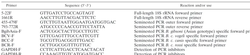
TABLE 1. Sequences for the oligonucleotide primers used in this study
Primer Sequence (5⬘-3⬘) Reaction and/or use
5-22F GTTGATCCTGCCAGTAGT Full-length 18S rRNA forward primer
1661R AACCTTGTTACGACTTCTC Full-length 18S rRNA reverse primer
455-479F GTCTTGTAATTGGAATGATGGTGAC Seminested PCR outer forward primer 793-772R ATGCCCCCAACCGTTCCTATTA Seminested PCR outer reverse primer
BgibAsia-F ACTCGGCTACTTGCCTTGTC Seminested PCRB. gibsoni(Asian genotype) specific forward primer BCV-F GTTCGAGTTTGCCATTCGTT Seminested PCRB. c. vogelispecific forward primer
BCC-F TGCGTTGACGGTTTGACC Seminested PCRB. c. canisspecific forward primer BCR-F GCTTGGCGGTTTGTTGC Seminested PCRB. c. rossispecific forward primer GAPDH-F CCTTCATTGACCTCAACTACAT Detection of PCR inhibitors
GAPDH-R CCAAAGTTGTCATGGATGACC Detection of PCR inhibitors
on May 15, 2020 by guest
http://jcm.asm.org/
outer primer pair was performed in a thermal cycler (PCR ExPress) at the following temperatures: initial denaturation at 95°C for 5 min, followed by 50 amplification cycles (95°C for 45 s, 58°C for 45 s, and 72°C for 45 s), and a final extension step at 72°C for 5 min.
Seminested PCRs (i.e., specific forward primers paired with the outer reverse primer) were each carried out in separate tubes under the same conditions as the outer primer pair, except for the following: 0.5l from the initial reaction was used as a DNA template, and the reactions were amplified for 30 cycles. In order to prevent PCR amplicon contamination, sample preparation, reaction setup, PCR amplification, and amplicon detection were all performed in separate areas. Positive and negative controls were used in all processing steps, including the DNA extraction. The presence of PCR inhibitors in DNA samples that tested negative in our PCR test was excluded by the amplification of a fragment of the GAPDH (glyceraldehyde-3-phosphate dehydrogenase) gene. The presence of
T. annaeDNA in the sample from Spain was confirmed by amplification of the nearly full-length 18S rRNA gene as described above.
Amplicon detection.All PCR products were visualized after electrophoresis in a 2% agarose gel containing 0.2g of ethidium bromide/ml by transillumination with an UV light.
Cloning and sequencing of PCR products.The PCR products were cloned into a plasmid vector (PCR 2.1; Invitrogen, Carlsbad, Calif.), andEscherichia coli
(TOP10⬘; Invitrogen) strain was transformed according to the protocol of the supplier. Recombinants were selected by the blue-white color of colonies, and plasmid DNA from at least three clones, for each isolate, were sequenced. Recombinant plasmid DNA was sequenced bidirectionally with the infrared fluorescence-labeled primers M13R-700 (5⬘-CAGGAAACAGCTATGACC ATG) and T7-800 (5⬘-TAATACGACTCACTATAGGGCGA) synthesized by LI-COR, Inc., Lincoln, Nebr. The previously described internal sequencing prim-ers 515F and 1391R were also used for full-length 18S ribosomal DNA sequenc-ing (18). The sequencsequenc-ing reaction conditions were as follows: 2 min at 92°C, followed by 30 amplification cycles (30 s at 92°C, 15 s at 55°C, and 30 s at 72°C) (Hybaid PCR ExPress). The sequencing reactions were analyzed by polyacryl-amide gel electrophoresis (3.75%) on an automated DNA sequencer (i.e., the LI-COR 4200 DNA sequencer).
Limit of detection.The DNA concentrations of the purified plasmids con-taining nearly full-length B. gibsoni(Asian genotype) (sequence identical to AF271081) andB. canissubsp.canis(sequence identical to AY072926) 18S rRNA genes were determined by spectrophotometry (Biospec-Mini; Shimadzu Corp., Columbia, Md.). These plasmid clones were individually serially diluted 10-fold in Tris-EDTA buffer to concentrations ranging from 1,000,000 to 10 plasmids/ml. One microliter of each dilution was then used as a template for the primary PCR.
To determine the practical limit of detection of the seminested PCR, leuko-cyte-reduced packed red blood cells fromB. gibsoni(Asian genotype) or whole blood from aB. canissubsp.canis-infected dog with parasitemias of known percentages were added to whole blood obtained from a noninfected dog. For theB. gibsoni(Asian genotype) mock-infected samples, 500 ml of whole blood was collected by sterile methods, using CPDA-1 as an anticoagulant, from the jugular vein of a splenectomized dog that had been experimentally infected with
B. gibsoni(Asian genotype) via intravenous injection. The blood was leukore-duced by using a commercially available leukocyte reduction filter. (Purecell; Pall Corp., East Hills, N.Y.). The leukoreduced blood was centrifuged at 4,000 rpm, and the plasma was removed. The red blood cell count was determined in an automated cell counter, and a thin smear was stained with a modified Wright stain to determine the percent parasitemia. After leukoreduction and concen-tration, the red blood cell count was 12.5⫻106cell/l with a parasitemia of 10%
based on a 1,000-cell count. Then, 40l (5⫻107organisms) of these
leukore-ducedB. gibsoni(Asian genotype)-infected concentrated red blood cells was added to 960l of noninfected EDTA anticoagulated canine whole blood. These samples were then serially diluted 10-fold by using the noninfected canine whole blood. The red blood cell count of the noninfected dog was 6.8⫻106cells/l. For
the mock-infectedB. canissubsp.canissamples, 3 ml of EDTA anticoagulated whole blood was collected via jugular venipuncture from a splenectomized dog that had been infected intravenously withB. canissubsp.canis. TheB. canis
subsp.canis-infected dog’s red blood cell count was 5.7⫻106cell/l with a
parasitemia of 9% based on a 1,000-cell count. Next, 100l (5⫻107organisms)
of theseB. canissubsp.canis-infected red blood cells was added to 900l of noninfected EDTA anticoagulated canine whole blood. These samples were then 10-fold serially diluted by using the noninfected canine whole blood. The red blood cell count of the noninfected dog was 6.8⫻106cells/l. The final test
concentrations of both mock-infected samples ranged from 500,000 to 5 organ-isms/ml. Total DNA was extracted from 200-l aliquots of these dilutions by
using a commercially available DNA extraction kit. A 5-l portion of each dilution was used as a template for the primary PCR.
Specificity.Portions (5l) of DNA extracted fromT. gondii-,N. caninum-,
C. parvum-, andL. infantum-infected canine whole blood and a noninfected dog were used as templates for the PCRs.
DNA sequence analysis.The DNA sequences generated in the present study were aligned and compared to the DNA sequences in GenBank (3) by using a computer program. (MegAlign; DNAStar, Inc., Madison, Wis.).
Animals.All work with live animals in the present study was conducted under the approval of and in accordance with the guidelines of the North Carolina State University Institute for Animal Care and Use Committee.
RESULTS
PCR. Nearly full-length (⬃1.7-kb) 18S rRNA genes were amplified from the experimentally infected B. gibsoni(Asian genotype), B. canis subsp. canis samples, and the naturally infectedT. annaesample from Spain (data not shown).
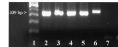
During the primary reaction of the seminested PCR, an ⬃340-bp product was amplified fromB. gibsoni(Asian geno-type),B. canissubsp.vogeli,B. canissubsp.canis, orB. canis
subsp.rossi-infected canine whole-blood samples. An⬃370-bp product was amplified from the B. gibsoni (USA/California genotype)-infected canine whole-blood samples. No amplicons were detected when DNA from theT. annae-infected canine whole-blood sample was used as a template for the PCR (Fig. 1). The PCR tests did not produce amplicons when canine DNA or DNA extracted fromT. gondii,N. caninum, orC. par-vumwas used as a template (data not shown).
During the secondary seminested reaction, the test was able to differentiate B. gibsoni (Asian genotype), B. canis subsp.
vogeli,B. canissubsp.canis, andB. canissubsp.rossiwhen the specific internal primers were paired with the reverse primer in the secondary reactions (Fig. 2). Occasionally, after the second round of amplification both the⬃340-bp and the 185- to 200-bp amplicons were visualized on the gel, but test interpretation was not affected (Fig. 2). An additional⬃300-bp amplicon of unknown significance was also detectable after the secondary seminested PCRs. In both the primary and secondary reac-tions, low-molecular-weight bands (⬃25 to 50 bp) were often detected in both positive and negative samples, including the no-DNA controls. These low-molecular-weight bands were presumed to be “primer dimers.”
[image:3.603.322.516.69.146.2]Amplicon contamination was not detected in any of the negative control samples at any time. As determined by am-plification of GAPDH, no PCR inhibitors were detected in any
FIG. 1. Analysis of seminested PCR products (outer primer pair only) by 2% agarose gel electrophoresis and ethidium bromide stain-ing. From left to right the lanes contain the following: molecular weight marker (lane 1),B. gibsoni(Asian genotype) (lane 2),B. canis
subsp.vogeli(lane 3),B. canissubsp.canis(lane 4),B. canissubsp.rossi
(lane 5),B. gibsoni(California/USA genotype) (lane 6),T. annae(lane 7), and negative (i.e., no-DNA) control (lane 8).
on May 15, 2020 by guest
http://jcm.asm.org/
of the DNA samples that were negative when our PCR test was used.
Limit of detection. The absolute limit of detection, with plasmid clones of nearly full-lengthB. gibsoni(Asian genotype) and B. canis subsp. canis 18S rRNA genes diluted in Tris-EDTA buffer, was one molecule/reaction (data not shown). The practical limit of detection ofB. gibsoni(Asian genotype) and B. canis subsp. canis organisms diluted in noninfected canine whole blood was 50 organisms/ml. The second round of amplification with a specific internal primer paired with the reverse primer did not improve the limit of detection but did improve ease of interpretation due to the enhanced visualiza-tion of bands on the gel.
Sequencing. The sequences of the nearly full-length 18S rRNA gene clones forB. gibsoni(Asian genotype) andB. canis
subsp.caniswere 100% identical to GenBank accession num-bers AF271081 and AY072926, respectively. The sequences of the⬃340-bp amplicons from the North American dogs natu-rally infected with small piroplasms were identical to GenBank accession numbers AF271081, AF271082, AF205636, AF175300, and AF175301 (B. gibsoniAsian genotype). The sequences of the⬃340-bp amplicons from the North American dogs natu-rally infected with large piroplasms were ⱖ99% identical to GenBank accession numbers AJ009795 and AY072926 (B.
ca-nis subsp. vogeli). The sequences of the ⬃340-bp amplicons from the dog experimentally infected withB. canissubsp.canis
was identical to GenBank accession number AY072926 (B. ca-nissubsp.canis). The sequences of the⬃340-bp amplicons from the South African dogs naturally infected with large piroplasms wereⱖ99% identical to GenBank accession number L19079 (B. canis subsp.rossi). The sequence of the 369-bp amplicon from the dog experimentally infected with B. gibsoni (USA/ California) was 98% identical to GenBank accession numbers AF158702 and AF231350 (B. gibsoni USA/California geno-type). The sequence of the nearly full-lengthT. annae18S rRNA amplicon wasⱖ99% identical to GenBank accession number AF188001 (T. annae).
DISCUSSION
For several reasons, a definitive diagnosis of canine babesi-osis can be difficult to achieve in the clinical setting. Light microscopic examination cannot consistently differentiate spe-cies or subspespe-cies. The lack of standardized serologic assays, the presence of cross-reactive antibodies, and recent changes in the geographic ranges of several canine piroplasms have also further complicated the diagnosis of babesiosis in dogs. In the present study, we describe a seminested PCR that has a
prac-FIG. 2. Analysis of the specificity of the seminested PCR by 2% agarose gel electrophoresis and ethidium bromide staining.(A) Analysis of seminestedB. gibsoni(Asian genotype)-specific secondary reactions; (B) analysis of seminestedB. canissubsp.vogeli-specific secondary reactions; (C) analysis of seminestedB. canissubsp.canisspecific secondary reactions; (D) analysis of seminestedB. canissubsp.rossi-specific secondary reactions. In panels A to D the lanes contain, from left to right, the following: molecular weight marker (lane 1),B. gibsoni(Asian genotype) (lane 2),B. canissubsp.vogeli(lane 3),B. canissubsp.canis(lane 4),B. canissubsp.rossi(lane 5),B. gibsoni(California/USA genotype) (lane 6),T. annae
(lane 7), canine genomic DNA (lane 8), negative (i.e., no-DNA) control (lane 9).
on May 15, 2020 by guest
http://jcm.asm.org/
tical limit of detection of 50 organisms/ml of canine whole blood. The PCR is specific for the diagnosis and differentiation ofB. gibsoni(Asian genotype),B. canissubsp.vogeli,B. canis
subsp. canis, and B. canis subsp. rossi. Based on the lowest number of detectable organisms/ml of whole blood, the limit of detection test is 180-fold lower than the reported limit of de-tection for a previously described PCR-based test forB. gibsoni
(Asian genotype) (1). Unfortunately, the majority of reports describing PCR-based tests for the diagnosis of canine babe-siosis have not reported a limit of detection or, in the case of one study (12), only reported the limit of detection in terms of the lowest detectable percent parasitemia (7, 10, 14–17, 26). Although the lowest detectable percent parasitemia is often reported for the detection of piroplasmosis, comparisons of tests based on the percent parasitemia can be difficult to in-terpret. For example, Babesia-infected animals can have red blood cell counts ranging from severely anemic to normal (1.5 ⫻106to 7.6⫻106red blood cells perl); therefore, the degree of anemia could result in as much as a fivefold difference in the total number of parasites/volume when samples from different animals with identical percent parasitemias are examined. The lowest estimated percent parasitemia that was detectable by our test was 0.00000073%, which is about 1,300-fold lower than the accepted limit of detection (0.001% parasitemia) for light microscopic examination of stained blood smears (5). Despite the aforementioned difficulty in interpreting differences in test sensitivities based on the percent parasitemia, it seems unlikely that differences in red blood cell counts would account for a 1,300-fold difference.
The clinical sensitivity of the PCR could not be determined in the present study and to our knowledge has not been de-scribed for any other PCR tests for canine babesiosis. In order to determine the clinical sensitivity, a population of animals in which the true prevalence of infection is known (i.e., experi-mental infection) is required. The detection limit of our test is superior to or comparable to the tests that have been described for the detection of piroplasmosis in other species (2, 8, 19). A blinded study comparing PCR and serologic testing has been performed evaluating the clinical sensitivity of a PCR test in cattle experimentally infected withB. bovis(8). The PCR test used in that study had an absolute sensitivity that was similar to our PCR for canine babesiosis. That study demonstrated false-negative PCR tests in 30% of the samples, especially when the samples were obtained from chronically infected cattle with a low percent parasitemia. However, the clinical sensitivity was improved to ⬎90% by retesting the cattle that had tested negative by PCR 10 to 14 days later. A similar clinical sensi-tivity would be expected for the PCR test described here.
The approach used to develop the outer primer pair of our seminested PCR should permit the detection of previously unrecognized and/or unsequenced piroplasms with genetic variation in this hypervariable region of the 18S rRNA gene. If the⬃340-bp amplicon is detected but species-specific amplifi-cation is not possible with the internal primers, then the⬃340 bp can be sequenced directly for genotype and species identi-fication. Surprisingly, despite a 4-bp difference on the 3⬘end of the forward primer compared to theB. gibsoni (USA/Califor-nia genotype) (AF216496 and AF175300) rRNA gene se-quences, an 18S rRNA gene amplicon was detected when a sample containing high numbers of parasites (30%
para-sitemia) was used as a template for the outer primer pair reaction. Despite an identical sequence in the primer regions, amplicons were not detected when a T. annae (AF188001)-infected sample with a lower percent parasitemia (0.01%) was used as a template. Since there were no detectable amplicons in the noninfected canine whole-blood samples under the test-ing conditions, we chose not to increase the annealtest-ing tem-perature to eliminate the mispriming of theB. gibsoni(USA/ California genotype). Although the test might be useful for identifying someB. gibsoni(USA/California genotype)- orT. annae-infected samples in which there are large numbers of organisms, we do not recommend the use of this test due to the limited sensitivity for the routine diagnosis ofB. gibsoni(USA/ California genotype) orT. annae. In these instances, i.e., when a 370-bp amplicon is detected, we recommend that either spe-cific tests forB. gibsoni(USA/California genotype) orT. annae
be performed or that the amplicons be sequenced to confirm the genotype of the organism.
In conclusion, we describe here a PCR test for the diagnosis of canine babesiosis that can detect and differentiateB. gibsoni
(Asian genotype),B. canissubsp.vogeli,B. canissubsp.canis, andB. canissubsp.rossiwith a defined limit of detection. This test should improve the diagnostic capabilities for the detec-tion and differentiadetec-tion of canineBabesiaspp. in clinical sam-ples and facilitate future research studies that assess canine infection with these organisms.
ACKNOWLEDGMENTS
This work was supported in part by a grant from North Carolina State University.
We thank our colleagues George E. Moore, Patricia Conrad, Nich-olas Sharp, and Lance Perryman for providing biological samples and isolates.
REFERENCES
1. Ano, H., S. Makimura, and R. Harasawa.2001. Detection of babesia species from infected dog blood by polymerase chain reaction. J. Vet. Med. Sci.63:
111–113.
2. Bashiruddin, J. B., C. Camma, and E. Rebelo.1999. Molecular detection of
Babesia equiandBabesia caballiin horse blood by PCR amplification of part of the 16S rRNA gene. Vet. Parasitol.84:75–83.
3. Benson, D. A., I. Karsch-Mizrachi, D. J. Lipman, J. Ostell, B. A. Rapp, and D. L. Wheeler.2002. GenBank. Nucleic Acids Res.30:17–20.
4. Birkenheuer, A. J., M. G. Levy, K. C. Savary, R. B. Gager, and E. B. Breitschwerdt.1999.Babesia gibsoniinfections in dogs from North Carolina. J. Am. Anim. Hosp. Assoc.35:125–128.
5. Bose, R., W. K. Jorgensen, R. J. Dalgliesh, K. T. Friedhoff, and A. J. de Vos.
1995. Current state and future trends in the diagnosis of babesiosis. Vet. Parasitol.57:61–74.
6. Breitschwerdt, E. B., J. B. Malone, P. MacWilliams, M. G. Levy, C. W. Qualls, Jr., and M. J. Prudich.1983. Babesiosis in the greyhound. J. Am. Vet. Med. Assoc.182:978–982.
7. Caccio, S. M., B. Antunovic, A. Moretti, V. Mangili, A. Marinculic, R. R. Baric, S. B. Slemenda, and N. J. Pieniazek.2002. Molecular characterisation ofBabesia canis canisandBabesia canis vogelifrom naturally infected Eu-ropean dogs. Vet. Parasitol.106:285–292.
8. Calder, J. A., G. R. Reddy, L. Chieves, C. H. Courtney, R. Littell, J. R. Livengood, R. A. Norval, C. Smith, and J. B. Dame.1996. Monitoring Babe-sia bovisinfections in cattle by using PCR-based tests. J. Clin. Microbiol.
34:2748–2755.
9. Camacho, A. T., E. Pallas, J. J. Gestal, F. J. Guitian, A. S. Olmeda, H. K. Goethert, and S. R. Telford.2001. Infection of dogs in north-west Spain with aBabesia microti-like agent. Vet. Rec.149:552–555.
10. Carret, C., F. Walas, B. Carcy, N. Grande, E. Precigout, K. Moubri, T. P. Schetters, and A. Gorenflot.1999.Babesia canis canis,Babesia canis vogeli,
Babesia canis rossi: differentiation of the three subspecies by a restriction fragment length polymorphism analysis on amplified small subunit ribosomal RNA genes. J. Eukaryot. Microbiol.46:298–303.
11. Conrad, P., J. Thomford, I. Yamane, J. Whiting, L. Bosma, T. Uno, H. J. Holshuh, and S. Shelly.1991. Hemolytic anemia caused byBabesia gibsoni
infection in dogs. J. Am. Vet. Med. Assoc.199:601–605.
on May 15, 2020 by guest
http://jcm.asm.org/
12. Fukumoto, S., X. Xuan, S. Shigeno, E. Kimbita, I. Igarashi, H. Nagasawa, K. Fujisaki, and T. Mikami.2001. Development of a polymerase chain reaction method for diagnosingBabesia gibsoniinfection in dogs. J. Vet. Med. Sci.
63:977–981.
13. Gaskin, A. A., P. Schantz, J. Jackson, A. Birkenheuer, L. Tomlinson, M. Gramiccia, M. Levy, F. Steurer, E. Kollmar, B. C. Hegarty, A. Ahn, and E. B. Breitschwerdt.2002. Visceral leishmaniasis in a New York foxhound kennel. J. Vet. Intern. Med.16:34–44.
14. Irizarry-Rovira, A. R., J. Stephens, J. Christian, A. Kjemtrup, D. B. DeNi-cola, W. R. Widmer, and P. A. Conrad.2001.Babesia gibsoniinfection in a dog from Indiana. Vet. Clin. Pathol.30:180–188.
15. Kjemtrup, A. M., A. A. Kocan, L. Whitworth, J. Meinkoth, A. J. Birkenheuer, J. Cummings, M. K. Boudreaux, S. L. Stockham, A. Irizarry-Rovira, and P. A. Conrad.2000. There are at least three genetically distinct small piro-plasms from dogs. Int. J. Parasitol.30:1501–1505.
16. Kocan, A. A., A. Kjemtrup, J. Meinkoth, L. C. Whitworth, G. L. Murphy, L. Decker, and M. Lorenz.2001. A genotypically uniqueBabesia gibsoni-like parasite recovered from a dog in Oklahoma. J. Parasitol.87:437–438. 17. Macintire, D. K., M. K. Boudreaux, G. D. West, C. Bourne, J. C. Wright, and
P. A. Conrad.2002.Babesia gibsoniinfection among dogs in the southeastern United States. J. Am. Vet. Med. Assoc.220:325–329.
18. Pitulle, C., D. M. Citron, B. Bochner, R. Barbers, and M. D. Appleman.1999. Novel bacterium isolated from a lung transplant patient with cystic fibrosis. J. Clin. Microbiol.37:3851–3855.
19. Salem, G. H., X. Liu, J. D. Johnsrude, J. B. Dame, and G. Roman-Reddy.
1999. Development and evaluation of an extra chromosomal DNA-based PCR test for diagnosing bovine babesiosis. Mol. Cell Probes13:107–113. 20. Uilenberg, G., F. F. Franssen, N. M. Perie, and A. A. Spanjer.1989. Three
groups ofBabesia canisdistinguished and a proposal for nomenclature. Vet. Q.11:33–40.
21. Wozniak, E. J., B. C. Barr, J. W. Thomford, I. Yamane, S. P. McDonough, P. F. Moore, D. Naydan, T. W. Robinson, and P. A. Conrad.1997. Clinical, anatomic, and immunopathologic characterization ofBabesia gibsoni infec-tion in the domestic dog (Canis familiaris). J. Parasitol.83:692–699. 22. Yamane, I., P. Conrad, and I. Gardner.1993.Babesia gibsoniinfections in
dogs. J. Protozool. Res.3:111–125.
23. Yamane, I., J. W. Thomford, I. A. Gardner, J. P. Dubey, M. Levy, and P. A. Conrad.1994. Serosurvey ofBabesia canis,Babesia gibsoni, andErhlichia canisin pound dogs in California. Prevent. Vet. Med.18:293–304. 24. Zahler, M., H. Rinder, E. Schein, and R. Gothe.2000. Detection of a new
pathogenicBabesia microti-like species in dogs. Vet. Parasitol.89:241–248. 25. Zahler, M., H. Rinder, E. Zweygarth, T. Fukata, Y. Maede, E. Schein, and R.
Gothe.2000. “Babesia gibsoni” of dogs from North America and Asia belong to different species. Parasitology120:365–369.
26. Zahler, M., E. Schein, H. Rinder, and R. Gothe.1998. Characteristic geno-types discriminate betweenBabesia canisisolates of differing vector speci-ficity and pathogenicity to dogs. Parasitol. Res.84:544–548.