Introduction
FSHD is now recognized as one of the most prevalent forms of muscular dystrophy in adults (http://www.orpha.net). Prominent features of this myopathy are the progressive weakening of the skeletal muscles in the face, shoulder gir-dle, and the upper arms, and these muscular aspects are often combined (>50% of patients) with retinal vasculopathy [1, 2]. The genetic lesion leading to the most prominent form of FSHD (FSHD1A), accounting for ~98% of FSHD patients, is an autosomal dominant contraction of the D4Z4 repeat array at chromosome 4q35 below 11 copies [3, 4]. This contraction leads to hypomethylation of the D4Z4 repeats, which has been proposed to lead downstream to the
misregulation of one or more of the 4q35 local-ized genes including FRG1, ANT1, FRG2, DUX4, and DUX4c [5]. However, none of these candi-date genes has consistently been shown to ex-hibit significantly altered RNA expression levels in affected FSHD muscle biopsies compared to unaffected controls [6-13]. Multiple issues com-plicate these expression analyses including large differences within an affected muscle and potentially at the site of biopsy, the bias focus-ing on FSHD gene misexpression exclusively in the skeletal muscle lineage, and the potential that FSHD gene misexpression occurs during cell differentiation [10, 14-16]. Thus, without knowing when and where in human muscle de-velopment gene misexpression leading to FSHD occurs, the cause of the FSHD pathophysiology
Original Article
Testing the effects of FSHD candidate gene expression in
vertebrate muscle development
Ryan D. Wuebbles1,2, Steven W. Long1, Meredith L. Hanel, Peter L. Jones
Department of Cell and Developmental Biology, University of Illinois at Urbana-Champaign, 601 S. Goodwin Ave, B107 Chemical and Life Sciences Laboratory, Urbana, IL 61801 USA.
1These authors contributed equally to this work. 2Present address: Department of Pharmacology, University of
Nevada, School of Medicine, 1664 N. Virginia St., Reno, NV 89557, USA.
Received March 2, 2010, accepted March 23, 2010, available online: March 28, 2010
Abstract: The genetic lesion leading to facioscapulohumeral muscular dystrophy (FSHD) is a dominant deletion at the 4q35 locus. The generally accepted disease model involves an epigenetic dysregulation in the region resulting in the upregulation of one or more proximal genes whose overexpression specifically affects skeletal muscle. However, mul-tiple FSHD candidate genes have been proposed without clear consensus. Using Xenopus laevis as a model for verte-brate development our lab has studied the effects of overexpression of the FSHD candidate gene ortholog, frg1 (FSHD region gene 1), showing that increased levels of frg1 systemically led specifically to an abnormal musculature and increased angiogenesis, the two most prominent clinical features of FSHD. Here we studied the overexpression effects of three other promising FSHD candidate genes, DUX4, DUX4c, and PITX1 using the same model system and methods for direct comparison. Expression of even very low levels of either DUX4or pitx1 early in development led to massive cellular loss and severely abnormal development. These abnormalities were not muscle specific. In contrast, elevated levels of DUX4c resulted in no detectable adverse affects on muscle and DUX4c levels did not alter the ex-pression of myogenic regulators. This data supports a model for DUX4 and PITX1 in FSHD only as pro-apoptotic fac-tors if their expression in FSHD is confined to cells within the myogenic pathway; neither could account for the vascu-lar pathology prevalent in FSHD. Taken together, increased frg1 expression alone leads to a phenotype that most closely resembles the pathophysiology observed in FSHD patients.
has remained controversial.
To circumvent the ambiguity of RNA expression analyses, we have taken a developmental ap-proach to the problem by first addressing the normal function of an FSHD candidate gene during development and then assaying the ef-fect of overexpression of an FSHD candidate gene on vertebrate development. The system for these studies is the early development of Xenopus laevis. Our initial analysis focused on understanding the function and expression of one candidate gene, frg1 [17, 18]. FRG1 is a highly conserved gene of unknown function that is overexpressed in FSHD patient derived myoblasts undergoing myogenic differentiation [15]. These studies found that frg1 is required for the normal development of the vertebrate musculature and vasculature [17, 18]. Consis-tent with a role in FSHD pathology, systemically elevated levels of frg1 led to phenotypes specifi-cally in the vertebrate musculature and vascula-ture which strongly correlated to the two most common symptoms of FSHD, dystrophic muscle and increased angiogenesis [17, 18]. Thus, de-velopmentally, FRG1 overexpression fits the criteria for being causal for FSHD pathology.
We have continued our analysis with three addi-tional FSHD candidate genes, DUX4, DUX4c, and PITX1 [6, 19, 20]. DUX4 and DUX4c are encoded within open reading frames (ORFs) of different 4q35 D4Z4 repeat units within or near the FSHD deletion [21, 22]. Although D4Z4 re-peat arrays exist in multiple loci in the genome [23], RNAs originating specifically from the 4q35 localized D4Z4/DUX4 and D4Z4/DUX4c loci are increased in certain FSHD patient-derived muscle cells [6, 19, 20]. A normal cellu-lar or developmental role for the 4q35 DUX4 protein, if any, has not been described; how-ever, expression of the currently accepted 4q35 derived DUX4 protein is highly toxic to all cells leading to a rapid onset of apoptosis [19, 24]. This apoptotic effect of DUX4 expression is pos-tulated to be from direct competition with the regulatory targets of PAX3/PAX7 and is inhibited by elevated expression of PAX3 or PAX7 [24]. Interestingly, in a cell culture system, DUX4 has been shown to bind the promoter and activate expression of PITX1, a non-4q35 localized FSHD candidate gene whose expression has been found to be upregulated in FSHD muscle, pro-viding an alternative mechanism for DUX4-mediated pathology [6]. DUX4c, located within a
partial D4Z4 unit 42 kb proximal to the FSHD-associated D4Z4 array, is identical to DUX4 through their N-terminal double homeobox do-mains however they have differing C-terminal amino acid sequences [22]. DUX4c expression has been detected in muscle cells where it is proposed to act as a myogenic regulator and inhibitor of myoblast differentiation [20, 25].
In this study, we assayed the effects of expres-sion of human DUX4 and DUX4c, as well as the X. laevis ortholog of PITX1 on early vertebrate development, with particular attention to mus-cle growth and differentiation. We show that DUX4 expression and pitx1 overexpression both lead to massive cellular loss that is not muscle specific. With DUX4 in particular the cellular loss occurred at extremely low expression levels and was cell-type independent indicating that this protein is highly toxic to all vertebrate cells and this toxic effect was not specific to muscle. DUX4c expression did not lead to any observ-able change in muscle development or differen-tiation or changes in the expression of the myo-genic regulators myf5 or myoD in Xenopus. Con-tradictory to what has been reported in cell cul-ture, we found that both DUX4 and DUX4c sig-nificantly reduced expression levels of pitx1 transcripts in our animal model. Together with our previous studies on frg1, this presents the first analysis for direct comparison of the effects of expression of the main FSHD candidate genes in a developing vertebrate system.
Materials and methods
Frog husbandry
Adult X. laevis were purchased from Xenopus Express. All procedures were carried out in ac-cordance with established UIUC IACUC approved protocols for animal welfare.
Plasmid constructs and RNA production
pcDNA3.1 (Invitrogen). Production of EGFP mRNA was performed as previously described [17]. For DUX4, DUX4c and pitx1 mRNA, con-structs were linearized and cappedmRNA was generated using T7 RNA polymerase and the mMessage mMachine kit (Ambion, Inc).
Xenopus embryo injections
In vitro fertilized embryos were generated as described [17]. Embryos were microinjected after completion of the two cell stage, as indi-cated by the beginning of the second cleavage, in 1X MMR with 3% Ficoll and incubated at 19° C. Between 3-6 hours after injection, embryos were transferred to 0.1X MMR with 3% Ficoll. After 24-36 hours embryos were either peeled and fixed for stage 18-22 embryos or cultured in 0.1X MMR until the desired stage. After neu-ral tube closure all injected embryos were sorted based on left, right or bilateral fluores-cence. DUX4 mRNA was injected at 500 pg, 250 pg, 100 pg, 10 pg, 1 pg, and 0.5 pg along with 500 pg EGFP mRNA. DUX4c mRNA was injected at 1 ng along with 500 pg EGFP. pitx1 was injected at 150 pg and 50 pg along with 500 pg EGFP. Control EGFP mRNA injections were performed at 500 pg.
TUNEL assay
TUNEL staining of whole-mount Xenopus em-bryos was carried out using a protocol adapted from Hensey and Gautier [26]. All procedures were carried out at room temperature unless noted otherwise. Embryos were fixed for 1 hr. in MEMFA, (100 mM MOPS (pH 7.4), 2 mM EGTA, 1 mM MgSO4, 4% formaldehyde). Embryos were washed in methanol 2 x 30 min. and stored in methanol at -20°C. For rehydration, half of methanol was replaced with PBS and washed 5 x 5min. The embryos were washed with PBT (0.2% Tween-20 in PBS), 2 x 15 min., followed by 2 x 15 min. washes in PBS. Embryo pigment was removed by treatment for 1-2 hours in 1% H2O2, 5% Formamide, and 0.5X SSC under bright light, and washed 3 x 15 min. in PBS. Embryos were transferred to terminal deoxynu-cleotidyl transferase, (TdT), buffer (Invitrogen) and washed for 30 min. End labeling was car-ried out overnight in TdT buffer containing 0.5 mM digoxygenin-dUTP (Roche Diagnostics), and 150 U/ml TdT (Invitrogen). Embryos were then washed 2 x 1 hr. in PBS/1 mM EDTA, at 65°C, followed by 4 x 1 hr. in PBS. Detection and
chro-mogenic reaction was carried out as previously described [27]. Embryos were viewed and stored following rehydration in 1X PBS.
In situ hybridizations
Embryos were staged according to Nieuwkoop and Faber [28], fixed 1-2 hrs in MEMFA, washed 2 x 30 min in 100% methanol and stored in 100% methanol at -20°C until use. The EGFP, Xenopus myoD, pax3, and myf5 antisense probes generated as previously described [17]. The pitx1 probe was generated by linearizing pGEM pitx1 with SalI and using T7 RNA poly-merase transcription to generate digoxigenin (DIG) -11-UTP (Roche Diagnostics) antisense RNA probes. In situ hybridizations were per-formed according to standard methods [27] and detected with alkaline phosphatase (AP) linked anti-DIG antibody (Roche Diagnostics) and the chromogenic substrates BCIP (5-Bromo-4-chloro -3-indolyl phosphate, toluidine salt) and NBT (Nitro blue tetrazolium chloride) (Roche Diag-nostics). Embryos were refixed overnight in Bouin’s fixative, followed by washing in 70% ethanol/30% PBS-Tween 0.1%, and pigment was removed by treatment for 1-2 hours in 1% H2O2, 5% Formamide, and 0.5X SSC under bright light. Embryos were then washed in methanol 10 minutes and transferred to 1mM EDTA in PBS or glycerol for analysis and photog-raphy.
Immunohistochemistry
by U. Rutishauser, were obtained from the De-velopmental Studies Hybridoma Bank devel-oped under the auspices of the NICHD and maintained at The University of Iowa, Depart-ment of Biological Sciences, Iowa City, IA 52242.
qRT-PCR
For each sample, total RNA was purified from 10 pooled embryos using Trizol reagent (Invitrogen) per manufacture’s protocol. RNA was then treated with 1U RQ1 DNase (Promega) per 1ug RNA for 30 min. at 37°C. cDNA synthe-sis was preformed using 1ug of total RNA, 50ng of random hexamer, and Superscript III (Invitrogen) per manufacture’s suggested method. Relative transcript levels were deter-mined using 1ul of (1:20 diluted) cDNA (in tripli-cate), iQ SYBR Supermix, and gene specific primers (myoD: 5’ TGCCAAGAGTCCAGATTTCC 3’, 5’ CAGGTCTTCAAAGAAACTCATGTC 3’; myf5: 5’ GCTTATCTAGTATTGTGGATCGG 3’, 5’ CTGGTT TGTTGGGTGTAAGG 3’; pitx1: 5’ CATGAGCA-GAAGTGATTGAC 3’, 5’ GTAAAGTGAGTCCTTTGTC TCC 3’; gapdh: 5’ GGTGAAGGTTGGAATTAACGG 3’, 5’ GATCAGCTTGCCATTCTCAG 3’) on a Bio-Rad iCycler IQ machine. Experiments were pre-formed at least 3 times. Data analyses were preformed using the comparative Ct method and error bars are + standard error of the mean. Changes were determined using the two tailed student’s t-test and considered significantly different at a P-value <0.05.
Results
Expression of DUX4 and pitx1 lead to cellular loss while DUX4c expression has minimal effect on development
The early development of X. laevis was used as a model system to determine the effects of FSHD candidate genes DUX4, DUX4c, and pitx1 expression levels on vertebrate development. In normal human tissues, DUX4 and DUX4c ex-pression is undetectable and the proteins are neither required for nor involved in any known normal cellular function. The human 4q35 DUX4 and DUX4c genes are not conserved out-side of their double homeobox domains and Xenopus do not possess any orthologs. There-fore, we assayed the effects of the presence of DUX4 or DUX4c during muscle development compared to a background of no expression.
Conversely, the pitx1 transcription factor, as was the case with frg1, is highly conserved be-tween mammals and Xenopus in protein se-quence, function, and developmental expres-sion [29]. For this gene, we assayed the effect of increasing its expression during development as well as expression outside its normal devel-opmental profile.
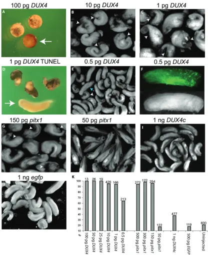
DUX4 is either cellular toxicity or nothing (Figure 1E). Even in these embryos, the fluorescence of the EGFP appeared patchy (Figure 1F), suggest-ing that though they appeared to have pro-gressed through development normally, DUX4 had still led to cellular loss. Therefore, consis-tent with other systems, DUX4 expression is extremely cytotoxic in developing Xenopus [19, 24].
DUX4 is capable of inducing PITX1 expression in mouse C2C12 cell culture suggesting PITX1 misexpression as a possible mechanism for DUX4-mediated pathology [6]. To determine the effect of aberrantly induced pitx1 expression, we bypassed DUX4 expression and microin-jected mRNA encoding Xenopus pitx1 into em-bryos as above. Similar to injections of DUX4 mRNA, we found that both 500 pg (n = 272) and 300pg (n = 122) of pitx1 mRNA led to early embryo arrest while injection of 150 pg pitx1 (n = 254) led cellular loss specifically on the in-jected side leading to a curled phenotype and gastrulation or neurulation defects (Figure 1G & 1K). Injections of 50 pg pitx1 mRNA (n = 122) had no obvious effects on development (Figure 1H & 1K). Thus, while pitx1 overexpression is cytotoxic, developing embryos are much more tolerant of pitx1 overexpression than DUX4 ex-pression suggesting that DUX4 cytotoxicity is not mediated through the activation of pitx1.
The analysis of the third FSHD candidate gene, DUX4c, produced results that were a stark con-trast to the effects of DUX4 and pitx1. Microin-jections of as much as 1ng of DUX4c mRNA (n=477) only lead to a modest increase over uninjected background levels of developmental abnormalities, yet still far fewer and less in se-verity than seen with a 2000-fold lower amount of DUX4 mRNA (Figure 1I & 1K). Because DUX4c shares an identical double homeobox domain with DUX4, the fact that a 2000-fold increase in mRNA produced only minor develop-mental problems indicates that a strictly com-petitive interaction with Pax3 and Pax7 for DNA regulatory targets is not responsible for the DUX4 phenotype.
Cytotoxicity mediated by DUX4 and pitx1 is not muscle specific
The symptoms associated with FSHD are pri-marily muscular and often combined with a less prominent vascular component. Taking into
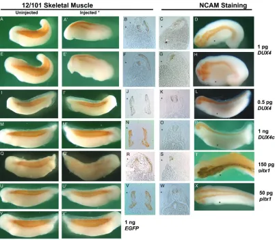
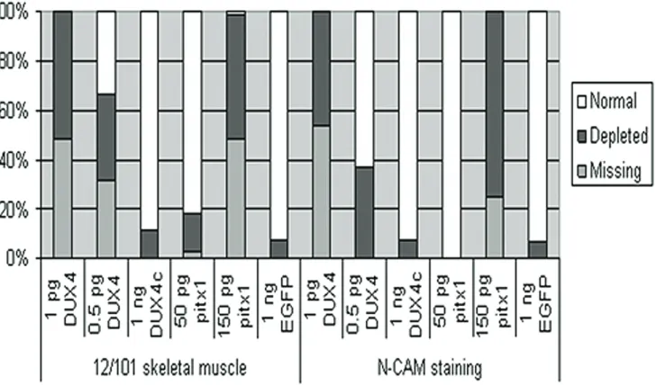
ac-count the generally accepted model whereby the FSHD1A deletion leads to an epigenetic upregulation of gene expression, one would expect the effect of a viable candidate gene’s systemic expression to be primarily seen in tis-sues affected in FSHD as is the case with frg1. In order to determine if muscle was specifically affected by DUX4, pitx1, or DUX4c expression, injected embryos were analyzed for differenti-ated muscle and neurologic tissue by immu-nostaining with the 12/101 or NCAM antibod-ies, respectively (Figure 2 & 3). Due to the se-vere loss of tissue in the 1 pg DUX4 and 150 pg pitx1 injected animals, the figures depict some of the best developing and staining animals, as most failed to form a complete neural tube clo-sure and were consequently too curled to cap-ture the staining pattern in photographs.
Immunostained animals were qualitatively de-termined to exhibit normal, depleted, or absent levels of immunostaining on the mRNA injected side (Figure 2). Embryos scored as depleted included those exhibiting highly dispersed but significant staining, missing somites and somite disruptions, and a significantly thinner somite area.
DUX4 injected embryos were first analyzed for affects on the developing muscle. Qualitative inspection of 1 pg DUX4 injected embryos stained with 12/101 (n = 45, missing n = 22, depleted n = 23) indicate a severe loss of mus-cle tissue by the lack of 12/101 immunostain-ing specifically on the injected side of the em-bryo (Figure 2A’ and 2B’ compared to Figure 2A and 2B). However, transverse paraffin section-ing revealed these animals had a weak appear-ance of staining due to a build up of cellular debris between the 12/101 stained muscle near the notochord/neural tube and the lateral edge of the injected side (Figure 2B and 2F). The area of the myotome on the injected side was significantly decreased due to cellular loss. We find a large increase in the number of em-bryos with normal 12/101 staining when we inject 0.5 pg DUX4 mRNA (n = 87, missing n = 27, depleted n = 31) and this increase in nor-mally stained embryos corresponded well to the number of normally developing embryos (Figure 2I’ compared to 2I, 2J, Figure 3).
as-sayed in 1 pg DUX4 injected embryos (n = 24). Immunostaining for NCAM, which at stage 34 of Xenopus development is confined to neural tis-sue, resulted in levels of missing (n = 13) and depleted (n = 11) NCAM staining similar to that of 12/101 (Figure 3). In several animals the NCAM staining was specifically depleted in the area of the eye (Figure 2D & 2H) and transverse sections revealed that often the neural tube staining was depleted as well (Figure 2C & 2H).
We conclude that the cellular loss mediated by DUX4 is not restricted to muscle.
[image:7.612.104.506.85.436.2]Embryos injected with pitx1 mRNA were simi-larly analyzed for muscle and neuronal defects. The injection of pitx1 led to developmental ab-normalities similar to those seen with DUX4 albeit at a much higher concentration of mRNA. As with the DUX4injections, 150 pg pitx1 mRNA injections (n = 64) led to depletion (n = 32) and
loss (n = 31) of 12/101 immunostaining from the injected side (Figure 2Q’ compared to 2Q, 2R). This effect was dose dependent, with 50 pg pitx1 mRNA (n = 71) causing almost no loss of 12/101 immunostaining (missing n = 2, de-pleted n = 11) (Figure 2U’ compared to 2U, 2V). Similarly, the 150 pg pitx1 injection (n = 8) led to depleted (n = 6) or missing (n = 2) NCAM im-munostaining (Figure 2S & 2T), while 50 pg pitx1 injection (n = 19) did not (Figure 2W &2X).
Despite the lack of any significant gross devel-opmental defects (Figure 1I), DUX4c injected embryos were assayed for muscular and neu-ronal alterations. Differences were expected in the myotome based on previous findings of DUX4c mediated inhibition of myoblast differen-tiation [20, 25]. However, when stained with 12/101, 1 ng DUX4c mRNA injected embryos (n = 89) still displayed normal immunostaining (Figure 2M’ compared to 2M, 2N) with abnor-malities (missing n = 0, depleted n = 10) com-parable to EGFP mRNA injected controls (n = 41, missing n = 0, depleted n = 3) (Figure 2Y, 2Y’, Figure 3). Similarly, the NCAM staining of DUX4c injected embryos (Figure 2O & 2P) had abnormalities (n = 14, missing n = 0, depleted n = 1) similar to NCAM stained EGFP mRNA injected controls (n = 15, missing n = 0, de-pleted n = 1) (Figure 3). We conclude that
DUX4c levels have no effect on the developing musculature or neurons.
DUX4 eliminates myoD, myf5, and pax3 expression profiles
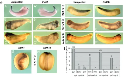
DUX4 expression led to the loss of 12/101 im-munostaining indicating differentiated muscle was degraded or missing. To determine if DUX4 expression leads to a loss of muscle cell precur-sors, in situ hybridizations with probes against myoD, myf5, or pax3 were performed (Figure 4). In stage 34 embryos from the 1 pg DUX4 mRNA injections, the expression of all three of these markers was missing in a large majority (myoD n = 60/62; myf5 n = 12/20; pax3 n = 23/28) (Figure 4A’, 4B’, & 4C’ compared to 4A, 4B, & 4C). Assaying earlier in development, stage 20 DUX4 injected embryos were missing expres-sion of pax3 (n = 11/20) (Figure 4G). Although the markers myf5 and pax3 were depleted, the entire tissue, including neural tissue was af-fected. When taken into consideration with our previous findings that the DUX4-meidated de-fects began at gastrulation (Figure 1A) and we conclude that the DUX4-mediated cellular loss occurs prior to stage 20 and therefore is clearly not muscle specific.
[image:8.612.126.490.86.300.2]Unlike DUX4, DUX4c had little to no observable
e f
-fect on the expression of myoD, myf5, and pax3 (myoD n = 5/31; myf5 n = 0/8; pax3 n = 0/30) (Figure 4D’, 4E’, & 4F’ compared to 4D, 4E, & 4F, respectively). Similarly, no change was ob-served in pax3 staining at stage 20 in embryos injected with 1 ng DUX4c mRNA (n = 0/12) (Figure 4H). Quantitative reverse transcription PCR (qRT-PCR) was used to determine the tran-script levels of the myogenic regulators myoD or myf5 when injected with 1 ng DUX4c mRNA. Neither stage 20 embryos (n = 30) nor stage 34 embryos (n = 20) have statistically significant differences in myoD or myf5 transcript levels when compared to EGFP mRNA injected con-trols normalized to gapdh (Figure 5). Thus, DUX4c has no effect on muscle precursors or myogenic regulators. Moreover, we found no significant changes of myogenic regulators by qRT-PCR on remaining intact tissue from 1 pg DUX4 stage 34 injected embryos, further
sug-gesting DUX4 has a strictly apoptotic role (Figure 5).
DUX4 and DUX4c expression reduce endogenous pitx1 expression
[image:9.612.86.514.82.347.2]FSHD affected muscle has elevated levels of PITX1 transcript and in cell culture assays DUX4 activates transcription of PITX1 [6]. Endogenous pitx1 expression was examined by in situ hy-bridization in 1 pg DUX4 and 1 ng DUX4c in-jected stage 34 X. laevis embryos. During early X. laevis development (prior to hind-limb devel-opment), observable pitx1 expression is con-fined to the cement and pituitary glands, and this expression pattern is not altered by expres-sion of either DUX4 (Figure 6A’ and 6A; n = 23) or DUX4c (Figure 6B’and 6B; n = 27). Although there is no statistically significant change in stage 20 embryos, surprisingly, analysis of
Figure 5. Global levels of myoD and myf5 mRNA in DUX4 and DUX4c injected embryos. Levels of mRNA were quanti-fied by qRT-PCR for myoD and myf5 in DUX4c (1 ng) or DUX4 (1 pg) injected embryos. Fold change is normalized to
gapdh levels and compared to EGFP mRNA at a relative level of 1; error bars are ± standard error of the mean.
[image:10.612.101.513.286.641.2]stage 34 transcript levels by qRT-PCR showed that pitx1 expression was actually reduced by DUX4c and DUX4 when compared to EGFP con-trols (p-value < 0.05) (Figure 6D). We conclude that neither DUX4 nor DUX4c expression acti-vates pitx1 expression in vivo.
Discussion
Multiple candidate genes have been proposed as causal of FSHD pathophysiology based on selective upregulation in FSHD patient-derived tissues or cell lines [6, 8, 10, 13, 30]. However inconsistent results, likely due in part to varia-tions within and between FSHD patient biopsies as well as culturing conditions, have made it unclear which gene(s) are misexpressed. Previ-ously we used X. laevis as a developing verte-brate model to analyze the effects of altering the expression levels of frg1. In the current study, we have used this same system to simi-larly analyze the effects of three additional FSHD candidate genes, DUX4, DUX4c and pitx1, on vertebrate muscle development.
X. laevis is a well-defined model system for mus-cle development with many advantages includ-ing external development, cells grow and differ-entiate under normal growth and environmental conditions giving rise to all tissues, and gene expression is easily manipulated through micro-injection or transgenesis. In addition, Xenopus and humans share high levels of conservation of tissue organization, developmental proc-esses, genes, and proteins. For example, the conservation of some muscle and FSHD associ-ated proteins between human and Xenopus are as follows: FRG1 (80% identity, 88% similar), PITX1 (77% identity, 84% similar), PAX3 (91% identity, 96% similar) including 100% conserva-tion of the homeodomain (aa 220-277), PAX7 (88% identity, 94% similar) including 100% con-servation of the homeodomain (aa 217-274), MYOD1 (65% identity, 75% similar), and MYF5 (69% identity, 84% similar), as determined by alignments using NCBI BLAST Alignp. In con-trast, DUX4 and DUX4c belong to the DUX family of double homeobox domain proteins but do not have Xenopus orthologs. In fact, when consider-ing the entire protein sequence, includconsider-ing that residing outside their homeodomains, they both completely lack evolutionary conservation and appear to be unique to humans. Even the most closely related DUX4 ORFs in mice [31-34] show levels of sequence similarity (31% amino acid
identity for the Duxbl protein aligned to DUX4 using the ClustalW function of BioEdit Sequence Alignment Editor software) far below what is expected for mouse to human conservation. Without any clear ortholog available, the human sequences for DUX4 and DUX4c were used for these studies.
We have confirmed the toxic effect of DUX4 ex-pression in the context of normal vertebrate development during stages of active myogene-sis. By differential staining of neuronal and mus-cle tissues in DUX4 injected Xenopus embryos, we observe massive cellular loss of both tissue types and thus toxicity to not be muscle specific. Moreover, DUX4 injected X. laevis embryos show heavy TUNEL staining, indicating the func-tion of DUX4 to induce apoptosis is conserved in Xenopus. Interestingly we have identified an extremely low threshold level of DUX4 mRNA (0.5 pg) required for developmental abnormali-ties in Xenopus, at which point embryos appear to be either apoptotic or normal on the injected side. Although “non-toxic levels” of DUX4 in myoblast cell culture has been shown to impair differentiation [24], we observed no change in myotome development unless it was accompa-nied with generalized tissue apoptosis. This ob-servation suggests that DUX4 has an “all or nothing” effect in Xenopus; we observe either severe developmental consequences or no ef-fect on normal development of the organism.
function-ing as a competitor of both PAX3 and PAX7 tar-get genes, and consequently an antipodal regu-lator of myogenic genes in myoblasts, we would expect this competition to also be conserved in our study. In phenotypic DUX4 injected em-bryos, we observed a reduced level of myoD and myf5 staining by in situ hybridization when com-pared to EGFP controls. Furthermore, DUX4 in-jected embryos showed that pax3 transcripts, the upstream regulator of myoD, was absent by stage 20 of Xenopus development indicating DUX4 toxicity precedes muscle development. These data lead us to conclude that the DUX4 injected Xenopus phenotype is likely due to massive apoptosis on the injected side of the individuals and not resulting from muscle cell specific competition for pax3/7 targets. Taking into account that an aberrant increase in apop-tosis is not generally considered to be part of the muscle pathology in FSHD [43, 44], this data could be consistent with DUX4 having a role in FSHD muscle pathology provided DUX4 is either only expressed at very low levels if at all under normal conditions and is only overex-pressed in the muscle cell precursors of FSHD patients and not any other cells.
DUX4c, located within a truncated and inverted D4Z4 repeat located just centromeric from the FSH1A locus, has been shown to be up-regulated in FSHD [20, 22]. The gene encodes an ORF identical to DUX4 except for differing in the last 82 amino acids which are substituted with 32 unrelated amino acids. Interestingly, the C-terminal substitution leaves DUX4c with the exact homeodomains found in DUX4. This, in theory, would enable DUX4c to interact with all of the same genetic targets of DUX4. Therefore, considering 100% conservation of pax3/7 ho-meodomains from human to Xenopus, expres-sion of DUX4c should also compete with the pax3 and pax7 for myogenic target genes and thus lead to myogenic abnormalities in Xenopus. Interestingly, 1ng DUX4c mRNA injec-tions (2000 fold over DUX4 threshold levels) produce only a slight increase in abnormal Xenopus development, further indicating the DUX homeodomains do not compete with pax3 or pax7 for myogenic target genes. In two previ-ous studies, DUX4c has been shown to inhibit myoblast differentiation and down-regulate MyoD [20, 25]. Oddly, both studies investigate effects of DUX4c on Myf5 expression in identical myoblast cell lines and find opposite results; one finding Myf5 is down-regulated [25], while
the other shows an up regulation of Myf5 [20]. Interestingly, differences between FSHD and control DUX4c levels were only observed in myo-tubes, after the effects on myoblast differentia-tion would have passed [20]. We observed no obvious changes in staining patterns for myoD or myf5 in DUX4c injected Xenopus embryos by in-situ hybridization and mRNA levels are not significantly different from that of EGFP injected controls by qRT-PCR. This study on the effects of DUX4c on myogenic regulators in a vertebrate going through muscle development leads us to conclude that DUX4c expression has no overt effects on muscle development and is not con-sistent with DUX4c expression having a role in FSHD pathology.
expression and cellular loss [54]. This result is consistent with PITX1 playing a role in FSHD assuming it is only overexpressed in muscle lineages of FSHD patients.
At this point, the mechanism of FSHD patho-physiology remains unknown. In total we tested the effects of systemic overexpression of four FSHD candidate genes on vertebrate develop-ment in our Xenopus system. We have found systemic overexpression of DUX4c has little ef-fect while DUX4 and pitx1 produce a general cytotoxicity to all cell types in developing em-bryos. FSHD is likely an epigenetic disorder but it is not known if the cause of FSHD is a mis-regulation of a gene specifically restricted to skeletal muscle and its precursors or if there is a global misregulation of a gene with skeletal muscle myogenesis being specifically suscepti-ble. Only if it is the former, and DUX4 and PITX1 were exclusively overexpressed in skeletal mus-cle precursors, could they have a role in FSHD pathology. We know no mechanism whereby DUX4 or PITX1 cytotoxicity could produce the vasculature phenotype strongly associated with FSHD. In respect to DUX4c we conclude it likely has no role in FSHD pathology, however, it is possible multiple candidates including DUX4c could function together to produce a synergistic effect ultimately resulting in FSHD-like pathol-ogy. This compares poorly to FRG1 from our previous studies where systemic overexpression of frg1 could recapitulate both major symptoms of FSHD in Xenopus, dystrophic muscle and increased angiogenesis [17, 18]. Taken to-gether, the functional and phenotypic data point to FRG1 as the most likely candidate whose misexpression, either systemically or specifically during myogenesis, leads to FSHD pathology.
Acknowledgements
We thank Daniel Perez and the FSH Society for making this work possible. RDW was funded by the FSH Society Kelly Family Post-Doctoral Re-search Fellowship Grant #FSHS-KF-001. PLJ is funded by grant #1RO1AR055877 from the National Institute of Arthritis and Musculoskele-tal and Skin Diseases.
Please address correspondence to: Peter L. Jones, PhD, Department of Cell and Developmental Biology, University of Illinois at Urbana-Champaign, 601 S. Goodwin Ave, B107 Chemical and Life Sciences Laboratory, Urbana, IL 61801 USA. Tel: (217)
265-6462, Fax: (217) 244-1648, E-mail: [email protected]
References
[1] Fitzsimons RB, Gurwin EB and Bird AC. Retinal vascular abnormalities in facioscapulohumeral muscular dystrophy. A general association with genetic and therapeutic implications. Brain 1987; 110 ( Pt 3): 631-648.
[2] Padberg GW, Brouwer OF, de Keizer RJ, Dijkman G, Wijmenga C, Grote JJ and Frants RR. On the significance of retinal vascular disease and hear-ing loss in facioscapulohumeral muscular dystro-phy. Muscle Nerve 1995; 2: S73-80.
[3] van Deutekom JC, Wijmenga C, van Tienhoven EA, Gruter AM, Hewitt JE, Padberg GW, van Om-men GJ, Hofker MH and Frants RR. FSHD associ-ated DNA rearrangements are due to deletions of integral copies of a 3.2 kb tandemly repeated unit. Hum Mol Genet 1993; 2: 2037-2042. [4] Wijmenga C, Hewitt JE, Sandkuijl LA, Clark LN,
Wright TJ, Dauwerse HG, Gruter AM, Hofker MH, Moerer P, Williamson R and et al. Chromosome 4q DNA rearrangements associated with facio-scapulohumeral muscular dystrophy. Nat Genet 1992; 2: 26-30.
[5] van Overveld PG, Lemmers RJ, Sandkuijl LA, Enthoven L, Winokur ST, Bakels F, Padberg GW, van Ommen GJ, Frants RR and van der Maarel SM. Hypomethylation of D4Z4 in 4q-linked and non-4q-linked facioscapulohumeral muscular dystrophy. Nat Genet 2003; 35: 315-317.
[6] Dixit M, Ansseau E, Tassin A, Winokur S, Shi R, Qian H, Sauvage S, Matteotti C, van Acker AM, Leo O, Figlewicz D, Barro M, Laoudj-Chenivesse D, Belayew A, Coppee F and Chen YW. DUX4, a candidate gene of facioscapulohumeral muscu-lar dystrophy, encodes a transcriptional activator of PITX1. Proc Natl Acad Sci U S A 2007; 104: 18157-18162.
[7] Gabellini D, D'Antona G, Moggio M, Prelle A, Zecca C, Adami R, Angeletti B, Ciscato P, Pelle-grino MA, Bottinelli R, Green MR and Tupler R. Facioscapulohumeral muscular dystrophy in mice overexpressing FRG1. Nature 2006; 439: 973-977.
[8] Gabellini D, Green MR and Tupler R. Inappropri-ate gene activation in FSHD: a repressor com-plex binds a chromosomal repeat deleted in dystrophic muscle. Cell 2002; 110: 339-348. [9] Jiang G, Yang F, van Overveld PG,
Vedanaraya-nan V, van der Maarel S and Ehrlich M. Testing the position-effect variegation hypothesis for facioscapulohumeral muscular dystrophy by analysis of histone modification and gene ex-pression in subtelomeric 4q. Hum Mol Genet 2003; 12: 2909-2921.
van der Maarel SM. FRG2, an FSHD candidate gene, is transcriptionally upregulated in differen-tiating primary myoblast cultures of FSHD pa-tients. J Med Genet 2004; 41: 826-836.
[11] Snider L, Asawachaicharn A, Tyler AE, Geng LN, Petek LM, Maves L, Miller DG, Lemmers RJ, Wi-nokur ST, Tawil R, van der Maarel SM, Filippova GN and Tapscott SJ. RNA transcripts, miRNA-sized fragments and proteins produced from D4Z4 units: new candidates for the pathophysi-ology of facioscapulohumeral dystrophy. Hum Mol Genet 2009; 18: 2414-2430.
[12] Winokur ST, Chen YW, Masny PS, Martin JH, Ehmsen JT, Tapscott SJ, van der Maarel SM, Hayashi Y and Flanigan KM. Expression profiling of FSHD muscle supports a defect in specific stages of myogenic differentiation. Hum Mol Genet 2003; 12: 2895-2907.
[13] Klooster R, Straasheijm K, Shah B, Sowden J, Frants R, Thornton C, Tawil R and van der Maarel S. Comprehensive expression analysis of FSHD candidate genes at the mRNA and protein level. Eur J Hum Genet 2009; 17: 1615-1624. [14] Barro M, Carnac G, Flavier S, Mercier J,
Vas-setzky Y and Laoudj-Chenivesse D. Myoblasts from affected and non affected FSHD muscles exhibit morphological differentiation defects. J Cell Mol Med 2008;
[15] Bodega B, Ramirez GD, Grasser F, Cheli S, Brunelli S, Mora M, Meneveri R, Marozzi A, Muel-ler S, Battaglioli E and Ginelli E. Remodeling of the chromatin structure of the facioscapulo-humeral muscular dystrophy (FSHD) locus and upregulation of FSHD-related gene 1 (FRG1) expression during human myogenic differentia-tion. BMC Biol 2009; 7: 41.
[16] Morosetti R, Mirabella M, Gliubizzi C, Broccolini A, Sancricca C, Pescatori M, Gidaro T, Tasca G, Frusciante R, Tonali PA, Cossu G and Ricci E. Isolation and characterization of mesoan-gioblasts from facioscapulohumeral muscular dystrophy muscle biopsies. Stem Cells 2007; 25: 3173-3182.
[17] Hanel ML, Wuebbles RD and Jones PL. Muscular dystrophy candidate gene FRG1 is critical for muscle development. Dev Dyn 2009; 238: 1502 -1512.
[18] Wuebbles RD, Hanel ML and Jones PL. FSHD region gene 1 (FRG1) is crucial for angiogenesis linking FRG1 to facioscapulohumeral muscular dystrophy-associated vasculopathy. Dis Model Mech 2009; 2: 267-274.
[19] Kowaljow V, Marcowycz A, Ansseau E, Conde CB, Sauvage S, Matteotti C, Arias C, Corona ED, Nu-nez NG, Leo O, Wattiez R, Figlewicz D, Laoudj-Chenivesse D, Belayew A, Coppee F and Rosa AL. The DUX4 gene at the FSHD1A locus en-codes a pro-apoptotic protein. Neuromuscul Disord 2007; 17: 611-623.
[20] Ansseau E, Laoudj-Chenivesse D, Marcowycz A, Tassin A, Vanderplanck C, Sauvage S, Barro M, Mahieu I, Leroy A, Leclercq I, Mainfroid V,
Figle-wicz D, Mouly V, Butler-Browne G, Belayew A and Coppee F. DUX4c is up-regulated in FSHD. It induces the MYF5 protein and human myoblast proliferation. PLoS One 2009; 4: e7482.
[21] Gabriels J, Beckers MC, Ding H, De Vriese A, Plaisance S, van der Maarel SM, Padberg GW, Frants RR, Hewitt JE, Collen D and Belayew A. Nucleotide sequence of the partially deleted D4Z4 locus in a patient with FSHD identifies a putative gene within each 3.3 kb element. Gene 1999; 236: 25-32.
[22] Wright TJ, Wijmenga C, Clark LN, Frants RR, Wil-liamson R and Hewitt JE. Fine mapping of the FSHD gene region orientates the rearranged fragment detected by the probe p13E-11. Hum Mol Genet 1993; 2: 1673-1678.
[23] Winokur ST, Bengtsson U, Vargas JC, Wasmuth JJ, Altherr MR, Weiffenbach B and Jacobsen SJ. The evolutionary distribution and structural or-ganization of the homeobox-containing repeat D4Z4 indicates a functional role for the ances-tral copy in the FSHD region. Hum Mol Genet 1996; 5: 1567-1575.
[24] Bosnakovski D, Xu Z, Gang EJ, Galindo CL, Liu M, Simsek T, Garner HR, Agha-Mohammadi S, Tas-sin A, Coppee F, Belayew A, Perlingeiro RR and Kyba M. An isogenetic myoblast expression screen identifies DUX4-mediated FSHD-associated molecular pathologies. EMBO J 2008; 27: 2766-2779.
[25] Bosnakovski D, Lamb S, Simsek T, Xu Z, Belayew A, Perlingeiro R and Kyba M. DUX4c, an FSHD candidate gene, interferes with myogenic regula-tors and abolishes myoblast differentiation. Exp Neurol 2008;
[26] Hensey C and Gautier J. A developmental timer that regulates apoptosis at the onset of gastrula-tion. Mech Dev 1997; 69: 183-195.
[27] Harland RM. In situ hybridization: an improved whole-mount method for Xenopus embryos. Methods Cell Biol 1991; 36: 685-695.
[28] Nieuwkoop PD and Faber J. Normal table of Xenopus laevis (Daudin) : a systematical and chronological survey of the development from the fertilized egg till the end of metamorphosis. New York: Garland Pub., 1994.
[29] Chang W, KhosrowShahian F, Chang R and Crawford MJ. xPitx1 plays a role in specifying cement gland and head during early Xenopus development. Genesis 2001; 29: 78-90.
[30] Reed PW, Corse AM, Porter NC, Flanigan KM and Bloch RJ. Abnormal expression of mu-crystallin in facioscapulohumeral muscular dystrophy. Exp Neurol 2007; 205: 583-586.
[31] Bosnakovski D, Daughters RS, Xu Z, Slack JM and Kyba M. Biphasic myopathic phenotype of mouse DUX, an ORF within conserved FSHD-related repeats. PLoS One 2009; 4: e7003. [32] Clapp J, Mitchell LM, Bolland DJ, Fantes J,
facioscapulohumeral muscular dystrophy. Am J Hum Genet 2007; 81: 264-279.
[33] Kawazu M, Yamamoto G, Yoshimi M, Yamamoto K, Asai T, Ichikawa M, Seo S, Nakagawa M, Chiba S, Kurokawa M and Ogawa S. Expression profiling of immature thymocytes revealed a novel homeobox gene that regulates double-negative thymocyte development. J Immunol 2007; 179: 5335-5345.
[34] Wu SL, Tsai MS, Wong SH, Hsieh-Li HM, Tsai TS, Chang WT, Huang SL, Chiu CC and Wang SH. Characterization of genomic structures and ex-pression profiles of three tandem repeats of a mouse double homeobox gene: Duxbl. Dev Dyn [35] Bopp D, Burri M, Baumgartner S, Frigerio G and
Noll M. Conservation of a large protein domain in the segmentation gene paired and in function-ally related genes of Drosophila. Cell 1986; 47: 1033-1040.
[36] Finkelstein R, Smouse D, Capaci TM, Spradling AC and Perrimon N. The orthodenticle gene en-codes a novel homeo domain protein involved in the development of the Drosophila nervous sys-tem and ocellar visual structures. Genes Dev 1990; 4: 1516-1527.
[37] Hewitt JE, Lyle R, Clark LN, Valleley EM, Wright TJ, Wijmenga C, van Deutekom JC, Francis F, Sharpe PT, Hofker M and et al. Analysis of the tandem repeat locus D4Z4 associated with faci-oscapulohumeral muscular dystrophy. Hum Mol Genet 1994; 3: 1287-1295.
[38] Simeone A, Puelles E and Acampora D. The Otx family. Curr Opin Genet Dev 2002; 12: 409-415. [39] Buckingham M. Myogenic progenitor cells and
skeletal myogenesis in vertebrates. Curr Opin Genet Dev 2006; 16: 525-532.
[40] Olguin HC and Olwin BB. Pax-7 up-regulation inhibits myogenesis and cell cycle progression in satellite cells: a potential mechanism for self-renewal. Dev Biol 2004; 275: 375-388.
[41] Relaix F, Montarras D, Zaffran S, Gayraud-Morel B, Rocancourt D, Tajbakhsh S, Mansouri A, Cumano A and Buckingham M. Pax3 and Pax7 have distinct and overlapping functions in adult muscle progenitor cells. J Cell Biol 2006; 172: 91-102.
[42] Tajbakhsh S, Rocancourt D, Cossu G and Buck-ingham M. Redefining the genetic hierarchies controlling skeletal myogenesis: Pax-3 and Myf-5 act upstream of MyoD. Cell 1997; 89: 127-138. [43] Winokur ST, Barrett K, Martin JH, Forrester JR,
Simon M, Tawil R, Chung SA, Masny PS and Figlewicz DA. Facioscapulohumeral muscular dystrophy (FSHD) myoblasts demonstrate in-creased susceptibility to oxidative stress. Neuro-muscul Disord 2003; 13: 322-333.
[44] Sandri M, El Meslemani AH, Sandri C, Schjerling P, Vissing K, Andersen JL, Rossini K, Carraro U and Angelini C. Caspase 3 expression correlates with skeletal muscle apoptosis in Duchenne and facioscapulo human muscular dystrophy. A po-tential target for pharmacological treatment? J
Neuropathol Exp Neurol 2001; 60: 302-312. [45] Tremblay JJ, Goodyer CG and Drouin J.
Transcrip-tional properties of Ptx1 and Ptx2 isoforms. Neu-roendocrinology 2000; 71: 277-286.
[46] Lanctot C, Moreau A, Chamberland M, Tremblay ML and Drouin J. Hindlimb patterning and man-dible development require the Ptx1 gene. Devel-opment 1999; 126: 1805-1810.
[47] Szeto DP, Rodriguez-Esteban C, Ryan AK, O'Con-nell SM, Liu F, Kioussi C, Gleiberman AS, Izpisua-Belmonte JC and Rosenfeld MG. Role of the Bicoid-related homeodomain factor Pitx1 in specifying hindlimb morphogenesis and pituitary development. Genes Dev 1999; 13: 484-494. [48] Logan M and Tabin CJ. Role of Pitx1 upstream of
Tbx4 in specification of hindlimb identity. Sci-ence 1999; 283: 1736-1739.
[49] Cole NJ, Tanaka M, Prescott A and Tickle C. Ex-pression of limb initiation genes and clues to the morphological diversification of threespine stick-leback. Curr Biol 2003; 13: R951-952.
[50] Shapiro MD, Marks ME, Peichel CL, Blackman BK, Nereng KS, Jonsson B, Schluter D and Kingsley DM. Genetic and developmental basis of evolutionary pelvic reduction in threespine sticklebacks. Nature 2004; 428: 717-723. [51] Tanaka M, Hale LA, Amores A, Yan YL, Cresko
WA, Suzuki T and Postlethwait JH. Developmen-tal genetic basis for the evolution of pelvic fin loss in the pufferfish Takifugu rubripes. Dev Biol 2005; 281: 227-239.
[52] Hollemann T and Pieler T. Xpitx-1: a homeobox gene expressed during pituitary and cement gland formation of Xenopus embryos. Mech Dev 1999; 88: 249-252.
[53] Schweickert A, Deissler K, Blum M and Stein-beisser H. Pitx1 and Pitx2c are required for ec-topic cement gland formation in Xenopus laevis. Genesis 2001; 30: 144-148.