Original Article
Correlation between IL-18 gene polymorphism and
vascular dementia and effects on Th1/Th2 equilibrium
Hua Zhang, Chen Luo
Department of Neurology, Zhejiang Chinese Medicine and Western Medicine Integrated Hospital, Hangzhou 310000, Zhejiang, China
Received November 15, 2018; Accepted April 10, 2019; Epub July 15, 2019; Published July 30, 2019
Abstract: Vascular dementia (VD) is a common reason of dementia. Oxidative damage and inflammation are closely related to VD. Th1/Th2 imbalance occurs in the process of VD. It has been shown that IL-18 is related to the patho-genesis of VD. However, the relationship of IL-18 gene polymorphism with degree of VD and Th1/Th2 balance has not been fully elucidated. VD patients in our hospital were divided into a mild group, a middle group, and a severe group. Healthy volunteers in the corresponding period were selected as control. Peripheral IL-18, IL-2, IL-4, IL-6, and TNF-α expressions were tested by ELISA. The correlation of IL-18 with disease degree and Th1/Th2 balance was analyzed. IL-18 gene rs187238 SNP distribution was tested by polymerase chain reaction restriction fragment length polymorphism (PCR-RFLP). The correlation relationship between IL-18 polymorphism and VD was analyzed. IL-18, IL-2, and TNF-α increased, while IL-4 and IL-6 reduced in VD patients compared with control (P < 0.05). They changed more obviously following aggravation of disease level. IL-18 was positively correlated with VD degree, IL-2, and TNF-α, whereas negatively correlated IL-4 and IL-6. Rs187238 was positively correlated with VD degree and significantly increased the risk of VD (OR 1.79, 95% CI 1.02-2.31) (P < 0.05). IL-18 was upregulated in VD patients and participated in regulating Th1/Th2 balance. IL-18 rs187238 mutation was the risk factor of VD.
Keywords: Vascular dementia, IL-18, gene polymorphism, Th1/Th2
Introduction
Vascular dementia (VD) is a type of brain tissue damage caused by cerebrovascular disease, such as ischemic and hemorrhagic stroke, leading to chronic, acquired, progressive mem-ory, cognitive, and behavioral disorders. It is a common cause of dementia [1, 2]. Recent research showed that the incidence of VD is only after Alzheimer’s disease, which seriously impacts the quality of life, brings huge econom-ic and mental stress, and generates heavy bur-den to the social and economic development [3]. VD prevention is a medical and social prob-lem needs to be solved by geriatrics and relat-ed disciplines [4, 5]. Following progress of aging society, the incidence of VD caused by athero-sclerosis, hypertension, or cerebrovascular dis-ease keeps rising [6]. The occurrence of VD is suffered from multiple risk factors, and the specific mechanism has not yet fully elucidat-ed. It was showed that oxidative stress injury and inflammation are closely related to VD [7, 8]. As a common type of senile dementia, VD is
the most promising type to be controlled. However, there is still lack of efficient drug for the treatment of VD [9]. Thus, investigation of the mechanism of VD is helpful to find effective therapy.
in patients with Alzheimer’s disease during two year follow up [16], whether IL-18 gene poly-morphism (rs187238) plays a role in the patho-genesis or development of VD has not been reported. T helper cells (Th) could be divided into two subgroups based on different cyto-kines section, including Th1 and Th2. It has been found that Th1 and Th2 regulate each other by secreting cytokines to maintain Th1 and Th2 balance [17]. However, the correlation relationship between IL18 and Th1/Th2 bal-ance in VD has not been elucidated.
Materials and methods
Object of study
A total of 120 VD patients between Oct 2015 and Oct 2016 were enrolled from Zhejiang Chinese Medicine and Western Medicine In- tegrated Hospital. VD was diagnosed accor- ding to the criteria published by the American Psychiatric Association’s Diagnostic and St- atistical Manual-V (DSM-V) [18]. There were 65 males and 55 females with mean age at 57.5 ± 6.7 (41-72) years old. Exclusion criteria: severe organic disease, severe aphasia or extremity disability that cannot complete neu-ropsychology test, deprementia or psychosis, thyroid dysfunction, renal dysfunction, or he- patic failure that affect cognitive function, auto-immune disease, brain trauma, encephalitis, and epilepsy. Another 120 cases of healthy vol-unteers in the corresponding period were selected as control, including 68 males and 52 females with mean age at 56.1 ± 7.1 (42-71) years old. No statistical difference was ob- served on gender and age between two groups. All the subjects were in Han race. The study was approved by the Ethics Committee of Zhejiang Chinese Medicine and Western Me- dicine Integrated Hospital and obtained in- formed consent from the subjects.
Main reagents and instruments
IL-2, IL-4, IL-6, TNF-α, and IL-18 ELISA kits were purchased from R&D company (USA). AU680 fully automatic biochemical analyzer
Labsystem Version 1.3.1 microplate reader was obtained from Bio-rad (USA).
General information and sample collection
VD patients were divided into mild, middle, and severe groups according to MMSE scoring. MMSE score 20-26 was defined as mild group including 35 cases, 10-19 was considered as middle group including 47 cases, and 0-9 was classified as severe group including 38 cases. Peripheral venous blood was collected in the fasting state.
ELISA
Serum IL-2, IL-4, IL-6, TNF-α, and IL-18 contents were tested by ELISA according to the manual. The standard substance was used to prepare the standard curve. A total of 50 μl sample was added to the well with three replicates. Then the plate was washed for five times and added with 50 μl conjugate reagent at 37°C for 30 minutes. Next, the plate was added with devel-oper A and B at 50 μl and incubated at 37°C avoid of light for 10 min, respectively. At last, the plate was added with 50 μl stop buffer and read at 450 nm immediately. The standard lin-ear regression equation was calculated based on the OD value of standard substance. The sample concentration was calculated accord-ing to the OD value.
PCR-RFLP
Total DNA was extracted using the blood DNA extraction kit and qualified on spectrophotom-eter. OD260/OD280 = 1.7-1.9 was considered as DNA. The primer sequence was designed by PrimerPremier 6.0 software and synthetized by Invitrogen (Shanghai, China) (Table 1). The PCR reaction system contained 10 pmol primers, 5 pmol dNTPs, 3 μl TaqDNA polymerase, and 1 μl DNA template. PCR product was digested by restriction endonuclease Nco I and Hinf I, re- spectively. The enzyme-digested product was tested by 3% agarose gel electrophoresis.
Statistical analysis
[image:2.612.89.289.83.125.2]All statistical analyses were performed on SPSS 19.0 software. The measurement data are presented as mean ± standard deviation and compared by t test. Allele and genotype fre-quencies were compared by Chi-square test to determine whether genotype distribution was
Table 1. PCR primers of IL-18 SNPs
SNPs Primer 5’-3’
analysis was performed using Spearman rank correlation. OR and 95% CI were calculated. P < 0.05 was depicted as statistical significance.
Results
General information analysis
General information and MMSE score in VD patients and healthy control were analyzed. No statistical difference was found on age, gender, and education between two groups. MMSE score significantly decreased, while the ratio of hypertension and diabetes obviously elevated in VD patients compared with control (P < 0.05) (Table 2).
IL-18 expression in the serum of VD patients
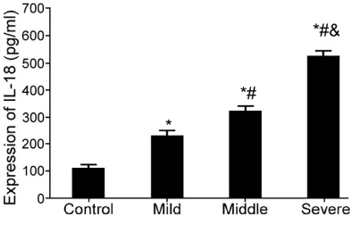
ELISA was applied to test IL-18 expression in the serum of VD patients. Serum IL-18 signifi-cantly increased in VD patients compared with control (P < 0.05). Its level kept rising following the aggravation of disease (Figure 1).
VD patients compared with healthy control and kept declining following aggravation (P < 0.05) (Figure 2).
The correlation between IL-18 and VD degree
The relationship between IL-18 and VD degree was further analyzed. IL-18 was negatively cor-related with MMSE score (P < 0.05), positively correlated with hypertension (P < 0.05), and showed no relationship with age, gender, or dia-betes (Table 3).
The relationship between IL-18 and Th1/Th2 balance in VD patients
The relationship between IL-18 and Th1/Th2 balance in VD patients was analyzed. IL-18 was positively correlated with IL-2 and TNF-α, whereas negatively correlated IL-4 and IL-6 (P < 0.05) (Table 4).
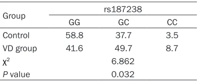
Correlation analysis of IL-18 SNP and VD pathogenesis
IL-18 gene rs187238 genotype was detected by PCR-RFLP. SNP genotyping success rate reached 99.2%. Allele frequency conformed to Hardy-Weinberg law. Rs187238 mutation ho- mozygote and heterozygote were CC and GC, respectively. Rs187238 frequency constituent ratio exhibited statistical difference (P < 0.05) (Table 5). IL-18 rs187238 variant allele (GC+CC; GC and CC) was related to VD pathogenesis. It obviously increased the risk of VD (1.79, 95% CI 1.02-2.31) (P < 0.05).
Discussion
[image:3.612.93.366.86.202.2]Immune responses and inflammation are key factors of VD occurrence and development [19,
Table 2. General information comparison
Index Control VD group
Mild Middle Severe
Cases 120 35 47 38
Age (year) 56.1 ± 7.1 55.5 ± 6.2 56.7 ± 7.4 57.2 ± 5.9
Male/female 68/52 18/17 25/22 22/16
Education (year) 7.2 ± 2.1 6.7 ± 3.8 6.8 ± 3.2 6.1 ± 2.5 Hypertension% 32 (26.7%) 22 (62.9%)* 40 (85.1%)*,# 35 (92.1%)*,# Diabetes% 12 (10.0%) 11 (31.4%)* 18 (28.2%)* 22 (57.9%)*,# MMSE 32.1 ± 2.1 24.0 ± 2.5* 17.2 ± 4.1*,# 7.1 ± 1.9*,#,&
*P < 0.05, compared with the control; #P < 0.05, compared with the mild group;
&P < 0.05, compared with the middle group.
Figure 1. IL-18 expression in the serum of VD pa-tients. *P < 0.05, compared with the control; #P < 0.05, compared with the mild group; &P < 0.05, com-pared with the middle group.
IL-2, TNF-α, IL-4, and IL-6
expression in the serum of VD patients
[image:3.612.90.287.246.372.2]20]. Th1 and Th2 cells regulate each other to maintain balance by regulating the secretion of cytokines, thus play a key role in maintaining normal immune function [21]. Th1 cells mainly secreted cytokines IL-2 and TNF-α, while Th2 cells majorly secrete IL-4 and IL-6 [22]. Th1/ Th2 homeostasis plays a crucial role in cellular immunity and humoral immunity through self-regulation and regulating cytokines secretion [23]. T cell subgroup Th1 mediated immune reaction is involved in the pathogenic process,
response [26]. IL-18 is an inflammatory factor with a wide range of immune regulatory effects to participate in tumor, inflammation, immune and other pathophysiologic processes [27]. This study found that serum IL-18 increased in VD patients and kept rising following aggrava-tion, which was consistent with previous report [15].
[image:4.612.90.375.74.215.2]A further study analyzed the impact of IL-18 expression on Th1/Th2 balance in VD patients. The results suggest that T1 cytokines IL-2 and TNF-α upregulated, while Th2 cytokines IL-4 and IL-6 reduced in VD patients, which was in accordance with previous report [25, 26]. Moreover, this study verified that Th1/Th2 cyto-kines changes more significantly following dis-ease aggravation. IL-18 was positively correlat-ed with disease condition, IL-2, and TNF-α, while negatively correlated with IL-4 and IL-6. IL-18 SNP was found in different populations. IL-18 SNP was not only related to gram-nega-tive bacteria and fungal infections, but also associated with cardiovascular and cerebro-vascular diseases [27]. However, the role of IL-18 gene polymorphism in VD has not been clarified. These results indicate that IL-18 was correlated with the occurrence and develop-ment of VD. Further in-depth investigation is
Table 3. Correlation analysis of IL-18 and clinical parameter in VD patients
Age Gender Education Hypertension Diabetes MMSE score
r value 0.198 0.147 0.341 0.782 0.356 -0.817
[image:4.612.91.374.303.351.2]P value 0.752 0.889 0.671 0.036 0.751 0.012
Table 4. Relationship between IL-18 and Th1/ Th2 balance in VD patients
IL-2 TNF-α IL-4 IL-6 r value 0.42 0.631 -0.941 -0.623 P value 0.026 0.031 0.028 0.033
Table 5. IL-18 SNP gene frequency analysis in VD patients
Group rs187238
GG GC CC
Control 58.8 37.7 3.5
VD group 41.6 49.7 8.7
χ2 6.862
[image:4.612.88.289.395.437.2]P value 0.032
Figure 2. IL-2, TNF-α, IL-4, and IL-6 expression in the serum of VD patients. *P < 0.05, compared with the control; #P < 0.05, compared with the mild group; &P < 0.05, compared with the middle group.
while Th2 cells mediate a pro-tective role by secreting cyto-kines [24].
[image:4.612.90.289.482.564.2]needed to explore the mechanism of IL-18 in regulating VD.
Conclusion
IL-18 was increased in VD patients and involved in regulating Th1/Th2 balance. IL-18 rs187238 mutation was the risk factor of VD. This study may provide a new theoretical basis for selec-tion of VD as a therapeutic target.
Acknowledgements
This work was supported by the Zhejiang Traditional Chinese Medicine Science and Technology Program Youth Talent Fund Project (No. 2015ZQ029).
Disclosure of conflict of interest
None.
Address correspondence to: Dr. Hua Zhang, De- partment of Neurology, Zhejiang Chinese Medicine and Western Medicine Integrated Hospital, No. 208, Huancheng East Road, Xiacheng District, Hangzhou 310000, Zhejiang, China. Tel: +86-0571-56109782; Fax: +86-0571-56109782; E-mail: [email protected]
References
[1] Janelidze S, Hertze J, Nägga K, Nilsson K, Nils-son C; Swedish BioFINDER Study Group, Wennström M, van Westen D, Blennow K, Zetterberg H, Hansson O. Increased blood-brain barrier permeability is associated with dementia and diabetes but not amyloid pathol-ogy or APOE genotype. Neurobiol Aging 2017; 51: 104-112.
[2] Usman R, Jamil M, Haq IU, Memon AA. Neuro-cognitive improvement in patients undergoing carotid endarterectomy for atherosclerotic oc-clusive carotid artery disease. Ann Vasc Dis 2016; 9: 307-311.
[3] van Opstal AM, van Rooden S, van Harten T, Ghariq E, Labadie G, Fotiadis P, Gurol ME, Ter-windt GM, Wermer MJ, van Buchem MA, Greenberg SM, van der Grond J. Cerebrovascu-lar function in presymptomatic and symptom-atic individuals with hereditary cerebral amy-loid angiopathy: a case-control study. Lancet Neurol 2017; 16: 115-122.
[4] Guo K, Yin G, Zi XH, Zhu HX, Pan Q. Effect of selective serotonin reuptake inhibitors on ex-pression of 5-HT1AR and neurotransmitters in rats with vascular dementia. Genet Mol Res 2016; 15.
[5] Lee JM, Park JM, Song MK, Oh YJ, Kim CJ, Kim YJ. The ameliorative effects of exercise on cog-nitive impairment and white matter injury from blood-brain barrier disruption induced by chronic cerebral hypoperfusion in adolescent rats. Neurosci Lett 2017; 638: 83-89.
[6] Cao WW, Wang Y, Dong Q, Chen X, Li YS, Zhou Y, Gao L, Deng Y, Xu Q. Deep microbleeds and periventricular white matter disintegrity are in-dependent predictors of attention/executive dysfunction in non-dementia patients with small vessel disease. Int Psychogeriatr 2017; 29: 793-803.
[7] Cheng MC, Pan TM. Prevention of hyperten-sion-induced vascular dementia by Lactobacil-lus paracasei subsp. Paracasei NTU 101-fer-mented products. Pharm Biol 2017; 55: 487-496.
[8] Sennik S, Schweizer TA, Fischer CE, Munoz DG. Risk factors and pathological substrates asso-ciated with agitation/aggression in alzheimer’s disease: a preliminary study using NACC data. J Alzheimers Dis 2017; 55: 1519-1528. [9] Mikkola TS, Savolainen-Peltonen H,
Tuomikos-ki P, Hoti F, Vattulainen P, Gissler M, Ylikorkala O. Lower death risk for vascular dementia than for alzheimer’s disease with postmenopausal hormone therapy users. J Clin Endocrinol Metab 2017; 102: 870-877.
[10] Donnellan C, Al Banna M, Redha N, Al Jishi A, Al Sharoqi I, Taha S, Bakhiet M, Abdulla F, Walsh P. Predictors of vascular cognitive im-pairment poststroke in a Middle Eastern (Bah-rain) cohort: a proposed case-control compari-son. JMIR Res Protoc 2016; 5: e223.
[11] Liao LX, Zhao MB, Dong X, Jiang Y, Zeng KW, Tu PF. TDB protects vascular endothelial cells ag- ainst oxygen-glucose deprivation/reperfusion-induced injury by targeting miR-34a to in-crease Bcl-2 expression. Sci Rep 2016; 6: 37959.
[12] Song KH, Kim MH, Kang SM, Jung SY, Ahn J, Woo HJ, Nam SY, Hwang SG, Ryu SY, Song JY. Analysis of immune cell populations and cyto-kine profiles in murine splenocytes exposed to whole-body low-dose irradiation. Int J Radiat Biol 2015; 91: 795-803.
[13] Vargas-Rojas MI, Solleiro-Villavicencio H, Soto-Vega E. Th1, Th2, Th17 and Treg levels in um-bilical cord blood in preeclampsia. J Matern Fetal Neonatal Med 2016; 29: 1642-5. [14] Tian T, Yu S, Liu L, Xue F, Yuan C, Wang M, Ji C,
Ma D. The profile of T helper subsets in bone marrow microenvironment is distinct for differ-ent stages of acute myeloid leukemia patidiffer-ents and chemotherapy partly ameliorates these variations. PLoS One 2015; 10: e0131761. [15] Di Rosa M, Dell’Ombra N, Zambito AM,
Chitotriosidase and inflammatory mediator lev-els in Alzheimer’s disease and cerebrovascular dementia. Eur J Neurosci 2006; 23: 2648-56. [16] Bossù P, Ciaramella A, Moro ML, Bellincampi L,
Bernardini S, Federici G, Trequattrini A, Mac-ciardi F, Spoletini I, Di Iulio F, Caltagirone C, Spalletta G. Interleukin 18 gene polymor-phisms predict risk and outcome of Alzheim-er’s disease. J Neurol Neurosurg Psychiatry 2007; 78: 807-11.
[17] Sudduth TL, Weekman EM, Price BR, Gooch JL, Woolums A, Norris CM, Wilcock DM. Time-course of glial changes in the hyperhomocyste-inemia model of vascular cognitive impairment and dementia (VCID). Neuroscience 2017; 341: 42-51.
[18] Ozturk C, Ozge A, Yalin OO, Yilmaz IA, Delialio-glu N, Yildiz C, Tesdelen B, Kudiaki C. The diag-nostic role of serum inflammatory and soluble proteins on dementia subtypes: correlation with cognitive and functional decline. Behav Neurol 2007;18: 207-215.
[19] Hakansson K, Bachert C, Konge L, Thomsen SF, Pedersen AE, Poulsen SS, Martin-Bertelsen T, Winther O, Backer V, von Buchwald C. Airway inflammation in chronic rhinosinusitis with na-sal polyps and asthma: the united airways con-cept further supported. PLoS One 2015; 10: e0127228.
[20] Ma L, Zeng J, Mo B, Wang C, Sun Y, Zhang M, Liu S, Xiang X, Wang CY. ANP/NPRA signaling preferentially mediates Th2 responses in favor of pathological processes during the course of acute allergic asthma. Int J Clin Exp Med 2015; 8: 5121-5128.
[21] Song C, Luchtman D, Kang Z, Tam EM, Yatham LN, Su KP, Lam RW. Enhanced inflammatory and T-helper-1 type responses but suppressed lymphocyte proliferation in patients with sea-sonal affective disorder and treated by light therapy. J Affect Disord 2015; 185: 90-96. [22] Tarkowski E, Tullberg M, Fredman P, Wikkelsö
C. Correlation between intrathecal sulfatide and TNF-alpha levels in patients with vascular dementia. Dement Geriatr Cogn Disord 2003; 15: 207-11.
[23] Calabrese V, Giordano J, Signorile A, Laura On-tario M, Castorina S, De Pasquale C, Eckert G, Calabrese EJ. Major pathogenic mechanisms in vascular dementia: roles of cellular stress response and hormesis in neuroprotection. J Neurosci Res 2016; 94: 1588-1603.
[24] Hempel P, Heinig B, Jerosch C, Decius I, Karc-zewski P, Kassner U, Kunze R, Steinhagen-Thiessen E, Bimmler M. Immunoadsorption of agonistic autoantibodies against alpha1-ad-renergic receptors in patients with mild to moderate dementia. Ther Apher Dial 2016; 20: 523-529.
[25] Wang XJ, Gao YP, Lu NN, Li WS, Xu JF, Ying XY, Wu G, Liao MH, Tan C, Shao LX, Lu YM, Zhang C, Fukunaga K, Han F, Du YZ. Endogenous polysialic acid based micelles for calmodulin antagonist delivery against vascular dementia. ACS Appl Mater Interfaces 2016; 8: 35045-35058.
[26] Cabrera-Fuentes HA, Aragones J, Bernhagen J, Boening A, Boisvert WA, Bøtker HE, Bulluck H, Cook S, Di Lisa F, Engel FB, Engelmann B, Fer-razzi F, Ferdinandy P, Fong A, Fleming I, Gn-aiger E, Hernández-Reséndiz S, Kalkhoran SB, Kim MH, Lecour S, Liehn EA, Marber MS, Mayr M, Miura T, Ong SB, Peter K, Sedding D, Singh MK, Suleiman MS, Schnittler HJ, Schulz R, Shim W, Tello D, Vogel CW, Walker M, Li QO, Yellon DM, Hausenloy DJ, Preissner KT. From basic mechanisms to clinical applications in heart protection, new players in cardiovascular diseases and cardiac theranostics: meeting report from the third international symposium on “new frontiers in cardiovascular research”. Basic Res Cardiol 2016; 111: 69.