Original Article
PIK3CA mutations and downstream effector
p-mTOR expression: implication for prognostic factors
and therapeutic targets in triple negative breast cancer
Jie Wang1, Xinxin Zhu2, Xin Xu3, Lei Guo1, Guihua Shen1, Xiuyun Liu1, Chen Chang3, Bingning Wang1, Hongying Yang1, Mingrong Wang3
1Department of Pathology, 3State Key Laboratory of Molecular Oncology, National Cancer Center/Cancer Hospital,
Chinese Academy of Medical Sciences and Peking Union Medical College, Beijing, China; 2Department of
Obstet-rics and Gynecology, Peking Union Medical College Hospital, Peking Union Medical College, Chinese Academy of Medical Sciences, Beijing, China
Received April 3, 2017; Accepted June 13, 2017; Epub July 1, 2017; Published July 15, 2017
Abstract: Although PIK3CA mutations and phosphorylated mTOR (p-mTOR) expression are two main events on PI3K/Akt/mTOR pathway, limited data has reported their roles in triple negative breast cancer (TNBC). Thus, we evaluated the associations of these two biomarkers and clinicopathological characteristics and prognosis in large Chinese TNBC patients. Immunohistochemistry (IHC) analysis was used to assess p-mTOR expression level in 218 TNBC patients. Direct sequencing was applied to detect the most important hotspot regions in exons 9 and 20 of PIK3CA gene. In the TNBC cohort, mutations were identified in 11.5% cases which were associated with basal-like subtype. The somatic point mutations were independently associated with worse overall survival (HR=0.400, 95% CI: 0.193-0.830, P=0.014). As for p-mTOR expression, 47.7% of the tumors were positive and the staining was shown in cytoplasm, nuclear and perinuclear areas. There were significant differences observed in tumor size, lymph node status, and clinical stage between p-mTOR positive and p-mTOR negative cases. Notably, we found a significant association between p-mTOR expression and PIK3CA mutations. Patients with p-mTOR staining also demonstrated shorter overall survival (HR=0.710, 95% CI: 0.514-0.980, P=0.037). Therefore, PIK3CA mutations and its downstream effector p-mTOR expression were two important regulators for activating the PI3K/Akt/mTOR pathway. Both of them could be served as adverse prognostic biomarkers and may contribute to the targeted therapy for TNBC patients with poor outcome.
Keywords: PIK3CA, p-mTOR, triple negative breast cancer, prognosis
Introduction
Triple negative breast cancer (TNBC) lacks the expression of estrogen-receptor (ER), proges-terone-receptor (PR) and epidermal growth fac-tor recepfac-tor 2 (HER2), accounting for approxi-mately 16% of all BC [1, 2]. A high proportion of TNBC comprises the basal-like breast cancer (BLBC) subtype which has been extensively characterized on the basis of gene expression profiling [3]. However, it is becoming increas-ingly common and facility to identify this pheno-type based on the immunohistochemistry (IHC) staining of basal-like markers. BLBC specifical-ly shows IHC expression of epidermal growth factor receptor (EGFR) and/or cytokeratin 5/6 (CK5/6), and is prone to have a poorer progno-sis and higher recurrence than non-BLBC as
reported [4]. The deficiency of target therapies and prognostic factors in TNBC patients is a critical clinical issue at present. Approximately 20% TNBC patients with family history carry an inherited BRCA1 or BRCA2 mutation. On the contrary, the sporadic TNBC patients do not have such genetic alterations [5]. The aggres-sive behavior and poor outcome of sporadic population might be controlled mainly by other genetic and molecular changes.
two subunits: regulatory subunit (p85) and cat-alytic subunit (p110). PIK3CA is located on chromosome 3q26.3 which encodes p110α catalytic subunit of class IA of PI3K [7, 8]. mTOR is a serine/threonine kinase that serve as a key downstream regulator on the path- way. The phosphorylation of mTOR (p-mTOR) stands for the hyperactivation of the molecule that can eventually lead to increased protein synthesis, cell growth and tumor development [9]. Emerging data has revealed that high PI3K pathway activity is the most frequent event in BLBC/TNBC when compared with TP53 and RB1 pathway. Among the aberrant activation pathway, PIK3CA mutations and downstream p-mTOR expression are two major events on the pathway [10]. Furthermore, Chen et al [11] recently demonstrated that PI3K/mTOR dual inhibitor BEZ235 and Trichostatin A (TSA) re- sult in synergistic growth inhibition and apop- tosis of BC cell lines. Hence, we postulated that the PIK3CA mutations and p-mTOR ex- pression might serve as two critical regulators and have a role in sporadic TNBC.
Based on the former reports, we systematically evaluated the mutation type of PIK3CA and staining forms of p-mTOR in a large cohort study of 218 TNBC population. We also ana-lyzed the associations of these two key mark-ers with clinicopathological features and prog-nostic impacts, which might have clinical signi- ficance for further development of drug combi-nations in the treatment of TNBC patients. Materials and methods
Patients and samples
A total of 218 patients who underwent surgery and diagnosed with TNBC were selected for this retrospective study during the period of January 1, 1999 - December 31, 2008 in the Department of Pathology, Cancer Hospital, Peking Union Medical College, Chinese Aca- demy of Medical Sciences, Beijing, China. The inclusion criteria were as follows: primary oper-able breast cancer, no family history for breast or ovary cancer, no prior radiotherapy and che-motherapy before surgery, mastectomies, or lumpectomies specimens with sufficient tis-sue. Clinicopathological information was avail-able for all the patients, including age, tumor size, grade, lymph node metastasis, tumor embolus, subtype, and prognosis. Survival da-
ta were last updated on August 24, 2014, and the median follow-up period was 77.25 months (range 2.13-168.47 months).
PCR and direct sequencing of PIK3CA
DNA extraction from formalin-fixed and paraf-fin-embedded (FFPE) TNBC tissue samples, with minimum of 75% malignant cells, was per-formed according to the manufacturer’s instruc-tions (ZEESAN, Xiamen, China). The PCR ampli-fications targeting PIK3CA exons 9 and 20 were conducted. Primers were designed using the Primer Design Tool from NCBI. The primers were as follows: 9-F: GTATTTGCTTTTTCTG- TAAATCATCTG, 9-R: CATGCTGAGATCAGCCAA- ATTC; 20-F: CTCTGGAATGCCAGAACTAC, 20- R: ATGCTGTTTAATTGTGTGGAAG. The amplified products were sequenced using an ABI 3730XL sequencer and the sequencing profiles were analyzed by Mutation Surveyor software v4.0.8 (SoftGenetics Corporation, America), in com-parison with the corresponding reference sequence (NM_006218) to identify mutations. Tissue microarray (TMA) and IHC assay
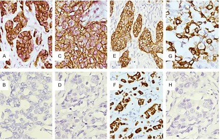
Figure 1. A. Cytoplasmic immunostaining of CK5/6; B. Negative control for CK5/6 by omission of the primary anti-body; C. Membranous immunostaining of EGFR; D. Negative control for EGFR by omission of the primary antianti-body; E. Cytoplasmic immunostaining of p-mTOR; F. Nuclear immunostaining of p-mTOR; G. Perinuclear immunostaining of p-mTOR; H. Negative control for p-mTOR by omission of the primary antibody. (Magnification 400×).
sity; 2+: > 50% with weak intensity or > 10% with moderate to strong intensity. These scores indicated, 0: negative; Score 1: low expression; Score 2: high expression; Both 1 and 2 scores were regarded as positive in the statistical analysis.
Statistical analysis
Statistical analysis was performed using the SPSS 17.0 software package. The associations between these two markers and clinicopatho-logical parameters were analyzed using the Pearson’s χ2-test. The overall survival time was described by Kaplan-Meier method and Cox proportional hazard regression analysis from the date of surgery till the date of death from BC. All P values were two-tailed and statistical significance was considered to be P < 0.05. Results
Sample cohort and clinical parameters
With regard to the complete cohort, a total of 218 TNBC patients could be further divide into
134 (61.5%) cases of BLBC and 84 (38.5%) cases of non-BLBC, based on IHC staining for EGFR and CK5/6 (Figure 1). The median age of all patients at the time of diagnosis was 50 years, with an age range of 24 to 82 years. According to the WHO Classification of Breast Tumors, 196 (89.9%) patients were histologi-cally classified as invasive carcinomas of no special type (IC-NST, including 78 cases of grade 2 and 118 cases of grade 3) and 2 (0.9%) of the cases had invasive lobular carcinoma (ILC). Tumors numbering 20 (9.2%) had carci-noma of other histologic types such as medul-lary carcinoma (n=13), metaplastic carcinoma (n=4), secretory carcinoma (n=1), and adenoid cystic carcinoma (n=2).
PIK3CA mutations and patients’ characteris-tics
Figure 2. Mutational analysis of PIK3CA exons 9 and 20 in TNBC. A and B. Mutant sequence E542V (A1625T) and corresponding wild-type sequence; C and D. Mutant sequence E545K (G1633A) and corresponding wild-type sequence; E and F. Mutant sequence E545A (A1634C) and corresponding wild-type sequence; G and H. Mutant sequence H1047R (A3140G) and corresponding wild-type sequence.
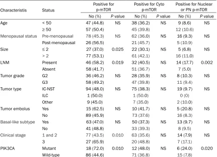
expression could be detected in the cytoplasm (38.1%, 83/218), the nuclear (7.8%, 17/218) and perinuclear (1.8%, 4/218) areas respec-tively. Typical staining patterns for p-mTOR were shown in Figure 1. As can be seen in Table 3, p-mTOR was more frequent in patients with lymph node metastasis than in those without lymph node metastasis; in tumors with size > 2 cm rather than in tumors with size ≤ 2 cm; and in advanced stage (3) compared with early stage (1 and 2). In contrast, p-mTOR was unre-lated to age, menopausal status, tumor grade, type, embolus, and basal-like subtype. Par- ticularly, p-mTOR-expressing tumors were more frequently detected in PIK3CA mutant than in PIK3CA wild-type patients. Moreover, we also noted a significant association between the protein present in the nuclear or perinuclear area and lymph node metastasis and PIK3CA mutations.
PIK3CA mutation and p-mTOR expression on prognosis
Patients with PIK3CA mutations or p-mTOR expression had significantly shorter overall sur-vival by Kaplan-Meier analysis (P=0.001 and P=0.001, respectively; Figure 3A and 3B). Univariate and multivariate analysis of 218 Table 1. PIK3CA mutations in TNBC patients
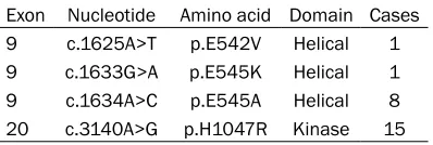
Exon Nucleotide Amino acid Domain Cases
9 c.1625A>T p.E542V Helical 1
9 c.1633G>A p.E545K Helical 1
9 c.1634A>C p.E545A Helical 8
20 c.3140A>G p.H1047R Kinase 15
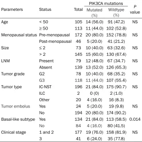
mutation was H1047R (6.9%), representing 100% of the exon 20 mutations. The exon 9 hotspot mutation was E545A (3.7%), corre-sponding to 80% of the exon 9 mutations. Other common helical mutations included were E542V and E545K which were found only in one case, respectively (Figure 2; Table 1). As shown in Table 2, there is no significant asso-ciation of PIK3CA mutations with age, meno-pausal status, size, lymph node metastasis, tumor grade, type, embolus, and clinical stage. However, it is worth noting that patients with gene mutations were more frequently detected in BLBC than in non-BLBC (84% versus 16%, P=0.014).
p-mTOR expression and patients’ characteris-tics
[image:4.612.90.289.398.465.2]Table 2. PIK3CA mutations and clinicopathological characteristics of 218 TNBC patients
Parameters Status Total
PIK3CA mutations P
value
Mutated (%)
Wildtype (%)
Age < 50 105 14 (56.0) 91 (47.2) NS
≥ 50 113 11 (44.0) 102 (52.8)
Menopausal status Pre-menopausal 172 20 (80.0) 152 (78.8) NS Post-menopausal 46 5 (20.0) 41 (21.2)
Size ≤ 2 73 10 (40.0) 63 (32.6) NS
> 2 145 15 (60.0) 130 (67.4)
LNM Present 79 12 (48.0) 67 (34.7) NS
Absent 139 13 (52.0) 126 (65.3)
Tumor grade G2 78 10 (40.0) 68 (35.2) NS
G3 118 11 (44.0) 107 (55.4)
Tumor type IC-NST 196 21 (84.0) 175 (90.7) NS
ILC 2 0 (0) 2 (1.0)
Other 20 4 (16.0) 16 (8.3)
Tumor embolus Yes 24 5 (20.0) 19 (9.8) NS
No 194 20 (80.0) 174 (90.2)
Basal-like subtype Yes 134 21 (84.0) 113 (58.5) 0.014
No 84 4 (16.0) 80 (41.5)
Clinical stage 1 and 2 177 19 (76.0) 158 (81.9) NS
3 41 6 (24.0) 35 (77.8)
Abbreviations: NS, not significant; IC-NST, invasive carcinoma of no special type; LNM, lymph nodes metastasis; ILC, invasive lobular carcinoma.
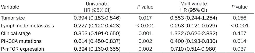
TNBC patients revealed that PIK3CA mutation and p-mTOR expression were independent pre-dictive factors for worse prognosis (HR=0.400, 95% CI: 0.193-0.830, P=0.014 and HR=0.710, 95% CI: 0.514-0.980, P=0.037, respectively), and also of lymph node metastasis (HR=0.253, P < 0.001; Table 4).
Discussion
In the TNBC cohort, 11.5% cases were identi-fied with PIK3CA mutations which were a little lower than the COSMIC databases with a total mutation frequency of 13%. The possible rea-son for the slight difference might be due to dif-ferent sequencing methods as reported pre- viously [14]. Other causes could be the differ-ences of patient cohorts, sample preservation and methods used for DNA isolation [15]. Somatic mutations of PIK3CA are clustered into the helical domain (exon 9, commonly E545K and E542K) and the kinase domain (exon 20, commonly H1047R) [16]. The major mutations of our study were present in exon 20 (60% vs 40% in exon 9), entirely in the hot spots of
H1047R. Wallin et al [17] detected that PIK3CA-H10- 47R mutation promoted PI3K pathway activity and indu- ced obvious epithelial-mes-enchymal transition (EMT) as well as invasive pheno-type, in comparison with the isogenic wild-type mammary epithelial cells. A previous study from the Peruvian BC patients showed that E54- 5A was the main mutation site in exon 9 (70%, 7/10) rather than E545K or E542K in usual [18], which were in accordance with our findings. It is possible that heteroge-neous feature of TNBC may lead to various mutated typ- es of oncogene in diverse ethnic population. Although there is no significant asso- ciation between PIK3CA mu- tations and most clinical pa- rameters, we found that PIK3CA mutations were sta-tistically correlated with BL- BC (P=0.014) which has not been reported in large TNBC cohort before. It has become clear that BLBC often induced unfavorable outcomes [4]. Thus, we assume that the relation of basal markers with oncogene mutations in TNBC can generate more aggressive biological behaviors and worse prognostic effects than wild-type.
Table 3. Relationship of p-mTOR expression with clinicopathological characteristics in 218 TNBC patients
Characteristis Status
Positive for
p-mTOR Positive for Cyto p-mTOR Positive for Nuclear or PN p-mTOR
No (%) P value No (%) P value No (%) P value
Age < 50 47 (44.8) NS 38 (36.2) NS 9 (8.6) NS
≥ 50 57 (50.4) 45 (39.8) 12 (10.6)
Menopausal status Pre-menopausal 78 (45.3) NS 62 (36.0) NS 16 (9.3) NS
Post-menopausal 26 (56.5) 21 (45.7) 5 (10.9)
Size ≤ 2 27 (37.0) 0.025 22 (30.1) NS 5 (6.8) NS
> 2 77 (53.1) 61 (42.1) 16 (11.0)
LNM Present 46 (58.2) 0.019 32 (40.5) NS 14 (17.7) 0.002
Absent 58 (41.7) 51 (36.7) 7 (5.0)
Tumor grade G2 36 (46.2) NS 28 (35.9) NS 8 (10.3) NS
G3 58 (49.2) 47 (39.8) 11 (9.4)
Tumor type IC-NST 94 (48.0) NS 75 (38.3) NS 19 (9.7) NS
ILC 1 (50.0) 1 (50.0) 0 (0)
Other 9 (45.0) 7 (35.0) 2 (10.0)
Tumor embolus Yes 15 (62.5) NS 10 (41.7) NS 5 (20.8) NS
No 89 (45.9) 73 (37.6) 16 (8.3)
Basal-like subtype Yes 63 (47.0) NS 50 (37.3) NS 13 (9.7) NS
No 41 (48.8) 33 (39.3) 8 (9.5)
Clinical stage 1 and 2 77 (43.5) 0.010 63 (35.6) NS 14 (7.9) NS
3 27 (65.9) 20 (48.8) 7 (17.1)
PIK3CA Mutant 18 (72.0) 0.010 12 (48.0) NS 6 (24.0) 0.020
Wild-type 86 (44.6) 71 (36.8) 15 (7.8)
Abbreviations: NS, not significant; IC-NST, invasive carcinoma of no special type; LNM, lymph nodes metastasis; ILC, invasive lobular carcinoma; Cyto, Cytoplasmic; PN, perinuclear.
tumor type usually lead to a pejorative effect on survival time [23]. Additionally, patients with PIK3CA mutations in HER2 overexpres- sion subtype also possessed an adverse effect on the outcome after treatment with trastu-zumab [24]. Our study confirmed that PIK3CA mutations could be served as an independent poor prognostic factor for overall survival in TNBC patients. From the above conclusions, specific BC samples based on different molec-ular subtypes which also include other genomic alterations (such as ER, HER2, EGFR, and so on) may contribute to the controversial results of PIK3CA mutations on prognosis.
Everolimus, as an oral inhibitor of mTOR, is the only US Food and Drug Administration (FDA)-approved drug for patients with hor- mone-receptor-positive, HER2-negative locally advanced or metastatic BC [25]. Nearly half of the TNBCs (47.7%) in our data showed positive immunostaining for p-mTOR, indicating a poten-tial role of mTOR inhibitors in TNBC targeted
therapy. Most of the previous studies regarded cytoplasm as the only positive form for p-mTOR expression without referring to other staining patterns [26-28]. Furthermore, few in-depth studies have been implemented on its expres-sion in TNBC patients. Our study found that the staining form of the protein was characterized in the cytoplasm, nucleus and perinuclear area. Moreover, there were no significant diffe- rences when identified tumors into high and low expression group in our cases. Thus, we consider any of the three staining forms as positive regardless of the staining intensity when analysis was performed. The p-mTOR positive group was related to bigger tumor size (≥ 2 cm), lymph node metastasis and advan- ced stage (3), all of which stand for aggressive biological behaviors and result in poor out- come.
Figure 3. Kaplan-Meier curves for overall survival according to PIK3CA mutation (A) and p-mTOR expression (B) in TNBC patients. (n=218).
Table 4. PIK3CA mutations and p-mTOR expression status in 218 TNBC patients
Variable HR (95% CI)Univariate P value HR (95% CI)Multivariate P value
Tumor size 0.394 (0.183-0.846) 0.017 0.553 (0.244-1.254) 0.156
Lymph node metastasis 0.227 (0.122-0.423) < 0.001 0.253 (0.121-0.529) < 0.001
Clinical stage 0.353 (0.191-0.650) 0.001 1.332 (0.626-2.832) 0.457
PIK3CA mutations 0.614 (0.450-0.837) 0.002 0.400 (0.193-0.830) 0.014
P-mTOR expression 0.324 (0.160-0.655) 0.002 0.710 (0.514-0.980) 0.037
levels of PIP3 lead to phosphorylation of Akt, which showed an impact on the cancer cell cycling, survival and growth [7]. Akt can acti-vate mTOR directly by phosphorylation at S2448 or indirectly, by phosphorylation and inactivation of tuberous sclerosis complex 2 (TSC2). When TSC2 loses its function, the GTPase Rheb is maintained in its GTP-bound state, allowing for increased activation of mTOR [29]. Hence, overexpression of p-mTOR (active form of mTOR) represents aberrant activation of this pathway and results in in- creased protein synthesis, proliferation and anti-apoptosis via its action on substrate 40S ribosomal protein S6 kinase 1 (S6K1) and eukaryotic initiation factor 4E binding protein (4EBP1) [30]. The expression of p-mTOR was expressed more frequently in PIK3CA mutant patients compared to PIK3CA wild-type pa- tients. To some extent, our finding reflects the activated associations between upstream and downstream effectors on the pathway. PIK3CA mutations have been shown to be associated with mTOR inhibitor sensitive in both cell lines and clinical studies [31, 32]. Therefore, it can
be speculated from our findings that TNBC patients harboring both PIK3CA mutations and p-mTOR expression are more likely to respond to mTOR inhibitors than those with either alter-ation alone.
[image:7.612.91.523.323.413.2]the perinuclear pattern. To sum up the above conclusions, along with our findings, the ex- pression form of p-mTOR is actually variable in BC with different meaning on specific subtypes. Although cytoplasmic positive is the main form, we believe that the nuclear p-mTOR plays a significant role in the progression of TNBC and potentially triggers an negative impact on the disease behaviors of patients. On the contrary, the perinuclear pattern is typically present in non-TNBC and appears to have an effect on tumors without triple-negative features.
The expression of p-mTOR induced different disease behaviors and outcomes through particular mechanisms in special subtypes of BC. Zhou et al [36] demonstrated the associa-tion of higher mTOR phosphorylaassocia-tion with sig-nificantly shorter disease-free survival (P < 0.01), and could lead to enhanced sensitivity to mTOR inhibitors in ErbB2-overexpressing cells. In contrast, one study found that p-mTOR expression was independently associated with longer disease-free survival and overall surviv-al in luminsurviv-al BC [26]. The protein expression has an unfavorable impact on survival in our TNBC patients. Zhang et al [37] found that mTOR inhibitor testing in TNBC xenografts showed significant tumor growth inhibition. Our finding emphasized the potential prognostic value for TNBC patients who were treated with mTOR inhibitor.
In summary, our findings indicated that PIK3CA mutations and p-mTOR expression were com-mon genetic-molecular events of the activated PAM pathway in TNBC. Both of the two markers were independently associated with poor over-all survival and patients who had both onco-gene mutations and downstream protein ex- pression seem to benefit from the PAM path-way inhibitors. While, clinical trials are required in future to clarify these two biomarkers as suitable and reliable drug targets.
Acknowledgements
This research did not receive any specific grant from funding agencies in the public, commer-cial, or not-for-profit sectors.
Disclosure of conflict of interest
None.
Address correspondence to: Dr. Hongying Yang, Department of Pathology, Cancer Institute and Cancer Hospital, Peking Union Medical College, Chinese Academy of Medical Sciences, 17 Pan-Jiayuan Nanli, Chaoyang District, Beijing 100021, China. Tel: +86-10-87787507; E-mail: [email protected]; Dr. Mingrong Wang, Department of State Key Laboratory of Molecular Oncology, Cancer Institute and Cancer Hospital, Peking Union Medical College, Chinese Academy of Medical Sciences, 17 Pan-Jiayuan Nanli, Chaoyang District, Beijing 100021, China. Tel: +86-10-87788454; E-mail: [email protected]
References
[1] Shah SP, Roth A, Goya R, Oloumi A, Ha G, Zhao Y, Turashvili G, Ding J, Tse K, Haffari G, Basha-shati A, Prentice LM, Khattra J, Burleigh A, Yap D, Bernard V, McPherson A, Shumansky K, Cri-san A, Giuliany R, Heravi-Moussavi A, Rosner J, Lai D, Birol I, Varhol R, Tam A, Dhalla N, Zeng T, Ma K, Chan SK, Griffith M, Moradian A, Cheng SW, Morin GB, Watson P, Gelmon K, Chia S, Chin SF, Curtis C, Rueda OM, Pharoah PD, Damaraju S, Mackey J, Hoon K, Harkins T, Tadi-gotla V, Sigaroudinia M, Gascard P, Tlsty T, Costello JF, Meyer IM, Eaves CJ, Wasserman WW, Jones S, Huntsman D, Hirst M, Caldas C, Marra MA and Aparicio S. The clonal and muta-tional evolution spectrum of primary triple-neg-ative breast cancers. Nature 2012; 486: 395-399.
[2] Schmadeka R, Harmon BE and Singh M. Triple-negative breast carcinoma: current and emerg-ing concepts. Am J Clin Pathol 2014; 141: 462-477.
[3] Lehmann BD, Bauer JA, Chen X, Sanders ME, Chakravarthy AB, Shyr Y and Pietenpol JA. Identification of human triple-negative breast cancer subtypes and preclinical models for se-lection of targeted therapies. J Clin Invest 2011; 121: 2750-2767.
[4] Tischkowitz M, Brunet JS, Begin LR, Huntsman DG, Cheang MC, Akslen LA, Nielsen TO and Foulkes WD. Use of immunohistochemical markers can refine prognosis in triple negative breast cancer. BMC Cancer 2007; 7: 134. [5] Afghahi A, Telli ML and Kurian AW. Genetics of
triple-negative breast cancer: implications for patient care. Curr Probl Cancer 2016; 40: 130-140.
[6] Yuan TL and Cantley LC. PI3K pathway altera-tions in cancer: variaaltera-tions on a theme. Onco-gene 2008; 27: 5497-5510.
[8] Zardavas D, Phillips WA and Loi S. PIK3CA mu-tations in breast cancer: reconciling findings from preclinical and clinical data. Breast Can-cer Res 2014; 16: 201.
[9] Ma XM and Blenis J. Molecular mechanisms of mTOR-mediated translation control. Nat Rev Mol Cell Biol 2009; 10: 307-318.
[10] Cancer Genome Atlas Network. Comprehen-sive molecular portraits of human breast tu-mours. Nature 2012; 490: 61-70.
[11] Chen L, Jin T, Zhu K, Piao Y, Quan T, Quan C and Lin Z. PI3K/mTOR dual inhibitor BEZ235 and histone deacetylase inhibitor trichostatin a synergistically exert anti-tumor activity in breast cancer. Oncotarget 2017; 8: 11937-11949.
[12] Zhu X, Shan L, Wang F, Wang J, Wang F, Shen G, Liu X, Wang B, Yuan Y, Ying J and Yang H. Hypermethylation of BRCA1 gene: implication for prognostic biomarker and therapeutic tar-get in sporadic primary triple-negative breast cancer. Breast Cancer Res Tr 2015; 150: 479-486.
[13] Ueng SH, Chen SC, Chang YS, Hsueh S, Lin YC, Chien HP, Lo YF, Shen SC and Hsueh C. Phos-phorylated mTOR expression correlates with poor outcome in early-stage triple negative breast carcinomas. Int J Clin Exp Pathol 2012; 5: 806-813
[14] Arsenic R, Treue D, Lehmann A, Hummel M, Dietel M, Denkert C and Budczies J. Compari-son of targeted next-generation sequencing and Sanger sequencing for the detection of PIK3CA mutations in breast cancer. BMC Clin Pathol 2015; 15: 20.
[15] Arsenic R, Lehmann A, Budczies J, Koch I, Prinzler J, Kleine-Tebbe A, Schewe C, Loibl S, Dietel M and Denkert C. Analysis of PIK3CA mutations in breast cancer subtypes. Appl Im-munohistochem Mol Morphol 2014; 22: 50-56.
[16] Janku F, Lee JJ, Tsimberidou AM, Hong DS, Na-ing A, Falchook GS, Fu S, Luthra R, Garrido-La-guna I and Kurzrock R. PIK3CA mutations fre-quently coexist with RAS and BRAF mutations in patients with advanced cancers. PLoS One 2011; 6: e22769.
[17] Wallin JJ, Guan J, Edgar KA, Zhou W, Francis R, Torres AC, Haverty PM, Eastham-Anderson J, Arena S, Bardelli A, Griffin S, Goodall JE, Grim-shaw KM, Hoeflich KP, Torrance C, Belvin M and Friedman LS. Active PI3K pathway causes an invasive phenotype which can be reversed or promoted by blocking the pathway at diver-gent nodes. PLoS One 2012; 7: e36402. [18] Castaneda CA, Lopez-Ilasaca M, Pinto JA,
Chiri-nos-Arias M and Doimi F. PIK3CA mutations in Peruvian patients with HER2-amplified and tri-ple negative non-metastatic breast cancers.
Hematol Oncol Stem Cell Ther 2014; 7: 142-148.
[19] Deng L, Chen J, Zhong XR, Luo T, Wang YP, Huang HF, Yin L, Qiu Y, Bu H, Lv Q and Zheng H. Correlation between activation of PI3K/AKT/ mTOR pathway and prognosis of breast cancer in Chinese women. PLoS One 2015; 10: e120511.
[20] Fu X, Osborne CK and Schiff R. Biology and therapeutic potential of PI3K signaling in ER+/ HER2-negative breast cancer. Breast 2013; 22 Suppl 2: S12-S18.
[21] Ellis MJ, Lin L, Crowder R, Tao Y, Hoog J, Snider J, Davies S, DeSchryver K, Evans DB, Steinseif-er J, Bandaru R, Liu W, GardnSteinseif-er H, Semiglazov V, Watson M, Hunt K, Olson J and Baselga J. Phosphatidyl-inositol-3-kinase alpha catalytic subunit mutation and response to neoadju-vant endocrine therapy for estrogen receptor positive breast cancer. Breast Cancer Res Treat 2010; 119: 379-390.
[22] Kalinsky K, Jacks LM, Heguy A, Patil S, Drobn-jak M, Bhanot UK, Hedvat CV, Traina TA, Solit D, Gerald W and Moynahan ME. PIK3CA muta-tion associates with improved outcome in breast cancer. Clin Cancer Res 2009; 15: 5049-5059.
[23] Creighton CJ, Fu X, Hennessy BT, Casa AJ, Zhang Y, Gonzalez-Angulo AM, Lluch A, Gray JW, Brown PH, Hilsenbeck SG, Osborne CK, Mills GB, Lee AV and Schiff R. Proteomic and transcriptomic profiling reveals a link between the PI3K pathway and lower estrogen-receptor (ER) levels and activity in ER+ breast cancer. Breast Cancer Res 2010; 12: R40.
[24] Cizkova M, Dujaric ME, Lehmann-Che J, Scott V, Tembo O, Asselain B, Pierga JY, Marty M, de Cremoux P, Spyratos F and Bieche I. Outcome impact of PIK3CA mutations in HER2-positive breast cancer patients treated with trastuzum-ab. Br J Cancer 2013; 108: 1807-1809. [25] Jerusalem G, Mariani G, Ciruelos EM, Martin
M, Tjan-Heijnen VC, Neven P, Gavila JG, Miche-lotti A, Montemurro F, Generali D, Simoncini E, Lang I, Mardiak J, Naume B, Camozzi M, Loriz-zo K, Bianchetti S and Conte P. Safety of evero-limus plus exemestane in patients with hor-mone-receptor-positive, HER2-negative locally advanced or metastatic breast cancer pro-gressing on prior non-steroidal aromatase in-hibitors: Primary results of a phase IIIb, open-label, single-arm, expanded-access mul- ticenter trial (BALLET). Ann Oncol 2016; 27: 1719-1725.
[27] Bakarakos P, Theohari I, Nomikos A, Mylona E, Papadimitriou C, Dimopoulos AM and Nako-poulou L. Immunohistochemical study of PTEN and phosphorylated mTOR proteins in familial and sporadic invasive breast carcinomas. His-topathology 2010; 56: 876-882.
[28] Bostner J, Karlsson E, Pandiyan MJ, Westman H, Skoog L, Fornander T, Nordenskjold B and Stal O. Activation of Akt, mTOR, and the estro-gen receptor as a signature to predict tamoxi-fen treatment benefit. Breast Cancer Res Treat 2013; 137: 397-406.
[29] Sarbassov DD, Guertin DA, Ali SM and Sabatini DM. Phosphorylation and regulation of Akt/ PKB by the rictor-mTOR complex. Science 2005; 307: 1098-1101.
[30] Sarbassov DD, Ali SM, Sengupta S, Sheen JH, Hsu PP, Bagley AF, Markhard AL and Sabatini DM. Prolonged rapamycin treatment inhibits mTORC2 assembly and Akt/PKB. Mol Cell 2006; 22: 159-168.
[31] Meric-Bernstam F, Akcakanat A, Chen H, Do KA, Sangai T, Adkins F, Gonzalez-Angulo AM, Rashid A, Crosby K, Dong M, Phan AT, Wolff RA, Gupta S, Mills GB and Yao J. PIK3CA/PTEN mutations and Akt activation as markers of sensitivity to allosteric mTOR inhibitors. Clin Cancer Res 2012; 18: 1777-1789.
[32] Gonzalez-Angulo AM and Blumenschein GJ Jr. Defining biomarkers to predict sensitivity to PI3K/Akt/mTOR pathway inhibitors in breast cancer. Cancer Treat Rev 2013; 39: 313-320. [33] Bakarakos P, Theohari I, Nomikos A, Mylona E,
Papadimitriou C, Dimopoulos AM and Nako-poulou L. Immunohistochemical study of PTEN and phosphorylated mTOR proteins in familial and sporadic invasive breast carcinomas. His-topathology 2010; 56: 876-882.
[34] Vazquez-Martin A, Oliveras-Ferraros C, Berna-do L, Lopez-Bonet E and Menendez JA. The serine 2481-autophosphorylated form of mammalian Target of Rapamycin (mTOR) is lo-calized to midzone and midbody in dividing cancer cells. Biochem Biophys Res Commun 2009; 380: 638-643.
[35] Walsh S, Flanagan L, Quinn C, Evoy D, McDer-mott EW, Pierce A and Duffy MJ. MTOR in breast cancer: differential expression in triple-negative and non-triple-triple-negative tumors. Breast 2012; 21: 178-182.
[36] Zhou X, Tan M, Stone HV, Klos KS, Lan KH, Yang Y, Yang W, Smith TL, Shi D and Yu D. Acti-vation of the Akt/mammalian target of rapamycin/4E-BP1 pathway by ErbB2 overex-pression predicts tumor progression in breast cancers. Clin Cancer Res 2004; 10: 6779-6788.