DOCKING STUDIES OF GLUCAN BASED COMPOUNDS ON
DECTIN-1 ENZYME
Varsha V. Joshi* and Sunil H. Ganatra
Department of Chemistry
Institute of Science, R. T. Road, Civil Lines, Nagpur-440008 (M.S.), India.
ABSTRACT
It is reported that Dectin-1 plays a crucial role in recognizing a process called tumor cell-associated glycosylation. It activates anti-tumor immune response. Hence extensive in-silico study executed to know how Dectin-1 interacts with certain known polysaccharides? Three basic polysaccharide class of compounds, β 1,3 glucan, β 1,6 glucan and β 1,3-1,6 glucan and their substituted derivatives were docked in-silico with Dectin-1. It is observed that substituted β 1,6 glucan compounds show better binding energy values with Dectin-1. Substitution at R2 by –CN group shows -10.5 kcal/mol. binding energy which is almost double than the natural ligand.
KEYWORDS: Polysaccharides, β glucan, Binding energy, in-silico, Cancer.
INTRODUCTION
Day by day, Cancer causes more deaths worldwide than any other diseases.[1] Cancer is caused due to various reasons. The main among them are tobacco chewing, pesticides in vegetable and food, environmental pollution, higher exposure to radiation, life style etc.[2-5]
Numbers of potent anti-cancer drugs are already available. These drugs are having high level of toxicity. High toxicity is the main reason for not recommending present drugs to old age cancer patients. There is always a need to identify non-toxic anti-cancer drugs.[6]
Chiba et. al. reported that cancer tumor shows excess production of Dectin-1.[7]. It is also reported that the Dectin-1 play a vital role in activating immunology responses by interacting with β-1,3 gulcan naturally. This interaction activates the immunology process and in further natural killer cells kill tumor cell, though the direct relationship is yet to be established.
Volume 5, Issue 11, 1467-1474. Research Article ISSN 2277– 7105
*Corresponding Author
Varsha V. Joshi
Department of Chemistry Institute of Science, R. T. Road, Civil Lines, Nagpur-440008 (M.S.), India. Article Received on 16 Sept. 2016,
The glucan classes of compounds are non toxic.[8] β-glucans are water soluble polysaccharides and consist of group of β-D-glucose. Naturally, β 1,3 glucans interacts with Dectin-1 enzyme. It is reported that this interaction activates the immune system.[7]
The aim of present study is to identify new β-glucan class of molecules which can interact with Dectin-1 having higher interaction and lower ∆G Kcal/mol. values.
MATERIALS AND METHODS
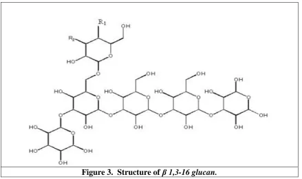
Three glucan classes of compounds were selected. They are β-1,3 glucan, β-1,6 glucan and β
-1,3-1,6 glucan. The selected classes of compounds are shown in figure 1,2 and 3 respectively.
Figure1. Structure of β 1,3 glucan.
Figure 3. Structure of β 1,3-16 glucan.
Total six compounds were designed for each class of glucans in two groups. Each group is having 3 compounds by substituting either at R1 or R2 positions by –OCH3, –Cl and –CN at a time, keeping other position intact.
[image:3.595.168.429.455.733.2]
The designed small molecules along with substituted functional groups are listed in table1, 2 and 3.
Table 1. Substituted β 1,6 glucan molecules
Molecule Number R1 R2
Basic Molecule B16G -OH -OH
B16G_ mol 1 -OCH3 -OH
B16G._mol 2 -Cl -OH
B16G._mol 3 -CN -OH
B16G _mol 4 -OH -OCH3
B16G_ mol 5 -OH -Cl
B16G_mol 6 -OH -CN
Table 2. Substituted β 1,3 glucan molecules
Molecule Number R1 R2
Basic Molecule B13G -OH -OH
B13G_mol1 -OCH3 -OH
B13G_mol 2 -Cl -OH
B13G_mol 3 -CN -OH
B13G_mol 4 -OH -OCH3
B13G_mol 5 -OH -Cl
Table 3. Substituted β 1,3-1,6 glucan molecules
Mol. No. R1 R2
Basic Molecule B1316G -OH -OH
B1316G_mol1 -OCH3 -OH
B1316G_mol 2 -Cl -OH
B1316G_mol 3 -CN -OH
B1316G_mol 4 -OH -OCH3
B1316G_mol 5 -OH -Cl
B1316G_mol 6 -OH -CN
The crystal structure of Dectin-1 enzyme was obtained from online database [9] having PDB ID 2CL8 [10-13].
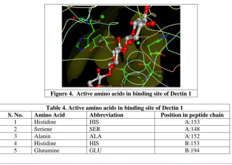
The obtained electronic structure cleaned for any ambiguities. All water molecules and surrounding ions were removed. This enzyme contains β-D-Glucose (three molecules). Using its position, the binding site were selected and used further for the docking purpose. The selected binding site along with natural inhibitor is shown in figure 4 whereas table 4 shows the list of amino acids in binding site.
The series of selected molecules and their substitutes were designed in-silico (virtually) using 2D design computer software CHEMDRAW [14-15] and their 3D structures were designed using molecular mechanics techniques.
[image:4.595.68.533.455.786.2]Figure 4. Active amino acids in binding site of Dectin 1
Table 4. Active amino acids in binding site of Dectin 1
S. No. Amino Acid Abbreviation Position in peptide chain
1 Histidine HIS A:153
2 Seriene SER A:148
3 Alanin ALA A:152
4 Histidine HIS B:153
Present days, In-silico methods are used to understand the basis of interactions. It provides the valuable information about the bonding between ligand and enzyme. Also provides the probable sites of actions, hydrophobic interactions and the confirmations of ligand inside the cavity of enzyme.
The Docking procedure is to prepare complex of Ligand (Small molecule) and Enzyme. The Autodock 4.0 program.[16] which is an automated docking program was used to dock all designed molecules, as well as parent glucan molecule in the active site of the Dectin -1 enzyme. The procedure for docking adopted as prescribed in Autodock manual.[17] and from our previous work. [18-19]. The mode of docking selected was genetical algorithm, which provides the most intelligent docking positions.
The docking process activated by docking the natural inhibitor in the selected active site to understand how natural inhibitor is placed and interacts in active site? Further each newly designed molecules were docked with selected enzyme. The process gives the binding energy as the major of strength of interactions between small molecules and enzyme.
[image:5.595.75.516.426.776.2]RESULTS AND DISCUSSION
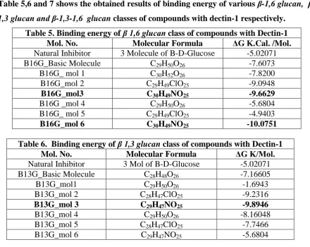
Table 5,6 and 7 shows the obtained results of binding energy of various 1,6 glucan,
β-1,3glucan and β-1,3-1,6 glucan classes of compounds with dectin-1 respectively. Table 5. Binding energy of β 1,6 glucan class of compounds with Dectin-1
Mol. No. Molecular Formula ΔG K.Cal. /Mol.
Natural Inhibitor 3 Molecule of B-D-Glucose -5.02071
B16G_Basic Molecule C29H50O26 -7.6073
B16G_ mol 1 C30H52O26 -7.8200
B16G_mol 2 C29H49ClO25 -9.0948
B16G_mol3 C30H49NO25 -9.6629
B16G _mol 4 C29H50O26 -5.6804
B16G_ mol 5 C29H49ClO25 -4.9403
B16G_mol 6 C30H49NO25 -10.0751
Table 6. Binding energy of β 1,3 glucan class of compounds with Dectin-1
Mol. No. Molecular Formula ΔG K/Mol.
Natural Inhibitor 3 Mol of B-D-Glucose -5.02071
B13G_Basic Molecule C28H48O26 -7.16605
B13G_mol1 C29H50O26 -1.6943
B13G_mol 2 C28H47ClO25 -9.2316
B13G_mol 3 C29H47NO25 -9.8946
B13G_mol 4 C29H50O26 -8.16048
B13G_mol 5 C28H47ClO25 -7.7466
Table 7. Binding energy of β 1,3-1,6 glucan class of compounds with Dectin-1
Mol. No. Molecular Formula ΔG K/Mol.
Natural Inhibitor 3 Mol of B-D-Glucose -5.02071
B1316G_Basic Molecule C34H58O31 -6.5095
B1316G_mol1 C35H60O31 -5.3257
B1316G_mol 2 C34H57ClO30 0.79816
B1316G_mol 3 C35H57NO30 -4.71303
B1316G_mol 4 C35H60O31 -5.2367
B1316G_mol 5 C34H57ClO30 -5.9051
B1316G_mol 6 C35H57NO30 -3.1068
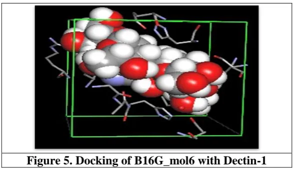
From the series β-1,6 glucan substituted small molecules docked, all molecules shows better binding energy than natural inhibitor. Whereas molecule B16G_mol 6 interact with highest possible binding energy -10.07 Kcal/mol. Figure 5 shows the complex of B16G_mol 6 with binding site of Dectin-1.
Figure 5. Docking of B16G_mol6 with Dectin-1
From the series β-1,3 glucan substituted small molecules docked, molecule B13G_mol2, B13G_mol3, B13G_mol4 show binding energy -9.2316, -9.8946 and -8.16048 respectively. These values are better than the natural inhibitor. Figure 6 shows the complex of B13G_mol 6 with binding site of Dectin-1.
From the series β-1,3-1,6 glucan substituted small molecules docked, no molecules shows better binding energy.
CONCLUSIONS
From this study, it is understood that Dectin-1 does interact with polysaccharides. In case of β
1,6 glucan class of compounds, substitution by Nitrile (–CN) at R1and R2 report highest possible binding energy. In case of β 1,3 glucan class of compounds, substitution by Nitrile (–CN) at R1 only report highest possible binding energy. Whereas, in case of β 1,3-1,6
glucan class of compounds, no one shows better interaction than natural inhibitor in Dectin-1. It is reported that among designed compounds of β 1,6 glucan class, the B16G._mol 1, B16G._mol 2, B16G._mol 3, B16G._mol 6 can be better Dectin-1 activator, particularly compounds having substitution at R2 position by Nitrile (-CN) group. Whereas, in case of β
1,3 glucan class of compounds, B13G_mol 2, B13G_mol 3, and B13G_mol 4 can be good Dectin-1 activator. The higher activity in case of nitril (-CN) group may be due to enhance polarity in the compound. Due to their higher binding energies, these compounds can be the best Dectin-1 activator. Higher binding energy can also provide more stability between these compounds and Dectin-1.
It is reported that Dectin-1 expressed by DCs and macrophages recognizes N-glycan structures on tumor cells. The complex of Dectin-1 with N-glycan signals to activate IRF5 pathway and other pathways, which further leads to activate Natural Killer (NK) cells. For the effective tumoricidal action by NK cells, it requires signaling by innate immune cells. [7]. Our computational docking study reports higher stability of Dectin-1 complex with β 1,6
glucan and β 1,3 glucan class of compounds particularly substituted with higher polar groups like Nitril (-CN) at R1 and R2 positions. The complex of these molecules with Dectin-1 show higher binding energy compares to natural N-glucan and hence must have higher stability and activity. Due to their enhance activity these molecules must have higher ability to activate NK cells.
Hence it is concluded that β 1,6 glucan and β 1,3 glucan class of compounds particularly substituted with higher polar functional groups at R1 and R2 positions can be the best NK cell activator by making stable complex with Dectin-1.
REFERENCES
2. Karagueuzian HS, White C, Sayre J, Norman A. Cigarette smoke radioactivity and lung cancer risk. Nicotine Tob Res., 2012; 14(1): 79-90.
3. Baker F, Ainsworth SR, Dye JT, Crammer C, Thun MJ, Hoffmann D et al. Health risks associated with cigar smoking. JAMA, 2000; 284(6): 735-740.
4. BE Henderson; L Bernstein; RK Ross. Holland-Frei Cancer Medicine. 6th edition, 2000. Hamilton, Ontario: BC Decker, ISBN-10: 1-55009-213-8.
5. Pagano JS, Blaser M, Buendia M-A, Damania B, Khalili K, Raab-Traub N, Roizman B. Infectious agents and cancer: criteria for a causal relation. Semin Cancer Biol, 2004; 14: 453–471.
6. Hedigan K, Cancer: Herbal medicine reduces chemotherapy toxicity. Nature Reviews Drug Discovery, 2010; 9: 765 | doi: 10.1038/nrd3280.
7. Chiba S et. al., Recognition of tumor cells by Dectin-1 orchestrates innate immune cells for anti-tumor responses, eLife, 2014; 3: e04177.
8. Delaney B et. al., Evaluation of the toxicity of concentrated barley β-glucan in a 28-day feeding study in Wistar rats. Food Chem Toxicol, 2003; 41(4): 477-87.
9. Berman HM; Westbrook J; Feng Z; Gilliland G; Bhat TN; Weissig H; Shindyalov IN; Bourne PE, The Protein Data Bank. Nucleic Acids Research, 2000; 28(1): 235-242. 10.Dutta S; Berman M; Bluhm F,Curr Protoc Bioinformatics Chapter 1: Unit 9, 2007.
11.Ananthalakshmi P; Samayamohan K; Chokalingam C; Mayilarasi C; Sekar K, Appl Bioinformatics, 2005; 4: 141-145.
12.National Center for Biotechnology Information. PubChem Compound
Database;CID=5988,https://pubchem.ncbi.nlm.nih.gov/compound /5988. 13.Brown J; O'Callaghan CA et. al., Protein Sci., 2007; 16: 1042-1052. 14.Nancy Mills, J. Am. Chem. Soc., 2006; 128(41): 13649–13650.
15.Chemdraw Software, Cambridge Corporation, Cambridge M.A. 02140 U.S.A. 16.Morris et. al. GM, J Comput Chem, 2009; 30: 2785-2791.
17.Morris GM, Goodsell DS, Halliday RS, Huey R, Hart WE et al., J Computational Chemistry, 1998; 19: 1639-1662.
18.Ganatra SH; Suchak AS, Inhibition studies of naturally occurring terpene based compounds with cyclin-dependent kinase 2 enzyme. J Comput Sci Syst Biol, 2012: 5: 068-073, doi:10.4172/jcsb.1000092.