High throughput screening for discovery of materials that control stem
cell fate
Asha K. Patel
a,b, Mark W. Tibbitt
a, Adam D. Celiz
c,d, Martyn C. Davies
d, Robert Langer
a,e,f,g,
Chris Denning
b, Morgan R. Alexander
d,⇑, Daniel G. Anderson
a,e,f,g,⇑a
David H. Koch Institute for Integrative Cancer Research, Massachusetts Institute of Technology, 500 Main Street, Cambridge, MA 02139, USA bWolfson Centre for Stem Cells, Tissue Engineering and Modeling, University of Nottingham, Nottingham NG7 2RD, UK
c
Wyss Institute for Biologically Inspired Engineering at Harvard University, Boston, MA 02115, USA d
Laboratory of Biophysics and Surface Analysis, School of Pharmacy, University of Nottingham, Nottingham NG7 2RD, UK e
Institute for Medical Engineering and Science, Massachusetts Institute of Technology, 500 Main Street, Cambridge, MA 02139, USA f
Harvard-MIT Division of Health Science and Technology, Massachusetts Institute of Technology, 500 Main Street, Cambridge, MA 02139, USA g
Department of Chemical Engineering, Massachusetts Institute of Technology, 500 Main Street, Cambridge, MA 02139, USA
a r t i c l e i n f o
Article history:
Received 14 August 2015 Revised 17 December 2015 Accepted 7 February 2016 Available online xxxx
Keywords: Stem cells Differentiation Biomaterials
High throughput screening 3D
a b s t r a c t
Insights into the complex stem cell niche have identified the cell–material interface to be a potent reg-ulator of stem cell fate via material properties such as chemistry, topography and stiffness. In light of this, materials scientists have the opportunity to develop bioactive materials for stem cell culture that elicit specific cellular responses. To accelerate materials discovery, high throughput screening platforms have been designed which can rapidly evaluate combinatorial material libraries in two and three-dimensional environments. In this review, we present screening platforms for the discovery of material properties that influence stem cell behavior.
Ó2016 The Authors. Published by Elsevier Ltd. This is an open access article under the CC BY license (http:// creativecommons.org/licenses/by/4.0/).
1. Introduction
The ability of stem cells to self-renew or to differentiate into specialized progeny makes them a valuable source for production of clinically relevant cells for regenerative medicine, disease mod-eling and biomedical applications. Stem cells broadly fall into two categories. The first, human pluripotent stem cells (hPSCs), include embryonic stem cells (hESCs) and have the potential to generate cells from any of the three germ layers that comprise all of the 200 cell types found in the body[1]. Also included in this group are induced pluripotent stem cells (hiPSCs), which bypass the need for cultivation from a blastocyst by reprogramming somatic cells into a stem cell state using a cocktail of transcription factors[2]. The second group encompasses tissue specific or ‘adult’ stem cells whose role is to assist in repair or renewal of tissue. These cells are generally considered multipotent meaning that their
differentia-tion potential is limited to the cell types of the tissue in which they reside.
The promise of stem cells in regenerative medicine is becoming reality with recent approval for the use of limbal stem cells for the treatment of ocular burns[3]and phase I clinical trials underway for the use of hPSC derivatives for spinal cord injury[4]macular degeneration[5]and heart failure[6]. To broaden the application of stem cells and their derivatives for wide ranging conditions there is a need for culture systems that enable controlled manipu-lation of these cells.
The first successful in vitropropagation of hESC was accom-plished in 1998, this was over a decade after the culture of mouse embryonic stem cells (mESC) was achieved[7]. The culture condi-tions found to maintain mESC pluripotency could not be translated to the human counterparts, where pluripotency could only be maintained when the cells were cultured on a feeder layer of mouse embryonic fibroblasts (MEFs) [1]. It was later discovered that this MEF layer could be replaced with a basement membrane matrix extracted from mouse sarcoma cells such as MatrigelTM
[8]. While these advances have enabled the culture of pluripotent stem cells outside of the body, the field requires culture conditions that are primed for clinical translation such as those that are scalable,
http://dx.doi.org/10.1016/j.cossms.2016.02.002
1359-0286/Ó2016 The Authors. Published by Elsevier Ltd.
This is an open access article under the CC BY license (http://creativecommons.org/licenses/by/4.0/). ⇑ Corresponding authors at: David H. Koch Institute for Integrative Cancer
Research, Massachusetts Institute of Technology, 500 Main Street, Cambridge, MA 02139, USA (D.G. Anderson). Laboratory of Biophysics and Surface Analysis, School of Pharmacy, University of Nottingham, Nottingham NG7 2RD, UK (M.R. Alexander).
E-mail addresses: [email protected] (M.R. Alexander), [email protected](D.G. Anderson).
Contents lists available atScienceDirect
Current Opinion in Solid State and Materials Science
j o u r n a l h o m e p a g e : w w w . e l s e v i e r . c o m / l o c a t e / c o s s m s
defined, reproducible and xeno-free. High throughput screening strategies have been adopted to search for substrates that achieve these goals[9].
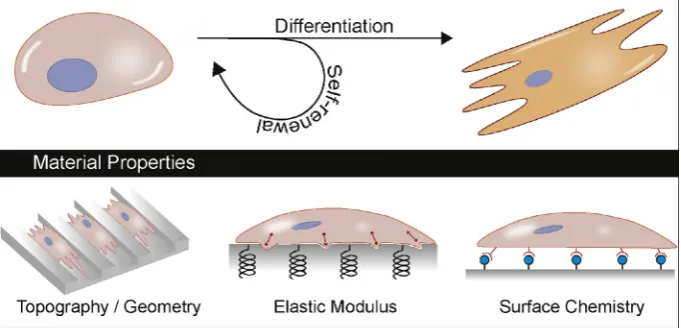
The critical role of the supporting substrate in maintaining pluripotency of human stem cellsin vitrohas been apparent since their derivation. Growth substrates to recapitulate the extra cellu-lar matrix (ECM) such as MatrigelTM or its components, such as laminin, have been commonly used[8]. More recently, substrates bearing epitopes that are capable of interacting with cells have been developed, for example SynthemaxTMis an acrylate substrate conjugated to RGD peptide derived from vitronectin that can sup-port self-renewal of hESCs[10]. The RGD ligand is a cell adhesive peptide that interacts with cell surface integrins [11]. Integrins and other cell adhesion molecules (CAMs), such as cadherins have all been implicated in regulating cellular behavior from maintaining pluripotency to directing differentiation [12]. Advances in the characterization of stem cell interaction with their environment has demonstrated that material physicochemical properties including chemistry, topography, geometry and stiff-ness also play an active role in modulating stem cell fate, particu-larly demonstrated with mesenchymal stem cells (MSCs)[13–16] (Fig. 1).
In the body, stem cells reside in a complex niche and receive a multitude of cues from the surrounding ECM, cell–cell contact and soluble factors contained within the aqueous milieu. In addition, the same stimuli may trigger a different biological response depending on the stem cell type. This and other complex struc-ture–function interrelationships, some of which are not fully known, hinder a rational approach in the design of stem cell cul-ture substrates, as it is difficult to predict how a given material property or combinations thereof will bias stem cell fate. More recently, the discovery of naïve states of hESCs and the difficulty in optimizing their culture conditions emphasizes the need for methods to keep pace with the rapidly evolving field[17]. There-fore, researchers have adapted high throughput screening (HTS) strategies to identify culture substrates that are appropriate for stem cell culture[9]. HTS has been utilized in a pharmaceutical set-ting facilitaset-ting early stage drug discovery since the 1980s. Libraries of compounds can be assayed for activity against a biolog-ical target to generate lead candidates essentially when structure based design is not possible. Such approaches rely on innovation in robotics for automation, robust biological assays to minimize false positives and high content analytical tools[18]. Adoption of the HTS strategy to accelerate the discovery of materials that can direct stem cell fate began around a decade ago [19,20]. By applying combinatorial methods used in conventional HTS, the
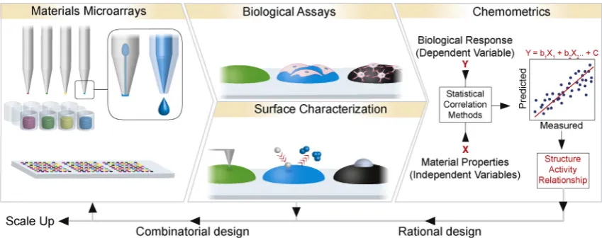
structural diversity of polymer libraries can be exponentially increased[20]. In addition, the design of material libraries can be guided by the outcome of biological activity. For this, a suite of high throughput materials characterization techniques is also required to generate comprehensive datasets that can be corre-lated to biological activity using statistical methods that identify structure activity relationships (SARs) in a systematic and unbiased fashion to enable a more rational approach to optimize materials identified from such screens[21–23](Fig. 2).
Synthetic materials allow for greater manipulation and control of physical and chemical properties compared to biological sub-strates, lending to design of modular systems that can be simpli-fied to uncouple substrate effects. In addition, for clinical applications, consistent material quality and function can be assured with fully characterized synthetic substrates, however it remains to be seen if these materials can recapitulate the complex nature of biological matrices. Nonetheless, HTS strategies can help to discover influential material properties to feedback into the design of robust differentiation systems, aid the isolation of rare or difficult to culture cell populations and begin to unravel com-plex molecular pathways underpinning the identified cell–material interaction.
This review will focus on an overview of the HTS systems designed to probe the interaction between material properties and stem cell phenotype, including surface chemistry, topography, elasticity and 3D micro-environments.
2. Substrate chemistry
[image:2.595.121.462.566.730.2]Tissue culture polystyrene (TCPS) is a widely used synthetic growth substrate suitable for various cell types including human mesenchymal stem cells (hMSCs). However, simple substrates such as TCPS have limited cellular interaction and usually require coating with ECM proteins and/or soluble factors from the culture medium to modulate the behavior of adherent cells[24]. This has led to the development of a new wave of synthetic growth sub-strates that have a broad range of surface chemistries to elicit a particular cell response. The surface chemistry of materials has been used to achieve the desired biomolecular adsorption from the culture medium to control cell response and/or act in itself as a ligand for cellular interaction. HTS of proteins, peptide frag-ments or chemical moieties presented at the substrate surface, to invoke a desired response (e.g., maintaining pluripotency or direct-ing differentiation toward a specific lineage) have been widely explored and will be discussed in this section.
Fig. 1.The culture substrate that stem cells adhere to can harness material properties such as topography, patterning, elastic modulus, surface chemistry and combinations thereof, to influence stem cell fate.
2.1. Proteins and peptides
HTS studies targeted toward understanding the necessary bio-molecules that direct stem cell fate were focused on ECM proteins. Solutions of fibronectin, laminin and collagen I, II and III in various combinations were robotically spotted onto acrylamide-coated slides[25]. Adhered mESCs cultured on these arrays were confined to the ECM environments and assayed for expression of an early hepatic fate marker, b-galactosidase, to identify conditions that encourage hepatic differentiation. Several hepatic promoting con-ditions were identified with nine of the ten highest signals mea-sured on cells adhered to collagen I containing matrices.
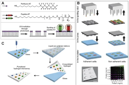
Smaller epitopes, such as peptides, can be easily handled and modified for immobilization using common chemistry techniques to increase the throughput of these screens. Surface modification strategies such as self-assembly are powerful tools to immobilize ligands capable of binding to cell surface integrins. The high spatial resolution with which self-assembling monolayers (SAMs) can be generated enables discrete chemical moieties to be screened in parallel (Fig. 3A). To facilitate SAM array preparation, gold surfaces have been modified with perfluoroalkanethiols that can be subse-quently removed via UV irradiation through a photo-mask to pro-duce high-resolution patterns. Arrays of peptide-substituted alkanethiols can then be prepared by spotting multiple solutions within the generated pattern, the fluorinated SAM prevents spreading of the spotted peptide–thiol solutions and also creates a low bio-fouling surface for cell screening. Phage display was employed for initial identification of cell binding peptides that could then be presented as a SAM to identify which binding pep-tides could also be used as a cell supportive substrate. Around 30,000 peptide-presenting phages were identified that were able to bind embryonic carcinoma (EC) cells. This initial pool was reduced to seven peptides with significant cell binding potential over peptide-free phages using a cell suspension enzyme-linked immunosorbent assay (CS-ELISA). When immobilized as a SAM two of these peptides, TVKHRPDALHPQ and LTTAPKLPKVTR, were able to support human embryonic stem cell (H9 cell line) growth at similar levels to MatrigelTMfor 20 days (3 passages)[26]. Inter-estingly, cell adhesion was not significantly reduced by addition of EDTA or heparin suggesting that adhesion to the phage-derived peptide surfaces was not mediated by integrins or proteo-glycans. This method of screening peptide motifs demonstrates the utility of HTS to identify novel functional peptides that could not have been predicted from what is currently known.
A similar approach has been applied to screening peptides that contain RGD and glycosaminoglycan binding epitopes, the most successful of which being a heparin-binding peptide derived from vitronectin (Table 1)[27]. At densities of 0.5–25%, this peptide was able to support self-renewal of hiPSCs and hESCs when combined with Rho-associated kinase (ROCK) inhibitor or cyclic RGD peptide. More recently, surfaces bearing this peptide in combination with RGD containing peptides have been used to promote ectoderm and neuronal differentiation. The cues for cell differentiation from this substrate were attributed to cell surface integrin engagement and subsequent stimulation of the Akt signaling pathway via inte-grin linked kinase[28]. Biotinylated-peptides were immobilized to streptavidin-coated TCPS dishes but scalability over large areas was not demonstrated limiting the applicability of using such pep-tides for stem cell expansion. SAMs have also proved to be a useful tool in investigating the relationship between surface chemistry mediated-adhesion protein binding and MSC differentiation[29].
To ease the manufacture of generating combinatorial peptide arrays, 384-well plates were prepared containing mixed solutions of alkanethiols bearing azide functionality and peptides modified with alkyne functionality. Conjugation of the peptides to the alka-nethiols was achieved using azide–alkyne ‘‘click” chemistry prior to spotting the solutions onto gold surfaces. Combinatorial mixtures of cell adhesion peptide, a bone morphogenetic protein 7 (BMP7) growth factor derived peptide, and a heparin binding peptide were printed [30]. The adhesion peptides allowed long-term (>1 week) observation of cell behavior with BMP peptide directing osteogenic differentiation of adipose derived stem cells. This was determined by the increased expression levels of osteogenic markers runt-related transcription fac-tor 2 (Runx2) and osteopontin (OPN) from cells cultured on BMP pep-tide surfaces versus adhesion peppep-tide only controls.
[image:3.595.88.513.66.234.2]Recently, a combinatorial bimolecular nano-patterned platform was designed to study the effects of cell behavior on nanoscale topography, controlled biomolecule conformation and identity in parallel over large areas (mm2per pattern). Vitronectin and Laminin 1 were mixed in 8 different ratios and immobilized on 8 nanoscale dimensions (100 nm – 150
l
m) in duplicate to create 128 combinato-rial dual protein patterns on a single surface. Adhesion profiles of human dental pulp stem cells were determined across the environ-ments. Line widths less than 500 nm encouraged focal adhesions and spreading across the patterns. However, widths greater than 700 nm guided adhesion and spreading along the patterns identifying the importance of nanoscale geometry on cell adhesion which will be discussed in further detail in Section3 [31].Fig. 2.Materials microarrays can be fabricated by automated robotics including contact and ink-jet printing shown inset in first panel, left and right, respectively. Characterization of cell response and corresponding surface properties generate large data sets that can be correlated to define structure activity relationships that inform the rational design of biomaterials. For this purpose, all data is input into statistical modeling to identify positive and negative relationships. However, for ‘hit’ material generation, the best performing materials can be taken forward to fabricate combinatorial libraries of materials to be investigated in further iterations of HTS platforms.
2.2. Polymers
Synthetic polymers offer an alternative to biological epitopes for the control of stem cell fate. The use of polymer microarrays to screen for their influence on the propagation of hESCs was achieved by Anderson et al. in 2004 and by applying principles of combinatorial materials design [32,33], demonstrated rapid syn-thesis of 576 acrylate co-polymers from a library of 24 monomers mixed in binary 70/30 % v/v ratios. Polymer microarrays are a pow-erful tool on which thousands of materials can be investigated in parallel on a single slide for desired cell responses[19,20,34]. Array fabrication can be performed using automated robotics. Ink-jet printing offers more flexibility and automation to enable easy drop volume control and drop in drop mixing of reagents to bypass manual pre-mixing. However, the wetting behavior and viscosity of the monomer solution highly influence droplet formation
therefore, uniform deposition of solutions with wide ranging sur-face energies or viscosities can be challenging. Contact printing generally affords greater control over spot size because it is pre-dominantly the geometry of the pin that determines the size of polymer spot enabling a wider range of solution viscosities to be printed uniformly[21](Fig. 2). Once optimized, both techniques enable rapid array fabrication of large numbers of polymers printed in discrete and identifiable regions. A strength of the microarray platform relative to casting polymers into microwells is the reduction in scale of materials required which transform impracticably large, laborious and expensive experiments into effi-cient and economical assays. To prevent background cell and bio-molecule adhesion between polymer spots that may lead to cross-talk, a bio-resistant polymer is used to coat the substrate that is to be printed upon such as poly(2-hydroxyethyl methacrylate) (pHEMA)[19], acrylamide[35]or poly(ethylene glycol)[36]. How-ever, due to this lack of spatial separation, it should be noted that any paracrine factors secreted by cells are free to diffuse through the shared culture medium for interaction with cells adhered to neighboring polymer islands. To partially abate such effects, poly-mer islands can be printed in replicate and in randomized regions on the array.
[image:4.595.76.506.65.348.2]Several groups have employed polymer microarrays as a means to discover suitable substrates to address a specific need. For pur-poses of gene targeting in stem cells, low seeding densities are required to allow clonal growth from single cells that have been correctly targeted. Mei et al. screened polymer microarrays for substrates that could support clonal propagation of hESCs from single cells[37]. Arrays were fabricated from 16 major monomers and 6 minor monomers mixed at 6 different ratios to generate 496 unique materials. A significant advance in this study was to per-form the screen using one of the first commercially available Fig. 3.High throughput surface modification strategies to probe stem cell behavior. (A) Self assembling monolayers of perfluoroalkanethiols can be photo-patterned to allow spotting of peptide-substituted alkanethiols (AT) presenting ligands for cellular interaction. (B) Microwells of unique protein microenvironments can be fabricated using contact printing to deposit solutions onto a silicon stamp to subsequently press into a PEG substrate to fabricate artificial niches. (C) Hydrogel microarrays are fabricated by first depositing substrates onto glass and which is embedded under a thiolated PEG and bis-acrylated PEG mixture that is UV cross-linked in the presence of a photoinitiator. The PEG hydrogel is peeled from the glass layer to expose the functionalized PEG surface. Adapted with permissions, (A)[26]2010ÓACS, (B)[46]2011ÓNPG, (C)[45]2009Ó
NPG.
Table 1
Peptide sequences found via HTS platforms to maintain pluripotency or promote differentiation.
Peptide sequence Origin Function References
TVKHRPDALHPQ Phage display Maintain pluripotency
[26]
LTTAPKLPKVTR Phage display Maintain pluripotency
[26]
GKKQRFRHRNRKG Vitronectin Maintain pluripotency
[27]
YIGSR Laminin Cell adhesion [30]
KPSSAPTQLN Bone morphogenetic protein 7
Osteogenic differentiation
[30]
KRSR Heparin-binding
protein
Cell adhesion [30]
[image:4.595.32.283.456.568.2]media, mTeSR1, made up of relatively defined components com-pared to conventional MEF conditioned medium. However, arrays required coating with fetal bovine serum (FBS) to overcome poor cell attachment. In an attempt to further refine the culture system, the authors investigated replacement of the FBS coating with three proteins, albumin, laminin or vitronectin. Integrin blocking exper-iments revealed that hit polymers enabled
a
Vb3anda
Vb5integrin binding of hESCs to adsorbed vitronectin and although vitronectin coated TCPS is known to support hESC growth, not all investigated polymer substrates coated with vitronectin supported clonal growth. This work was extended to quantify how roughness, elas-tic modulus, wettability and surface chemistry of the microenvi-ronment regulate self-renewal in ESCs concluding that polymer surface chemistry plays a dominant role.Microarrays have also facilitated discovery of a poly(4-vinyl) phenol substrate that can support proliferation of hPSC derived neural progenitor cells[38]and a methacrylamide polymer that can maintain hPSC pluripotency for over 20 passages in mTeSR1TM medium. Using quartz crystal microbalance (QCM) experiments it was found that bovine serum albumin (BSA) was critical for cell adhesion to the polymer [39]. However, BSA-containing media such as mTeSR1TM and StemProÒ although better defined than MEF conditioned medium, may still contain other undefined fac-tors that bind to BSA (e.g., Immunoglobulin E (IgE) antibodies), which can confound results in assays that may vary from batch to batch[40]. Developments in the propagation of hPSCs in fully defined media such as serum-free and xeno-free E8 medium[41], which contains only 8 components including transforming growth factor beta (TGFb) and fibroblast growth factor 2 (FGF2) aids unraveling the precise role of isolated material properties without confounding sources of unsolicited cues from, for example FBS and BSA. The prospect that simplified culture media such as E8 can be used reproducibly to maintain pluripotency and generate termi-nally differentiated cells provides encouragement to the materials science community that physicochemical cues from substrate materials can be developed to differentiate stem cells ‘‘on-demand” through stimuli supplied via substrate chemistry[42].
To demonstrate the ability of materials found by HTS to not only propagate hPSCs but to also support media induced differen-tiation of all three germ layers, Celiz et al. investigated the largest starting library to date of 141 (meth)acrylate and (meth)acry-lamide monomers in multi generational contact printed arrays of evolving libraries based on previous hit materials. Three genera-tions of arrays were investigated, the first to identify homopoly-mers that could support hPSC in Stempro medium, the second co-polymerized hits in major/minor ratios and the third expanded different ratios of hit combinations identified from the second gen-eration to optimize cell response. Using this approach, 909 differ-ent substrates in 4356 individual assays were analyzed and lead to the discovery of a co-polymer poly(HPhMA-co-HEMA) that could be scaled up to coat common culture-ware, support hPSC expansion through 5 serial passages and allow directed differenti-ation to cardiomyocytes, hepatocytes and neural cells[43].
With the scale of HTS increasing, automated processes are required to assist fabrication of these platforms. Hansen et al. recently demonstrated the capability of ink jet printing forin situ
mixing to facilitate rapid combinatorial polymer preparation [44]. Varying ratios of co-polymers were generated from a library of 26 acrylates and acrylamides. Elegant drop in drop mixing of monomers, photoinitiator and cross-linker was achieved to fabri-cate microarrays comprising of 7316 polymer islands. The low-fouling substrate was synthesized using a two-step fabrication procedure consisting of arrayed aqueous sucrose solution to serve as a mask followed by immersion in a cytophobic fluorinated silane. Removal of the mask and subsequent immersion in a methacrylated silane enabled the arrayed biomaterials to resist
spreading prior to photopolymerization. This screening platform
was able to identify a polymer, a copolymer of
2-(dimethylamino)ethyl methacrylate and ethylene glycol dicy-clopentenyl ether acrylate, that could maintain pluripotency of RH1 cells (hESC) for 5 passages in mTeSR1TMmedium.
The fabrication of microarrays on softer substrates with a Young’s modulus in the kPa range, that is more physiologically rel-evant than the GPa moduli displayed by conventional glass slides, has been demonstrated by Gupta and co-workers [45]. This was achieved by arraying thiol- or alkene-functionalized biomolecules onto a glass substrate that was subsequently coated with a liquid PEG pre-polymer. Curing via UV irradiation enabling coupling of the arrayed biomolecules to the PEG hydrogels which were peeled off the glass substrates to expose the hydrogel microarray (Fig. 3C). The orthogonal nature of thiol–ene chemistry was used to immobi-lize a large selection of molecules at the surface of the PEG hydrogel.
Thus far, while chemical moieties have been identified that can improve maturity of hESC derived cardiomyocytes in serum-free medium[47]and sustain the differentiation of hPSC derived hep-atic progenitors to hepatocytes using a maturation medium, no polymer has been shown to initiate the formation of the three germ layers, or to completely replace a biological substrate in the multi-step differentiation process from pluripotent to terminally-differentiated cell [48]. The identification, characterization and scale-up of such a substrate are major challenges for future investigation.
3. Material physical properties
In addition to the biochemical cues discussed earlier, stem cells also respond to biophysical cues from their surrounding environ-ment. Forces exerted via cues such as stiffness, topography, ligand density and stretch lead to activation of intra-cellular pathways via focal adhesions that connect the external ECM to the cellular cytoskeleton. This conversion of biophysical cues to biochemical signals is termed mechanotransduction[49]. Significant progress has been made in uncovering some of the transducing pathways relevant to guiding stem cell fate decisions. For example, phospho-rylation of focal adhesion kinase, has been implicated in modulat-ing response of hMSCs to nanotopography [50]. Whereas, the RhoA-ROCK signaling pathway was shown to be activated by geo-metric cues that influence hMSC shape and spread which subse-quently determined adipogenic (rounded, less spread shape) or osteogenic (spread) commitment [15]. However, without prior knowledge of precisely defined ECM-cellular interaction it is diffi-cult to predict how discrete presentations of biophysical cues determine cell fate. This section will focus on physical material properties that are amenable to HTS platforms such as surface topography and substrate stiffness to aid investigation of bio-physics on cellular behavior and guide the design of biophysical features.
3.1. Topography
Topographically patterned substrates have been shown to direct stem cell fate of MSCs[51], hESCs[52,53]and hiPSCs[54]. Dalby et al. used electron beam lithography to precisely fabricate nanopits of 120 nm diameter and 100 nm depth arranged in 5 dif-ferent patterns to compare varying degrees of disorder with the aim to re-create the natural disorder observed in the ECM protein, collagen. They fabricated polymethylmethacrylate (PMMA) arrays embossed with the various patterned topographies and found that a disordered topography, where nanopits 300 nm apart in a square array were randomly displaced by up to 50 nm, was able to induce
osteogenic differentiation of hMSCs that was comparable to cells that were chemically induced using dexamethasone on planar sub-strates. Interestingly, using ingenuity pathway analysis and canon-ical analysis the authors reported signaling events that were specifically triggered when cells were cultured on the patterned surfaces including fibroblast growth factor signaling and epithelial growth factor signaling which are associated with bone develop-ment [51]. Applying such in depth analysis to HTS provides a means of uncovering complex pathways of successfully identified material properties. The effect of topographical disorder on hESCs was later investigated using the same nanopit topography hot embossed onto polycarbonate. The study revealed that mesoder-mal lineage specification of hESCs could be encouraged using topo-graphical cues alone in basal medium. Despite the observation that planar surfaces could also direct mesodermal differentiation with-out chemical factors, the authors report that the additional topog-raphy enhanced up-regulation of a panel of mesenchymal specific markers that could give rise to mesodermal derivatives such as bone and cartilage and down regulation of non-mesenchymal markers[53].
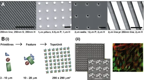
Ankam et al. investigated the influence of topography on glial or neuronal specification during neural differentiation of hESC. They fabricated a multi-architecture (MARC) chip using nano-imprint lithography that was used as a master mold to pattern PDMS. The 22 cm MARC chip is assembled with 18 topographies in duplicate (Fig. 4A). On planar surfaces, hESC differentiation in a neuro-basal medium (N2B27) resulted in a mixture of neuronal and glial cells. A higher neuronal population was found on aniso-tropic lines whereas culture on isoaniso-tropic patterns resulted in a greater proportion of the glial sub-type[52].
For exploration of a large topographical design space, the de Boer group developed a mathematical algorithm that could design over 150 million different topographies by combinatorial mixing of
three primitive shapes. Shapes ranging from 3 to 10
l
m were arranged to form features ranging from 10 to 28l
m and 5l
m in height (Fig. 4B.i). From this library 2176 features were selected to make a silicon master etched using photolithography. Polylactic acid (PLA) films were patterned with the master using hot emboss-ing to fabricate ‘Topochips’. Cell material interactions were screened using high content imaging to identify topographical pat-terns that were able to maintain pluripotency of hMSCs or induce osteogenesis (Fig. 4B.ii)[55]. Identification of such topographies will enable future investigation into the mechanisms underpinning their ability to control stem cell fate.3.2. Substrate stiffness
Cells are continuously assessing substrate stiffness through mechano-sensing and similar to soluble factors, matrix stiffness can influence stem cell lineage specification[56]. Engler et al. ele-gantly highlighted the profound effect that substrate stiffness can have on stem cell fate by culturing MSCs on polyacrylamide gels of three physiologically relevant moduli that mimicked brain (1 kPa), muscle (10 kPa) and bone (100 kPa) for 1 week. It was found that cells displayed morphology and RNA profiles indicative of neurogenesis, myogenesis and osteogenesis respectively[57].
[image:6.595.44.542.427.704.2]To address the need for a high throughput platform that would allow multifactorial investigation of varying substrate stiffness and biochemical composition on stem cell differentiation, Gobaa et al. fabricated soft hydrogel microwell arrays where the protein micro-environment of each well could be varied [46]. Protein was deposited by contact printing onto each micro pillar of a sili-con stamp and then pressed into a thin film of PEG hydrogel to cre-ate individual microwells of a confined protein niche. The proteins were tethered using thiol terminated PEG macromers in the hydro-gel which were available to react with maleimide functionalized
Fig. 4.Topographical assays developed for stem cell screening. (A) Scanning electron microscopy images (SEM) show patterned PDMS. The geometries were transferred from master molds using soft lithography and used for investigation of hESC neural differentiation. (S = spacing, H = height, pr = perpendicular). (B) i. TopoChip pattern generation using combinatorial design with three types of primitive shapes. ii. SEM of one TopoUnit pattern. hMSCs were cultured on this topography and displayed the highest immunostaining for alkaline phosphatase, a marker for early osteogenic differentiation. Scale bar = 20lm, height of feature is 5lm. Reproduced with permissions, (A)[52] 2013Ó, (B)[55]2011Ó.
proteins. Using this strategy an array with dimensions of a stan-dard slide, 2.5 cm7.5 cm, could contain up to 2016 microwells that could be seeded with cells in a single well of a rectangular four well plate (Fig. 3B). The authors found that the microarrays could be reproduced on PEG hydrogels with a Young’s modulus ranging from 3 to 150 kPa. Stiffness was achieved by adjusting the ratio of thiol and vinyl terminated PEG macromers, due to this the authors note that gels of varying stiffness also changes the number of thiol groups available for the maleimide protein conjugation which will vary the density of ligand presentation and that the molar concentration of free groups for ligand functionalization should be kept constant to overcome this. The study used the hydrogel microwell array to investigate the influence of substrate stiffness on hMSC differentiation and found that increasing the stiffness of the substrate increased osteogenic differentiation.
Mih et al. fabricated polyacrylamide arrays of varying substrate stiffness’s that were spatially separated by casting the gels in glass bottomed 96 and 384 multi well plates. A range of Young’s moduli from 0.3 to 55 kPa could be investigated in one multi well format by varying the bisacrylamide cross-linking ratio. The assay format was used to demonstrate applications for analysis of 7 different cell lines including hMSCs[58]. The authors aimed for PA films that were at least 3 times the threshold 20
l
m thickness that has been reported to prevent MSCs from sensing a rigid polystyrene sub-strate underlying a 1 kPa PA coating[59].4. Materials for surveying 3D micro-environments
While 2D platforms have been indispensable in constructing an understanding of stem cell fate control outside of the body, the native stem cell niche is inherently three-dimensional (3D). Recently, it has become clear that cell function often depends directly on the dimensionality of their surrounding matrix and that studying biology exclusively in 2D is insufficient. For example, freshly isolated murine hepatocytes de-differentiate when plated on tissue-culture polystyrene (TCPS) and fail to maintain liver-specific function[60]. In addition, while it is known that hMSC dif-ferentiate toward an osteogenic lineage on stiff 2D substrates[61], it has been observed that stiff and non-degradable matrices favor an adipogenic lineage in 3D[62]. In this work, adipogenesis was favored because cells were not able to spread; whereas, osteogenic differentiation was favored in degradable 3D matrices as they enabled hMSCs to spread and generate contractile force, which was necessary for osteogenesis and echoes the response to bio-physical geometric cues in 2D reported by McBeath[15]. Yet, all of the attributes of the micro-environment that discriminate 3D and 2D culture from the viewpoint of the cell still remain unknown.
2D environments present a highly polarizing environment with monolayer ligand presentation in which only a fraction of the cell membrane can interact with the surrounding ECM and other cells while the remainder of the cell is exposed to bulk culture media [63]. This diverges from the native ECM in which cells are immersed in a fibrous network of proteins with sequestered and diffusing growth factors as well as interactions with other cells in all directions via cell–cell contacts or paracrine signaling[64]. Individual signals within the complex signaling milieu of the ECM combine synergistically and antagonistically in a non-linear manner[65]. Therefore, 3D HTS techniques enable researchers to understand more completely the bidirectional and multimodal interactions between cells and the 3D ECM in a systematic manner. Many 3D cell culture platforms are composed of natural protein hydrogels (e.g., collagen or MatrigelTM) into which cells can be encapsulated. These fibrillar protein matrices are useful but they are challenged by batch-to-batch variability while providing the
user little control over individual cues such as ligand presentation, elasticity, and topography. Fortunately, the toolbox for 3D cell cul-ture has expanded significantly in the recent years as researchers have developed synthetic hydrogels with tunable mechanical and biochemical properties [66]. Much of this work has focused on the covalent cross-linking of hydrophilic polymers in the presence of cells. Polymer and cross-linking density control hydrogel mechanics while biochemical ligands can be incorporated into the network as whole proteins or peptide mimics[67]. Encapsula-tion of cells within synthetic hydrogels is often achieved using photopolymerization via chain polymerization of (meth)acrylates or thiol–ene click chemistry[68,69]. Highly efficient, water soluble, and cytocompatible photoinitiators exist for rapid fabrication of gels with visible light[70]. In parallel, several bio-orthogonal che-mistries (e.g., copper-free click chemistry or tetrazine–norbornene coupling) have been utilized for the fabrication of 3D ECM mimics that proceed without a catalyst at ambient conditions in cell cul-ture medium[71,72]. In each case, a liquid precursor of polymeric substituents, biochemical ligands, and cells is transformed into a gel for the 3D encapsulation of cells.
While rationally-designed 3D biomaterials have been used pre-dominantly in systematic studies of individual cell–matrix interac-tions they are generally amenable to HTS. Sala et al. leveraged liquid handling robotics to form arrays of artificial extracellular matrix (
a
ECM) through the selective, enzyme-mediated crosslink-ing of peptide-functionalized PEG to screen cell function within a range ofa
ECMs [73]. The complexity of cell–matrix signaling requires experimental design that investigates non-linear interac-tions between multiple cues, which even with a limited number of inputs, generates a large number of experimental conditions, making HTS an attractive strategy.Robotic handling of living cells presents unique challenges. Crit-ically, the precursor solution (viscosity, chemical composition, and additives), cross-linking strategies, as well as the handling tech-niques must not be cytotoxic or mutagenic. Pioneering groups have developed a range of suitable ‘bio-inks’ (biologically-compatible printing solutions) based on advances in the field of 3D stem cell culture using auto-gelation, photoinitiation, or thermal gelation for the fabrication of 3D microarrays. Notably, Lutolf and cowork-ers pioneered the fabrication of modular 3D hydrogel arrays using liquid handling robotics to investigate the dynamic interplay between mechanical and biochemical signals in 3D ECM mimics [74](Fig. 5A). Specifically, the authors created defined microenvi-ronments through selective enzyme-mediated crosslinking of fac-tor XIIIa (FXIIIa) with PEG modified with substrates for FXIIIa. Additional modification of PEG to include protease susceptible peptides, enables degradation and remodeling similar to native environments through matrix-metalloproteinase (MMP) cell secre-tion. This approach enables defined control over initial mechanical properties (E, Young’s moduli) by tuning polymer concentration; degradability by altering the MMP-sensitive peptide linkage; cell density down to single cells; ECM components (e.g., collagen IV, fibronectin, laminin); cell-cell signals (e.g., E-cadherin, Jagged, EpCAM); and soluble factors (e.g., FGF4, BMP4, LIF). Additionally, this strategy facilitates real-time monitoring of cell function as the microgels are optically clear and can be formed in standard 1536-well plates for complete isolation of the 1024 possible unique microenvironments. Oct4-GFP expressing mESCs were encapsulated within each microenvironment for facile readout of mESC pluripotency. Systematic analysis of the effects of microenvi-ronment on mESC proliferation and self-renewal was achieved using generalized linear models (GLM). The initial screen demon-strated that soluble factors (FGF4, BMP4, and LIF) were the stron-gest predictors of mESC self-renewal and proliferation with an additional strong effect of matrix mechanics. Gels with low elastic-ity led to increased self-renewal and proliferation.
In another approach, Khademhosseini and coworkers developed a combinatorial hydrogel microarray to investigate matrix effects on the osteogenesis of hMSCs[75]. Here, hMSCs were combined with liquid precursor solutions of a photoinitiator, methacrylated gelatin (GelMA), ECM proteins (fibronectin, laminin, and/or osteo-calcin), and BMPs (BMP2 and/or BMP5) (Fig. 5B). The precursor solutions were contact printed onto functionalized glass slides and UV polymerized to form hydrogels. Alkaline phosphatase expression (a marker of osteogenesis) and alizarin red staining (an indicator of mineralization) were used as measures of the osteogenic potential of each material. In this study, hydrogels with all three ECM proteins (fibronectin, laminin, and osteocalcin) demonstrated improved osteogenesis as compared to controls and encapsulated ECM proteins had a more pronounced effect than soluble factors added to the medium.
5. Challenges accompanying 3D HTS
As 3D HTS techniques are adopted, there are several major chal-lenges to consider. As cell biology moves to the third-dimension, new tools to understand and assess changes in material properties are required. As cells are increasingly cultured in degradable matri-ces, it is essential to develop imaging techniques to understand precisely how cells are altering the local environment over time. For example, micro-rheology has been used to monitor, in real-time, how a PEG based ECM mimic is remodeled during cell culture and migration[78]. In addition, cells push and pull on materials as they degrade and move through them and advances in quantifying these traction forces are needed. Toward this aim, imaging sensors have been designed to quantitate and understand cell-generated forces at the integrin level during adhesion and migration[79]. A DNA hairpin FRET-based probe was also developed to image in real-time cell generated forces at the cell–material interface[80].
Further, the biochemical signatures of the matrices are often altered during culture by deposition of proteins, from the cell cul-ture medium and those expressed by the encapsulated cells. In order to quantify how cells interact with and sense their microen-vironment, a more precise and comprehensive understanding of protein fouling and the evolution of the materials biochemical
landscape is needed. For example, improved mass spectrometry and elemental analysis methods are improving the detection of individual protein species on and within materials[81].
Simultaneously, improved techniques are necessary to eluci-date cell phenotype in 3D. Advances in reporter cell lines have aided[74], but there is still a need to develop easy readouts of cell function that can be used to correlate material properties to cell behavior that are amenable to HTS. This includes new probes for monitoring intracellular signaling, cytokine secretion, differentia-tion, surface receptor presentation and binding, as well as tran-scriptional landscapes. Many of the standard molecular biology techniques become significantly more complicated in 3D and dur-ing HTS. In order for 3D HTS to be adopted beyond very prelimi-nary screening, the field will need to adapt molecular biology approaches to these more complex settings.
Finally, the ECM is not a static material and many cues that are important in development and disease progression alter in time [61]. Dynamic biomaterials that enable user-defined control over material properties to study how a stiffening matrix influences fibrosis or growth factor secretion regulates neurogenesis are needed[82]. Ultimately, matrices that enable reversible biophysi-cal and biochemibiophysi-cal properties will be needed to capture the full dynamics of the native ECM as seen in development and disease. This has been captured using photoresponsive azobenzene cross-links[83]and sequential photoaddition and photocleavage reac-tions [84]. All of these techniques become increasingly difficult to incorporate into HTS approaches, but the chemical advances in the biomaterials field are bringing these concepts into reality.
6. Summary
[image:8.595.65.534.64.269.2]The studies discussed in this review have identified peptide ligands, chemical moieties, elastic moduli and topographical pat-terns that influence the behavior of stem cells. It is envisioned that future HTS platforms will advance further to incorporate dynamic biomaterials for investigation of spatiotemporal effects. As we look ahead, combinations of 2D and 3D HTS approaches will be indis-pensible in providing critical information as to how the environ-ment informs stem cell function and fate. HTS studies have Fig. 5.Fabrication strategies of 3D microenvironments for stem cells. (A) Liquid handling robots dispense into 1536 microwell plates to generate a HTS platform of enzyme mediated cross-linked PEG gels that encompass tunable 3D microenvironments consisting of mESCs, degradable metalloproteinase (MMP) components, ECM proteins, cell– cell interaction proteins and soluble factors. (B) A gelatin methacrylamide solution containing hMSCs were contact printed and UV cross-linked to create 3D encapsulated hMSCs islands containing varying protein environments consisting of fibronectin (FN), osteocalcin (OCN) and laminin (LN). Adapted with permissions, (A)[76]2014Ó, NPG (B)[77]2014Ó, NPG.
helped to gain retrospective mechanistic insights and it is hoped that further development of high content analytical tools will allow elucidation of the role of material properties identified during screening to help build roadmaps that will instruct materials development for regenerative medicine applications such as
ex vivo tissue growth, stem cell maintenance and controlled
expansion.
Acknowledgements
A.K.P. gratefully acknowledges the Engineering and Physical Sciences Research Council (EPSRC) for the Engineering, Tissue
Engi-neering and Regenerative Medicine (E-TERM) award (EP/
I017801/1). M.W.T. gratefully acknowledges funding from the National Institutes of Health (NIH) through a Ruth L. Kirschstein National Research Service Award (F32HL122009). A.D.C. gratefully acknowledges the European Commission for funding under FP7 IOF project Stem Cell Hydrogels (agreement no. 629320). C.D. would like to thank the EPSRC, British Heart Foundation (BHF), Heart Research UK, Medical Research Council (MRC) and National Centre for the Replacement, Refinement and Reduction of Animals in Research (NC3Rs). M.R.A. gratefully acknowledges funding from the EPSRC (EP/H045384/1) and the Wellcome Trust and The Royal Society for provision of the Wolfson Research Merit Award. D.G.A. would like to acknowledge support from the NIH (R01 DE016516).
References
[1] J.A. Thomson, J. Itskovitz-Eldor, S.S. Shapiro, et al., Embryonic stem cell lines derived from human blastocysts, Science 282 (1998) 1145–1147,http://dx.doi. org/10.1126/science.282.5391.1145.
[2] K. Takahashi, K. Tanabe, M. Ohnuki, et al., Induction of pluripotent stem cells from mouse embryonic and adult fibroblast cultures by defined factors, Cell 126 (2006) 663–676,http://dx.doi.org/10.1016/j.cell.2006.07.024.
[3]E. Dolgin, Next-generation stem cell therapy poised to enter EU market, Nat. Biotechnol. 33 (2015) 224–225.
[4] F. Bretzner, F. Gilbert, F. Baylis, R.M. Brownstone, Target populations for first-in-human embryonic stem cell research in spinal cord injury, Cell Stem Cell 8 (2011) 468–475,http://dx.doi.org/10.1016/j.stem.2011.04.012.
[5] S.D. Schwartz, C.D. Regillo, B.L. Lam, D. Eliott, et al., Human embryonic stem cell-derived retinal pigment epithelium in patients with age-related macular degeneration and Stargardt’s macular dystrophy, Lancet 385 (2015) 509–516, http://dx.doi.org/10.1016/S0140-6736(14)61376-3.
[6] P. Menasché, V. Vanneaux, A. Hagège, et al., Human embryonic stem cell-derived cardiac progenitors for severe heart failure treatment: first clinical case report, Eur. Heart J. 36 (2015) 2011–2017,http://dx.doi.org/10.1093/ eurheartj/ehv189.
[7]M.J. Evans, M.H. Kaufman, Establishment in culture of pluripotential cells from mouse embryos, Nature 292 (1981) 154–156.
[8]C.H. Xu, M.S. Inokuma, J. Denham, K. Golds, et al., Feeder-free growth of undifferentiated human embryonic stem cells, Nat. Biotechnol. 19 (2001) 971– 974.
[9] A.D. Celiz, J.G.W. Smith, R. Langer, D.G. Anderson, et al., Materials for stem cell factories of the future, Nat. Mater. 13 (2014) 570–579, http://dx.doi.org/ 10.1038/nmat3972.
[10] Z. Melkoumian, J.L. Weber, D.M. Weber, A.G. Fadeev, et al., Synthetic peptide– acrylate surfaces for long-term self-renewal and cardiomyocyte differentiation of human embryonic stem cells, Nat. Biotechnol. 28 (2010) 606–610,http://dx. doi.org/10.1038/nbt.1629.
[11] R.O. Hynes, The extracellular matrix: not just pretty fibrils, Science 326 (2009) 1216–1219,http://dx.doi.org/10.1126/science.1176009.
[12] L. Li, S. Bennett, L. Wang, Role of E-cadherin and other cell adhesion molecules in survival and differentiation of human pluripotent stem cells, Cell Adhes. Migr. 6 (2012) 59–73,http://dx.doi.org/10.4161/cam.19583.
[13] P.-Y. Wang, W.-T. Li, J. Yu, W.-B. Tsai, Modulation of osteogenic, adipogenic and myogenic differentiation of mesenchymal stem cells by submicron grooved topography, J. Mater. Sci. – Mater. Med. 23 (2012) 3015–3028, http://dx.doi.org/10.1007/s10856-012-4748-6.
[14] W.L. Murphy, T.C. McDevitt, A.J. Engler, Materials as stem cell regulators, Nat. Mater. 13 (2014) 547–557,http://dx.doi.org/10.1038/nmat3937.
[15] R. McBeath, D.M. Pirone, C.M. Nelson, K. Bhadriraju, et al., Cell shape, cytoskeletal tension, and RhoA regulate stem cell lineage commitment, Dev. Cell 6 (2004) 483–495,http://dx.doi.org/10.1016/s1534-5807(04)00075-9. [16] K.A. Kilian, B. Bugarija, B.T. Lahn, M. Mrksich, Geometric cues for directing the
differentiation of mesenchymal stem cells, PNAS 107 (2010) 4872–4877, http://dx.doi.org/10.1073/pnas.0903269107.
[17] T.W. Theunissen, B.E. Powell, H. Wang, M. Mitalipova, et al., Systematic identification of culture conditions for induction and maintenance of naive human pluripotency, Cell Stem Cell 15 (2014) 471–487,http://dx.doi.org/ 10.1016/j.stem.2014.07.002.
[18] A.L. Hook, D.G. Anderson, R. Langer, P. Williams, et al., High throughput methods applied in biomaterial development and discovery, Biomaterials 31 (2010) 187–198,http://dx.doi.org/10.1016/j.biomaterials.2009.09.037. [19] D.G. Anderson, S. Levenberg, R. Langer, Nanoliter-scale synthesis of arrayed
biomaterials and application to human embryonic stem cells, Nat. Biotechnol. 22 (2004) 863–866,http://dx.doi.org/10.1038/nbt981.
[20] J. Kohn, New approaches to biomaterials design, Nat. Mater. 3 (2004) 745–747, http://dx.doi.org/10.1038/nmat1249.
[21] M.C. Davies, M.R. Alexander, A.L. Hook, J. Yang, et al., High throughput surface characterization: a review of a new tool for screening prospective biomedical material arrays, J. Drug Target. 18 (2010) 741–751,http://dx.doi.org/10.3109/ 1061186x.2010.521941.
[22] F.R. Burden, D.A. Winkler, Optimal sparse descriptor selection for QSAR using bayesian methods, QSAR Comb. Sci. 28 (2009) 645–653,http://dx.doi.org/ 10.1002/qsar.200810173.
[23] V.C. Epa, J. Yang, Y. Mei, A.L. Hook, et al., Modelling human embryoid body cell adhesion to a combinatorial library of polymer surfaces, J. Mater. Chem. 22 (2012) 20902–20906,http://dx.doi.org/10.1039/c2jm34782b.
[24] M.M. Mahlstedt, D. Anderson, J.S. Sharp, R. McGilvray, et al., Maintenance of pluripotency in human embryonic stem cells cultured on a synthetic substrate in conditioned medium, Biotechnol. Bioeng. 105 (2010) 130–140,http://dx. doi.org/10.1002/bit.22520.
[25] C.J. Flaim, S. Chien, S.N. Bhatia, An extracellular matrix microarray for probing cellular differentiation, Nat. Methods 2 (2005) 119–125,http://dx.doi.org/ 10.1038/NMETH736.
[26] R. Derda, S. Musah, B.P. Orner, J.R. Klim, et al., High-throughput discovery of synthetic surfaces that support proliferation of pluripotent cells, J. Am. Chem. Soc. 132 (2010) 1289–1295,http://dx.doi.org/10.1021/ja906089g.
[27] J.R. Klim, L.Y. Li, P.J. Wrighton, M.S. Piekarczyk, et al., A defined glycosaminoglycan-binding substratum for human pluripotent stem cells, Nat. Methods 7 (2010) 989–996,http://dx.doi.org/10.1038/nmeth.1532. [28] P.J. Wrighton, J.R. Klim, B.A. Hernandez, C.H. Koonce, et al., Signals from the
surface modulate differentiation of human pluripotent stem cells through glycosaminoglycans and integrins, PNAS 111 (2014) 18126–18131,http://dx. doi.org/10.1073/pnas.1409525111.
[29] J.E. Phillips, T.A. Petrie, F.P. Creighton, A.J. Garcia, Human mesenchymal stem cell differentiation on self-assembled monolayers presenting different surface chemistries, Acta Biomater. 6 (2010) 12–20, http://dx.doi.org/10.1016/j. actbio.2009.07.023.
[30] D. Zhang, K.A. Kilian, Peptide microarrays for the discovery of bioactive surfaces that guide cellular processes: a single step azide–alkyne ‘‘click” chemistry approach, J. Mater. Chem. B 2 (2014) 4280–4288,http://dx.doi.org/ 10.1039/C4TB00375F.
[31] Y.Y.I. Amin, K. Runager, F. Simoes, A. Celiz, et al., Combinatorial biomolecular nanopatterning for high-throughput screening of stem cell behavior, Adv. Mater. (2015),http://dx.doi.org/10.1002/adma.201504995.
[32]X.D. Xiang, X.D. Sun, G. Briceno, Y.L. Lou, et al., A combinatorial approach to materials discovery, Science 268 (1995) 1738–1740.
[33] S. Brocchini, K. James, V. Tangpasuthadol, J. Kohn, A combinatorial approach for polymer design, J. Am. Chem. Soc. 119 (1997) 4553–4554,http://dx.doi. org/10.1021/ja970389z.
[34] R. Zhang, A. Liberski, R. Sanchez-Martin, M. Bradley, Microarrays of over 2000 hydrogels – identification of substrates for cellular trapping and thermally triggered release, Biomaterials 30 (2009) (2000) 6193–6201,http://dx.doi.org/ 10.1016/j.biomaterials.2009.07.055.
[35] D.A. Brafman, C.W. Chang, A. Fernandez, K. Willert, et al., Long-term human pluripotent stem cell self-renewal on synthetic polymer surfaces, Biomaterials 31 (2010) 9135–9144,http://dx.doi.org/10.1016/j.biomaterials.2010.08.007. [36] E.A. Appel, B.L. Larson, K.M. Luly, J.D. Kim, et al., Non-cell-adhesive substrates
for printing of arrayed biomaterials, Adv. Healthcare Mater. (2014),http://dx. doi.org/10.1002/adhm.201400594.
[37] Y. Mei, K. Saha, S.R. Bogatyrev, J. Yang, et al., Combinatorial development of biomaterials for clonal growth of human pluripotent stem cells, Nat. Mater. 9 (2010) 768–778,http://dx.doi.org/10.1038/NMAT2812.
[38] Y. Tsai, J. Cutts, A. Kimura, D. Varun, A chemically defined substrate for the expansion and neuronal differentiation of human pluripotent stem cell-derived neural progenitor cells, Stem Cell Res. 15 (2015) 75–87,http://dx.doi. org/10.1016/j.scr.2015.05.002.
[39] E.E. Irwin, R. Gupta, D.C. Dashti, K.E. Healy, Engineered polymer-media interfaces for the long-term self-renewal of human embryonic stem cells, Biomaterials 32 (2011) 6912–6919,http://dx.doi.org/10.1016/j.biomaterials.2011.05.058. [40] P. Restani, C. Ballabio, A. Cattaneo, P. Isoardi, et al., Characterization of bovine serum
albumin epitopes and their role in allergic reactions, Allergy 59 (2004) 21–24. [41] G. Chen, D.R. Gulbranson, Z. Hou, J.M. Bolin, et al., Chemically defined
conditions for human iPSC derivation and culture, Nat. Methods 8 (2011) 424– 429,http://dx.doi.org/10.1038/nmeth.1593.
[42] J.E. Dixon, D.A. Shah, C. Rogers, S. Hall, et al., Combined hydrogels that switch human pluripotent stem cells from self-renewal to differentiation, PNAS 111 (2014) 5580–5585,http://dx.doi.org/10.1073/pnas.1319685111.
[43] A.D. Celiz, J. Smith, A.K. Patel, A.L. Hook, et al., Discovery of a novel polymer for human pluripotent stem cell expansion and multilineage differentiation, Adv. Mater. (2015),http://dx.doi.org/10.1002/adma.201501351.
[44] A. Hansen, R. Zhang, M. Bradley, Fabrication of arrays of polymer gradients using inkjet printing, Macromol. Rapid Commun. 33 (2012) 1114–1118,http:// dx.doi.org/10.1002/marc.201200193.
[45] N. Gupta, B.F. Lin, L.M. Campos, M.D. Dimitriou, et al., A versatile approach to high-throughput microarrays using thiol–ene chemistry, Nat. Chem. 2 (2009) 138–145,http://dx.doi.org/10.1038/nchem.478.
[46] S. Gobaa, S. Hoehnel, M. Roccio, A. Negro, et al., Artificial niche microarrays for probing single stem cell fate in high throughput, Nat. Methods 8 (2011) 949– 955,http://dx.doi.org/10.1038/nmeth.1732.
[47] A.K. Patel, A.D. Celiz, D. Rajamohan, D.G. Anderson, et al., A defined synthetic substrate for serum-free culture of human stem cell derived cardiomyocytes with improved functional maturity identified using combinatorial materials microarrays, Biomaterials 61 (2015) 257–265, http://dx.doi.org/10.1016/j. biomaterials.2015.05.019.
[48] D.C. Hay, S. Pernagallo, J.J. Diaz-Mochon, C.N. Medine, et al., Unbiased screening of polymer libraries to define novel substrates for functional hepatocytes with inducible drug metabolism, Stem Cell Res. 6 (2011) 92– 102,http://dx.doi.org/10.1016/j.scr.2010.12.002.
[49] A.W. Holle, A.J. Engler, More than a feeling: discovering, understanding, and influencing mechanosensing pathways, Curr. Opin. Biotechnol. 22 (2011) 648– 654,http://dx.doi.org/10.1016/j.copbio.2011.04.007.
[50] B.K.K. Teo, S.T. Wong, C.K. Lim, T.Y.S. Kung, et al., Nanotopography modulates mechanotransduction of stem cells and induces differentiation through focal adhesion kinase, ACS Nano 7 (2013) 4785–4798,http://dx.doi.org/10.1021/ nn304966z.
[51] M.J. Dalby, N. Gadegaard, R. Tare, A. Andar, et al., The control of human mesenchymal cell differentiation using nanoscale symmetry and disorder, Nat. Mater. 6 (2007) 997–1003,http://dx.doi.org/10.1038/nmat2013.
[52] S. Ankam, M. Suryana, L.Y. Chan, A.A.K. Moe, et al., Substrate topography and size determine the fate of human embryonic stem cells to neuronal or glial lineage, Acta Biomater. 9 (2013) 4535–4545, http://dx.doi.org/10.1016/j. actbio.2012.08.018.
[53] E. Kingham, K. White, N. Gadegaard, M.J. Dalby, et al., Nanotopographical cues augment mesenchymal differentiation of human embryonic stem cells, Small 9 (2013) 2140–2151,http://dx.doi.org/10.1002/smll.201202340.
[54] F. Pan, M. Zhang, G. Wu, Y. Lai, et al., Topographic effect on human induced pluripotent stem cells differentiation towards neuronal lineage, Biomaterials 34 (2013) 8131–8139,http://dx.doi.org/10.1016/j.biomaterials.2013.07.025. [55] H.V. Unadkat, M. Hulsman, K. Cornelissen, B.J. Papenburg, et al., An
algorithm-based topographical biomaterials library to instruct cell fate, PNAS 108 (2011) 16565–16570,http://dx.doi.org/10.1073/pnas.1109861108.
[56] S.V. Plotnikov, A.M. Pasapera, B. Sabass, C.M. Waterman, Force fluctuations within focal adhesions mediate ECM-rigidity sensing to guide directed cell migration, Cell 151 (2012) 1513–1527, http://dx.doi.org/10.1016/ j.cell.2012.11.034.
[57] A.J. Engler, S. Sen, H.L. Sweeney, D.E. Discher, Matrix elasticity directs stem cell lineage specification, Cell 126 (2006) 677–689, http://dx.doi.org/10.1016/ j.cell.2006.06.044.
[58] J.D. Mih, A.S. Sharif, F. Liu, A. Marinkovic, et al., A multiwell platform for studying stiffness-dependent cell biology, PLoS ONE 6 (2011),http://dx.doi. org/10.1371/journal.pone.0019929. e19929–10.
[59] A. Buxboim, K. Rajagopal, A.E.X. Brown, D.E. Discher, How deeply cells feel: methods for thin gels, J. Phys.: Condens. Matter 22 (2010),http://dx.doi.org/ 10.1088/0953-8984/22/19/194116. 194116–11.
[60] Y. Chen, P.P. Wong, L. Sjeklocha, C.J. Steer, et al., Mature hepatocytes exhibit unexpected plasticity by direct dedifferentiation into liver progenitor cells in culture, Hepatology 55 (2012) 563–574,http://dx.doi.org/10.1002/hep.24712. [61] C. Yang, M.W. Tibbitt, L. Basta, K.S. Anseth, Mechanical memory and dosing influence stem cell fate, Nat. Mater. 13 (2014) 645–652, http://dx.doi.org/ 10.1038/NMAT3889.
[62] S. Khetan, M. Guvendiren, W.R. Legant, D.M. Cohen, Degradation-mediated cellular traction directs stem cell fate in covalently crosslinked three-dimensional hydrogels, Nat. Mater. 12 (2013) 458–465, http://dx.doi.org/ 10.1038/NMAT3586.
[63] M.W. Tibbitt, K.S. Anseth, Hydrogels as extracellular matrix mimics for 3D cell culture, Biotechnol. Bioeng. 103 (2009) 655–663,http://dx.doi.org/10.1002/ bit.22361.
[64] W.P. Daley, S.B. Peters, M. Larsen, Extracellular matrix dynamics in development and regenerative medicine, J. Cell Sci. 121 (2008) 255–264, http://dx.doi.org/10.1242/jcs.006064.
[65] F. Yang, S.-W. Cho, S.M. Son, S.P. Hudson, et al., Combinatorial extracellular matrices for human embryonic stem cell differentiation in 3D, Biomacromolecules 11 (2010) 1909–1914,http://dx.doi.org/10.1021/bm100357t.
[66] M.W. Tibbitt, K.S. Anseth, Dynamic microenvironments: the fourth dimension, Sci. Trans. Med. 4 (2012), http://dx.doi.org/10.1126/scitranslmed.3004804. 160ps24.
[67] A.M. Kloxin, C.J. Kloxin, C.N. Bowman, K.S. Anseth, Mechanical properties of cellularly responsive hydrogels and their experimental determination, Adv. Mater. 22 (2010) 3484–3494,http://dx.doi.org/10.1002/adma.200904179. [68]J.A. Burdick, K.S. Anseth, Photoencapsulation of osteoblasts in injectable
RGD-modified PEG hydrogels for bone tissue engineering, Biomaterials 23 (2002) 4315–4323.
[69] B.D. Fairbanks, M.P. Schwartz, A.E. Halevi, C.R. Nuttelman, et al., A versatile synthetic extracellular matrix mimic via thiol–norbornene photopolymerization, Adv. Mater. 21 (2009) 5005–5010,http://dx.doi.org/ 10.1002/adma.200901808.
[70] B.D. Fairbanks, M.P. Schwartz, C.N. Bowman, K.S. Anseth, Photoinitiated polymerization of PEG-diacrylate with lithium phenyl-2,4,6-trimethylbenzoylphosphinate: polymerization rate and cytocompatibility, Biomaterials 30 (2009) 6702–6707, http://dx.doi.org/10.1016/j. biomaterials.2009.08.055.
[71] C.A. DeForest, B.D. Polizzotti, K.S. Anseth, Sequential click reactions for synthesizing and patterning three-dimensional cell microenvironments, Nat. Mater. 8 (2009) 659–664,http://dx.doi.org/10.1038/nmat2473.
[72] D.L. Alge, M.A. Azagarsamy, D.F. Donohue, K.S. Anseth, Synthetically tractable click hydrogels for three-dimensional cell culture formed using tetrazine– norbornene chemistry, Biomacromolecules 14 (2013) 949–953,http://dx.doi. org/10.1021/bm4000508.
[73] A. Sala, P. Hänseler, A. Ranga, M.P. Lutolf, J. Vörös, Engineering 3D cell instructive microenvironments by rational assembly of artificial extracellular matrices and cell patterning, Int. Biol. (2011), http://dx.doi.org/10.1039/ c1ib00045d.
[74] A. Ranga, S. Gobaa, Y. Okawa, K. Mosiewicz, et al., 3D niche microarrays for systems-level analyses of cell fate, Nat. Commun. 5 (2014) 1–10,http://dx.doi. org/10.1038/ncomms5324.
[75] A. Dolatshahi-Pirouz, M. Nikkhah, A.K. Gaharwar, B. Hashmi, et al., A combinatorial cell-laden gel microarray for inducing osteogenic differentiation of human mesenchymal stem cells, Sci. Rep. 4 (2014),http:// dx.doi.org/10.1038/srep03896.
[76] A. Ranga, S. Gobaa, Y. Okawa, K. Mosiewicz, et al., 3D niche microarrays for systems-level analyses of cell fate, Nature (2014),http://dx.doi.org/10.1038/ ncomms5324.
[77] A. Dolatshahi-Pirouz, M. Nikkhah, A.K. Gaharwar, B. Hashmi, et al., A combinatorial cell-laden gel microarray for inducing osteogenic differentiation of human mesenchymal stem cells, Sci. Rep. 4 (2014) 1–9, http://dx.doi.org/10.1038/srep03896.
[78] K.M. Schultz, K.A. Kyburz, K.S. Anseth, Measuring dynamic cell–material interactions and remodeling during 3D human mesenchymal stem cell migration in hydrogels, PNAS 112 (2015) E3757–E3764,http://dx.doi.org/ 10.1073/pnas.1511304112.
[79] M. Morimatsu, A.H. Mekhdjian, A.S. Adhikari, A.R. Dunn, Molecular tension sensors report forces generated by single integrin molecules in living cells, Nano Lett. 13 (2013) 3985–3989,http://dx.doi.org/10.1021/nl4005145. [80] B.L. Blakely, C.E. Dumelin, B. Trappmann, L.M. McGregor, et al., A DNA-based
molecular probe for optically reporting cellular traction forces, Nat. Methods 11 (2014) 1229–1232,http://dx.doi.org/10.1038/nmeth.3145.
[81] Q. Wei, T. Becherer, S. Angioletti-Uberti, J. Dzubiella, et al., Protein interactions with polymer coatings and biomaterials, Angew. Chem. – Int. Ed. 53 (2014) 8004–8031,http://dx.doi.org/10.1002/anie.201400546.
[82] J.L. Young, A.J. Engler, Hydrogels with time-dependent material properties enhance cardiomyocyte differentiation in vitro, Biomaterials 32 (2011) 1002– 1009,http://dx.doi.org/10.1016/j.biomaterials.2010.10.020.
[83] A.M. Rosales, K.M. Mabry, E.M. Nehls, K.S. Anseth, Photoresponsive elastic properties of azobenzene-containing poly(ethylene-glycol)-based hydrogels, Biomacromolecules 16 (2015) 798–806, http://dx.doi.org/10.1021/ bm501710e.
[84] C.A. DeForest, K.S. Anseth, Photoreversible patterning of biomolecules within click-based hydrogels, Angew. Chem. – Int. Ed. 51 (2011) 1816–1819,http:// dx.doi.org/10.1002/anie.201106463.