Title: Structural elements of an NRPS cyclization domain and its intermodule docking domain Authors:
Daniel P. Dowling1,2,3, Yan Kung4, Anna K. Croft5, Koli Taghizadeh6, Wendy L. Kelly7, Christopher T. Walsh8, and Catherine L. Drennan1,2,6,9,$
Affiliations:
1Howard Hughes Medical Institute, Departments of Chemistry2 and Biology9, Massachusetts Institute of Technology, Cambridge, Massachusetts 02139; [email protected]
3Current Address, Department of Chemistry, University of Massachusetts, Boston, MA 02125 4Current Address, Department of Chemistry, Bryn Mawr College, Bryn Mawr, PA 19010
5Department of Chemical and Environmental Engineering, University of Nottingham, University Park, Nottingham NG7 2RD
6Center for Environmental and Health Sciences, Massachusetts Institute of Technology, Cambridge, Massachusetts 02139
7Current Address, School of Chemistry and Biochemistry and the Parker H. Petit Institute for Bioengineering and Bioscience, Georgia Institute of Technology, Atlanta, Georgia 30332
8Stanford University Chemistry, Engineering, and Medicine for Human Health, Stanford University, Stanford, California, 94305
$Correspondence to: [email protected]
Running title: Structure of an NRPS cyclization domain Keywords:
ABSTRACT
Epothilones are natural products with anticancer activity that are biosynthesized by polyketide synthase (PKS)-nonribosomal peptide synthetase (NRPS) enzymes EpoA-F. A cyclization domain of EpoB (Cy) assembles the thiazole functionality from an acetyl group and L-cysteine
via condensation, cyclization and dehydration. The PKS carrier protein of EpoA contributes the acetyl moiety, guided by a docking domain, whereas an NRPS EpoB carrier protein contributes L-cysteine. To visualize the structure of a cyclization domain with an accompanying docking domain, we have solved a 2.03 Å resolution structure of this bidomain EpoB unit, comprising residues M1-Q497 (62 kDa) of the 160 kDa EpoB protein. We find that the N-terminal docking domain is connected to the V-shaped Cy domain by a twenty-residue linker, but otherwise makes no contacts to Cy. Molecular dynamic simulations and additional crystal structures reveal a high degree of flexiblity for this docking domain, emphasizing the modular nature of the components of PKS-NRSP hybrid systems. These structures further reveal two 20-Å-long channels that run from distant sites on the Cy domain to the active site at the core of the enzyme, allowing two carrier proteins to dock with Cy and deliver their substrates simultaneously. Through mutagenesis and activity assays, catalytic residues N335 and D449 have been identified. Surprisingly these residues do not map to the location of the conserved HHxxxDG motif in the structurally homologous NRPS condensation (C) domain. Thus, although both C and Cy domains have the same basic fold, their active sites appear distinct.
Introduction
Epothilones are hybrid polyketide/nonribosomal peptide natural products that are indicated for treatment of metastatic or locally advanced breast cancers that are taxane-resistant (1, 2). They contain a large macrocycle with a thiazole-containing side chain, which is important for stabilizing microtubules and impairing cell division (Fig. 1A) (3–5). Azole heterocycles, such as thiazole, are commonly found in many natural products, and in different oxidation states (i.e. azolines and azolidines). They are more resistant to hydrolysis than a peptide bond, and their incorporation can increase a compound’s affinity for a target biomolecule (6). Despite their importance, there is still much to learn about the structure and mechanism of the enzymes involved in their biosynthesis (7, 8). Here we explore the structural basis of activity of the catalytic domain that generates the 2-methylthiazoline precursor of epothilone natural products.
The epothilone biosynthetic gene cluster from Sorangium cellulosum is encoded by EpoA-K (Fig. 1A). EpoA, a polyketide synthase (PKS), and EpoB, a nonribosomal peptide synthetase (NRPS), are responsible for making the methylthiazole functionality (9–12). 2-methylthiazole derives from the condensation of an acetyl group with L-cysteine (Fig. 1B).
Before condensation, the acetyl moiety and L-cysteine are covalently attached to the carrier T
12). PKS-NRPS docking domains (15), referred to as EpoAdd and EpoBdd for the upstream donor and the downstream acceptor proteins, respectively, serve to localize the T domains to the appropriate intermodule junction, facilitating what has often been referred to as an assembly line biosynthetic process.
There is much that is unknown about Cy domains; the molecular basis for the differentiation of C and Cy domain activity is not established and the key catalytic residues have not been identified. Keating et al. predicted that the C and Cy domains would adopt similar structures (16), and early sequence alignments identified a DxxxxDxxS Cy domain sequence that replaces the C domain HHxxxDG catalytic motif (Fig. S2). Although conservation of the DxxxxDxxS sequence within known Cy domains suggests its importance, mutational analyses have been inconclusive as to which residues are critical for catalysis (14, 17, 18).
Results
Structure of an NRPS Cy and docking domain protein complex
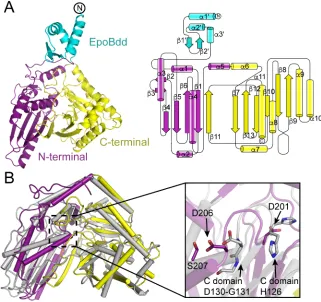
We determined two structures of EpoBcy in two different space groups. A 2.6-Å resolution structure of EpoBcy was solved, with two molecules in the asymmetric unit, in space group R32, by multiple isomorphous replacement techniques using data from five different heavy atom derivatives (Tables S1 and S2). A 2.03-Å resolution structure was solved in space group P21, using R32-EpoBcy as a molecular replacement search model, with one molecule per asymmetric unit (Table S2). The overall protein fold of the Cy domain (D76 – Q497) of EpoB is “V”-shaped, with the N- and C-terminal segments each comprising approximately one half of the “V” (Fig. 2A). The N- and C-terminal segments (D76 – K247 and S248 – Q497, respectively) contain aba sandwich folds, resulting in a structure that loosely resembles a pseudodimer. The N-terminal segment of the Cy domain consists of a 5-stranded mixed b-sheet in which the last b -strand is donated from the adjacent C-terminal half of the protein, and the C-terminal segment contains a mixed 6-stranded b-sheet positioned almost perpendicular to the N-terminal b-sheet. This protein fold is similar to that of both NRPS condensation (16) and epimerization domains (19); EpoBcy aligns with the condensation domain of VibH from vibriobactin synthetase (PDB accession code 1L5A)(16), with an overall RMSD of 3.9 Å for 392 Ca atoms (Fig. 2B), and the epimerase domain of TycA from tyrocidine synthetase (PDB accession code 2XHG)(19), with an overall RMSD of 3.6 Å for 400 Ca atoms, as determined by the DALI server (20).
docking domain structure is similar to the NMR structure of a docking domain at an NRPS-NRPS junction in the tubulysin system from Angiococcus disciformis, sharing an RMSD of 3.6 Å for 52 Ca atoms (PDB accession code 2JUG) (Fig. S3) (15).
Structures and MD simulations reveal conformational flexibility of docking domain
Three different conformations of the docking domain are observed in our structures, consistent with the presence of a flexible rather than rigid linker between the docking and Cy domains, and the existence of very little buried surface at the domain-domain interface in any of the structures (Fig. 3A and Fig. S4). The R32 crystal form reveals two conformations of the docking domain, with each molecule in the asymmetric unit adopting a different conformation. A third orientation is visible in the P21-EpoBcy structure (Fig. 3A). To further explore the flexibility of the docking domain, a 20 ns molecular dynamics (MD) simulation of the fully hydrated protein was run. Very little movement of the Cy domain was observed, whereas the docking domain sampled multiple positions, none of which were observed to make substantial interactions with the Cy domain (Fig. 3B, Movie S1). Thus, the connection between domains appears largely dependent on the covalent linker.
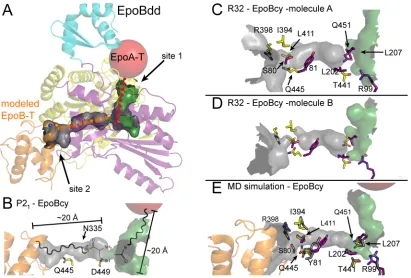
Structures suggest binding sites for T-domains of EpoA and EpoB
comes from a structure of a NRPS T domain bound to the terminal surfactin A module (22). Structural superposition places the EpoB-T binding site on the C-terminal half of the Cy domain (Fig. S5) (referred to as site 2 in Fig. 4A). These putative T domain binding sites are non-overlapping, consistent with the proposal that both T domains bind EpoB at the same time (16). Intriguingly, the P21-EpoBcy structure shows an extended L shaped channel that connects putative T domain binding site 1 to the active site, and the active site to putative T domain binding site 2 (Fig. 4A). The distance between each putative T domain binding site and the active site is ~20 Å, the length of an extended Ppant arm. This physical relationship suggests an acetyl moiety tethered via a Ppant arm from the EpoA-T domain would sit juxtaposed to a L-cysteinyl
moiety tethered via a Ppant arm from the EpoB T domain (Fig. 4B). Similar channels are found in both molecules that are present in the asymmetric unit of the R32 structure, however movement of surface loops at site 2 (the loops prior to b1 andb10), in addition to protein side chain differences near the active site result in channel constrictions (Fig. 4C,D and Figs. S2 and S6).
Structure and mutagenesis reveal unexpected location for Cy Active Site
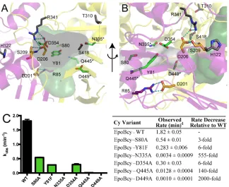
The N- and C-terminal halves of the EpoB Cy domain form a stable interface, which in turn forms the putative substrate-binding channels that were described above. In addition to several hydrophobic residues that may mediate favorable interactions with the hydrophobic Ppant arm and acetyl moiety of substrate, the structure reveals a set of previously uncharacterized polar residues that may be involved in catalysis, including S80, Y81, D354, Q445 and D449 (Fig. 5A,B and Fig. S7). We mutated these residues and also N335, which was studied previously in the homologous BacA system (Table S3) (14). These six residues were mutated individually and assayed for activity using LC/MS-MS detection, monitoring formation of 2-methylthiazole-4-carboxlic acid (2MTCA) (Fig. S1). Three active site variants have severely compromised rates of product formation: D449A, N335A and Q445A have 2000-, 555- and 140-fold decreased activities compared to wild-type EpoBcy (Fig. 5C). The S80A, Y81F and D354A variants of EpoBcy display only moderate effects with 3-, 6- and 6-fold decreased activities, respectively. Putting these results in context with the structure has allowed us to localize the active site to the C-terminal half of the Cy structure, where the channel is lined with residues N335, Q445, and D449 (Fig 5A,B).
maintaining the integrity of the substrate-channels. Importantly, neither Asp is free to interact with substrate. Rather, D201 is involved in a salt bridge with R85, which provides structure to one side of the channel, and D206 forms a salt bridge with R341 and a hydrogen bond to S209, supporting another side of the channel (Fig. 5A,B).
Discussion
Hybrid PKS/NRPSs are remarkable macromolecular assembly lines with carrier proteins delivering substrates from one enzyme module to the next and docking domains providing the intermodular communication that allows for the proper directionality (23, 15, 24). Our structures provide a visualization of the interactions between an N-terminal docking domain and a downstream enzyme within a NRPS module, and we find a bead-on-a-string type arrangement. Covalent attachment by a twenty-residue linker is all that is involved; EpoBdd makes no other contacts to the Cy domain. Thus, any docking domain could be substituted with no reengineering of the Cy domain protein surface required. Although EpoBdd is highly flexible, allowing it to search for its partner proteins (23), its attachment point to Cy appears key to its function. When EpoBdd localizes the EpoA-T to Cy through interaction with EpoAdd, this T domain will end up positioned near to one of two channel openings on Cy (site 1), allowing a substrate linked by a Ppant arm to reach down into the core of the Cy domain.
acetyl moiety from EpoA-T and L-cysteinyl moiety from EpoB-T will end up juxtaposed in the
active site and also proximal to catalytic residues D449 and N335 (Fig. 5B). Notably, the EpoA channel appears a bit longer than necessary for an acetyl moiety to be accommodated (Fig. 5A,B), perhaps explaining the observation that larger substrates (propionyl-, isobutryl-, and benozyl-EpoA-T) can be turned over by Cy, albeit more slowly (11).
In our structures and MD simulations, we observe snapshots of more open and more closed states of the channel leading to the active site, and we expect that the binding of EpoA-T and EpoB-T domains at their respective binding sites will shift the equilibrium towards the more open state, and that T domain departure will shift toward the closed state, thus restricting active site access in the absence of substrates. Although conformational changes of protein backbone have been observed for C domain proteins that could contribute to channel opening and closing (25), here we see little to no movement of the backbone atoms of the three structures that display various degrees of channel openness. Also MD simulations show that side chain motion is sufficient to open and close the internal protein cavities. Thus for this Cy domain, there appears to be no need to invoke domain hinge motions in catalysis.
location of the highly conserved sequence motifs (HHxxxDG for C domains and DxxxxDxxS for Cy domains). Importantly, residues of the DxxxxDxxS sequence motif are not free to interact with substrate, instead forming salt bridges and hydrogen bonding networks that stabilize the elaborate channels that run through the core of the protein fold. Although substrate binding might cause residues of the DxxxxDxxS motif to break their interactions and be available for catalysis, we instead propose that D449 is key to catalysis, potentially serving as a catalytic base. This proposal is consistent with the 2000-fold effect on 2MTCA production when D449 is mutated to alanine.
Inspection of the proposed mechanisms in Fig. 1B shows a number of base-catalyzed steps that might be involved in this three step reaction: deprotonation of the amino group of cysteine for the peptide bond formation step with the acetyl moiety; deprotonation of the cysteine side chain for the cyclization reaction; and deprotonation of the ring NH for the dehydration reaction. It is possible that one residue may catalyze all three deprotonations, since the protonation state of the base could be reset after each step. Notably, the number of deprotonations equals the number of protonations, with the Ppant sulfur accepting one proton and the acetyl moiety oxygen accepting two protons as it is first reduced to a hydroxy group and then to water (Fig. 1B). Thus from the perspective of stoichiometry, D449 could assist in all three reactions, but a structure with substrate bound would help to evaluate the geometric prospects of a single residue catalyzing all three different steps.
titratable side chain, we do not believe that it is a catalytic base. Instead, it may serve to position the substrates appropriately for cyclization or stabilize intermediates through hydrogen bonding. In short, the structures of EpoBcy explain much of the previous biochemical work and also reveal D449 as a key residue.
METHODS
The EpoBcy protein construct was expressed and purified as described (18), with minor modifications detailed in the SI Materials and Methods. EpoBcy site specific mutagenesis was performed using standard protocols, and activity assays (18) were adapted for product detection by LC/MS-MS (SI Materials and Methods). Purified wild-type EpoBcy was crystallized using the vapor diffusion method, and X-ray diffraction experiments, model building and refinement are detailed in the SI Materials and Methods. A 20 ns MD simulation was performed in GROMACS, and parameters are described in the SI Materials and Methods.
Acknowledgements: This work was supported by the CEHS Center core grant (P30-ES002109); and C.L.D is a Howard Hughes Medical Institute Investigator. This work is based upon research conducted at beamline 9-2 at the Stanford Synchrotron Radiation Lightsource (SSRL) and at the Advanced Photon Source (APS) on the Northeastern Collaborative Access Team (NE-CAT) beamlines. NECAT at APS is supported by grants from the National Center for Research Resources (5P41RR015301-10) and the National Institute of General Medical Sciences at the National Institutes of Health (8 P41 GM103403-10). APS is an Office of Science User Facility operated for the U.S. Department of Energy (DOE) Office of Science by Argonne National Laboratory, and is also supported by the U.S. DOE under Contract No. DE-AC02-06CH11357.
References
1. Rivera E, Gomez H (2010) Chemotherapy resistance in metastatic breast cancer: the evolving role of ixabepilone. Breast Cancer Res 12(Suppl 2):S2.
2. Rivera E, Lee J, Davies A (2008) Clinical development of ixabepilone and other epothilones in patients with advanced solid tumors. Oncologist 13(12):1207–1223. 3. Khrapunovich-Baine M, et al. (2011) Hallmarks of molecular action of microtubule
stabilizing agents: effects of epothilone B, ixabepilone, peloruside a, and laulimalide on microtubule conformation. J Biol Chem 286(13):11765–11778.
4. Kumar A, et al. (2010) Interaction of epothilone B (patupilone) with microtubules as detected by two-dimensional solid-state NMR spectroscopy. Angew Chemie Int Ed 49(41):7504–7507.
5. Thompson CA (2007) FDA approves new breast cancer treatment. Am J Heal Pharm 64(23):2406.
6. Walsh CT, Nolan EM (2008) Morphing peptide backbones into heterocycles. Proc Natl Acad Sci U S A 105(15):5655–5656.
7. Hur GH, Vickery CR, Burkart MD (2012) Explorations of catalytic domains in non-ribosomal peptide synthetase enzymology. Nat Prod Rep 29(10):1074.
8. Arnison PG, et al. (2013) Ribosomally synthesized and post-translationally modified peptide natural products: overview and recommendations for a universal nomenclature. Nat Prod Rep 30(1):108–160.
9. Julien B, et al. (2000) Isolation and characterization of the epothilone biosynthetic gene cluster from Sorangium cellulosum. Gene 249(1-2):153–160.
10. Tang L, et al. (2000) Cloning and heterologous expression of the epothilone gene cluster. Science (80- ) 287(5453):640–642.
11. Chen H, O’Connor S, Cane DE, Walsh CT (2001) Epothilone biosynthesis: assembly of the methylthiazolylcarboxy starter unit on the EpoB subunit. Chem Biol 8(9):899–912. 12. Molnár I, et al. (2000) The biosynthetic gene cluster for the microtubule-stabilizing agents
epothilones A and B from Sorangium cellulosum So ce90. Chem Biol 7(2):97–109. 13. Gehring AM, Mori I, Perry RD, Walsh CT (1998) The nonribosomal peptide synthetase
14. Duerfahrt T, Eppelmann K, Müller R, Marahiel MA (2004) Rational design of a bimodular model system for the investigation of heterocyclization in nonribosomal peptide biosynthesis. Chem Biol 11(2):261–271.
15. Richter CD, Nietlispach D, Broadhurst RW, Weissman KJ (2008) Multienzyme docking in hybrid megasynthetases. Nat Chem Biol 4(1):75–81.
16. Keating TA, Marshall CG, Walsh CT, Keating AE (2002) The structure of VibH
represents nonribosomal peptide synthetase condensation, cyclization and epimerization domains. Nat Struct Biol 9(7):522–526.
17. Marshall CG, Hillson NJ, Walsh CT (2002) Catalytic mapping of the vibriobactin biosynthetic enzyme VibF. Biochemistry 41(1):244–250.
18. Kelly WL, Hillson NJ, Walsh CT (2005) Excision of the epothilone synthetase B
cyclization domain and demonstration of in trans condensation/cyclodehydration activity. Biochemistry 44(40):13385–13393.
19. Samel S a., Czodrowski P, Essen LO (2014) Structure of the epimerization domain of tyrocidine synthetase A. Acta Crystallogr Sect D Biol Crystallogr 70(5):1442–1452. 20. Holm L, Rosenström P (2010) Dali server: conservation mapping in 3D. Nucleic Acids
Res 38:W545–W549.
21. Liu F, Garneau S, Walsh CT (2004) Hybrid nonribosomal peptide-polyketide interfaces in epothilone biosynthesis. Chem Biol 11(11):1533–1542.
22. Tanovic A, Samel SA, Essen LO, Marahiel MA (2008) Crystal structure of the
termination module of a nonribosomal peptide synthetase. Science (80- ) 321(5889):659– 663.
23. Weissman KJ, Müller R (2008) Protein–Protein Interactions in Multienzyme Megasynthetases. ChemBioChem 9(6):826–848.
24. Shen B, et al. (2001) The biosynthetic gene cluster for the anticancer drug bleomycin from Streptomyces verticillus ATCC15003 as a model for hybrid peptide-polyketide natural product biosynthesis. J Ind Microbiol Biotechnol 27(6):378–385.
25. Bloudoff K, Rodionov D, Schmeing TM (2013) Crystal structures of the first
Biotechnol 17(6):538–9.
27. Khosla C, Herschlag D, Cane DE, Walsh CT (2014) Assembly line polyketide synthases: mechanistic insights and unsolved problems. Biochemistry 53(18):2875–2883.
28. Walsh CT (2008) The chemical versatility of natural-product assembly lines. Acc Chem Res 41(1):4–10.
29. Strieker M, Tanović A, Marahiel MA (2010) Nonribosomal peptide synthetases: structures and dynamics. Curr Opin Struct Biol 20(2):234–240.
30. Bond CS (2003) TopDraw: a sketchpad for protein structure topology cartoons. Bioinformatics 19(2):311–312.
Figure Legends
Fig. 1. Scheme for epothilone biosynthesis, focusing on thiazol(in)e formation by the NRPS cyclization domain. (A) Epothilones (right) are large macrocycles with a thiazole-containing side chain (red), which are produced by a hybrid PKS/NRPS EpoA-K. EpoA and EpoB are responsible for producing the thiazole side chain and have the following domains (9–12): KSy, ketosynthase-like domain; AT, acyltransferase domain; ER, enoyl reductase; and T, an acyl carrier protein. The NRPS EpoB domains include: A, adenylation domain; Cy, cyclization; Ox, a flavin-dependent oxidase domain; and T, a carrier protein domain (28, 29). The Ppant group of each T domain is represented as , and residue numberings for the EpoA-T, and EpoBcy constructs are indicated. (B) Proposed mechanisms for condensation and cyclodehydration. The EpoA-T domain and EpoB-T domains are represented as red and blue spheres, respectively.