Halogen-substituted ureas for anion binding: solid state and solution
studies.
Arianna Casula,
aMarco Fornasier,
aRiccardo Montis,*
a,bAlexandre
Bettoschi,
aStephen P. Argent,
cAlexander J. Blake,
cVito Lippolis,
aLaura
Marongiu,
aGiacomo Picci,
aJeremiah P. Tidey,
cClaudia Caltagirone.*
aa Dipartimento di Scienze Chimiche e Geologiche, Università degli Studi di Cagliari, S.S. 554 Bivio per Sestu, 09042 Monserrato (CA), Italy. [email protected]
b Present address: Department of Materials, Imperial College London, South Kensington Campus, London, SW7 2AZ, UK, [email protected]
Halogen-substituted ureas for anion binding: solid state and solution
studies.
Herein, we report the synthesis and the anion binding properties of a family of N,N’-diphenylureas L1-L15, bearing on the aromatic ring(s) halogens (chlorine
and iodine) and/or nitro or trifluoromethyl electron-withdrawing groups. The analysis of the crystal structures obtained from single crystal X-ray diffraction experiments shows that self-assembled chains or tapes connected via N-H···O hydrogen bonds are the most commonly adopted arrangements for this type of molecules in the crystal lattice. In the presence of anion guests or solvent molecules with competing hydrogen bond donors and acceptors, other supramolecular arrangements can be observed. Solution studies conducted in DMSO-d6/0.5% H2O by means of 1H-NMR titrations show the formation of 1:1
adducts with all receptors. The different observed affinities of the receptors for the anion guests were rationalised in terms of steric hindrance of the substituents on the phenyl rings and their electron-withdrawing properties.
Keywords: anion binding, phenylurea
Introduction
The development of synthetic receptors for anion binding, sensing, catalysis and
transport is one of the most active area of Supramolecular Chemistry.1 In particular, the
design and synthesis of neutral receptors capable of recognizing anions in competitive
solvent mixture, and possibly in water, is rather challenging because of competition
issues. Urea and thiourea-based receptors have been widely studied for anion binding
because of their synthetic accessibility and also their ability to interact through strong,
directional hydrogen bonds.2 Recently, selenoureas have also been proposed for anion
binding and sensing.3 The urea (or thiourea) moiety bearing two N-H groups can bind
ligand with a single acceptor atom to yield a six-membered chelate ring. They may also
bind as a bidentate ligand with two adjacent oxygen atoms in an oxyanion to form an
eight-membered chelate ring. Among the different type of urea derivatives developed
over recent years, N,N’-diphenylurea represents one of the simplest and most popular
receptor for anion binding.4
In the solid state, this class of compounds have been extensively investigated.5N,N
’-diphenylurea forms robust and predictable self-assembled chains or tapes connected via
N-H···O hydrogen bonds. Etter et al. demonstrated that the presence of electron-withdrawing groups in diaryl urea decreases the tendency to form self-assembled 1-D
chains.6 This is due to the increased acidity of the ortho aromatic C-H that forms intramolecular hydrogen bonds with the urea C=O, reducing its ability to interact with
adjacent urea NHs. Therefore, the disruption of these 1-D chains is often associated to a
coplanar conformation of the phenyl rings with respect to the urea plane.
A similar behaviour was described by Nangia and collaborators who investigated a
family of substituted N-X-phenyl-N′-p-nitrophenyl urea compounds (X = H, F, Cl, Br, I, CN, C≡CH, CONH2, COCH3, OH, Me).7 The results allowed the authors to classify the
family of structures into two main categories: (i) urea tapes structures, formed by classic
urea N-H···O hydrogen bonds, in which phenyl rings adopt a twisted conformation with
respect to the urea plane, and (ii) non-urea tape structures in which the phenyl groups
adopt a coplanar conformation and the classical urea N-H···O hydrogen bonds are
replaced by interactions with NO2 groups or solvent molecules.
Recently, Gale, Coles, et al. described a systematic structural analysis on a series of urea-based anion receptor complexes including high-resolution, experimental an
electron density study.8 The authors demonstrated that by systematically altering the
scaffold, it is possible to modulate the strength of the interaction between the receptor
and anion. By geometric analysis of the hydrogen bonding interactions they also
suggested that moving from meta to para to 3,5-dinitro substitution the hydrogen bond strength increases.
In recent years, beside hydrogen bond-, also halogen bond- based receptors have been
developed for anion binding. The term “halogen bonding (XB)” was officially defined
by IUPAC in 2013 as a non-covalent interaction between a halogen bond donor R-X
(where X is a halogen atom with an electrophilic region and a R is any organic group)
and a halogen bond acceptor Y (where Y is a nucleophilic molecular entity).9 Halogen
bonds RX···Y are almost linear and they have an energy comparable with the hydrogen
bonds (5-180 kJ mol-1).
Taylor et al. have reported a family of urea based receptors for anion recognition that contain iodoperfluoro-arene groups.10 These systems are able to interact with anions via
both hydrogen and halogen bonds.
An example of simple symmetric N,N’-diphenylurea receptors para substituted with
halogens and able to bind anions forming both hydrogen and halogen bonds in solution
and in the solid state was reported by Das et al.11
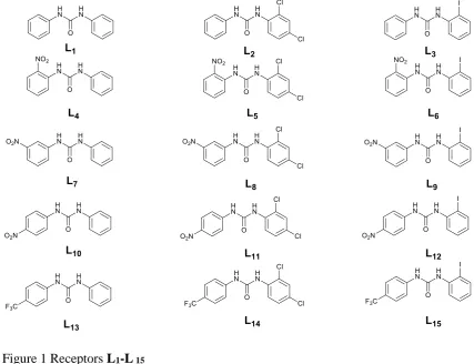
Inspired by these results we decided to synthesize a new family of simple asymmetric
N,N’-diphenylurea receptors L1-L15 for anionrecognition. Thesereceptors are
substituted on one of the phenyl ring with iodine and chlorine in various position (ortho
and para for chlorine and ortho for iodine), and with nitro or a trifluoromethyl moiety on the other (Figure 1).
These different combinations of substituents on the two phenyl groups were chosen in
order to evaluate the effect of electron-withdrawing groups and halogens on the anion
in the literature,6b, 12 were used as control molecules for each series of receptors with the
same substituents. We tested receptors L1-L 15 with a set of anions of different
geometries [(Y-shape (AcO- and BzO-), spherical (Cl- and F-) and tetrahedral (H2PO4-)]
by means of 1H-NMR spectroscopy and, where possible, single crystal X-ray
[image:5.595.83.511.211.539.2]diffraction.
Figure 1 Receptors L1-L 15
Results and discussion
Synthesis
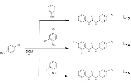
Receptors L1-L15 were designed and successfully synthesized according to Scheme 1-3.
The synthesis are based on the simple nucleophilic addition of an isocyanate (phenyl
isocyanate, nitro-phenyl isocyanate or trifluoromethyl-phenyl isocyanate for receptors
introduction, the synthesis of receptors L1,L4, L7,L10,and L13,had been reported
before.6b, 12 After two hours of reflux in DCM, all the products were obtained as pure
solids by precipitation, in a widly variable yields depending on the substituents
introduced in the systems (20- 96%).
Scheme 1 Reaction scheme adopted for the synthesis of L1, L2, and L3.
Scheme 3 Reaction scheme adopted for the synthesis of L13, L14, and L15.
Single Crystal X-Ray Diffraction
To investigate binding properties in solid state of L1-L15, all the receptors were
crystallised by slow evaporation from various solvents and in the presence of different
anion guests. Surprisingly, we could isolate crystals suitable for single crystal X-ray
diffraction only for the adduct L6-tetrabutylammonium benzoate (L6-BzO-).
Crystallisations of free receptors L1-L15 produced single crystals only for L1, L5, L8,
L14, and L15. In the case of receptor L2, crystallisations in presence of
tetrabutylammonium fluoride or tetrabutylammonium iodide produced two distinct
polymorphic phases, designated L2 and L2, respectively. L8 and L11 crystallised as
solvate forms, a DMSO solvate L8•DMSO and a mixed solvate L11•2DMSO•DMF,
[image:7.595.123.394.71.244.2]respectively.
Table 1. Unit cell parameters for the crystal structures of L1, L2, L2, L5, L8, L14, L15,
L8•DMSO, L11•2DMSO•DMF, and L6-BzO-.
L1
CCDC 1561823
L2
CCDC1561826
L2
CCDC1562645
L5
CCDC1561828
L8
Formula C13H12N2O C13H10N2OCl2 C13H10N2OCl2 C13H9N3O3Cl2 C13H9N3O3Cl2
FW 212.25 281.13 281.13 326.13 326.13
Crystal
System
orthorhombic triclinic triclinic monoclinic orthorhombic
Space
Group
Pna21 P-1 P-1 P21/n Pna21
a /Ǻ 9.0641(3) 4.6123(14) 4.5612(3) 4.6027(7) 42.4563(6)
b /Ǻ 10.3509(3) 11.9420(5 11.5202(11) 48.5814(8) 6.5738(1)
c /Ǻ 11.7422(3) 22.8508(7) 12.1448(9) 5.9207(14) 4.7887(1)
α / º 90 93.005(3) 103.972(7) 90 90
β / º 90 92.645(3) 94.249(5) 95.7193(17) 90
γ / º 90 97.764(3) 95.458(6) 90 90
V /Ǻ3 1101.68(5) 1243.57(8) 613.35(8) 1317.32(4) 1336.52(4)
T / K 120(2) 293(2) 120(2) 120(2) 120(2)
Z 4 4 2 4 4
L14
CCDC 1562644
L15
CCDC 1561819
L8•DMSO
CCDC 1561821
L11•2DMSO•DMF
CCDC 1561822
L6-BzO-
CCDC1561827
Formula C14H9N2OF3Cl2 C14H10N2OF3I C30H30Cl4N6O8S2 C15.68H15.67Cl2N3.68O4S0.31 C36H51IN4O5
FW 349.13 406.14 808.52 400.66 746.70
Crystal
System
monoclinic orthorhombic triclinic monoclinic monoclinic
Space
Group
Cc Pca21 P-1 P21/n P21/n
a /Ǻ 11.4548(2) 29.971(5) 12.0136(4) 21.6216(9) 8.8751(2)
b /Ǻ 13.5410(2) 4.5599(7) 12.6801(4) 3.8114(1) 22.2235(3)
c /Ǻ 9.0285(2) 10.4038(14) 13.8642(5) 22.9689(10) 18.3822(3)
β / º 92.4156(16) 90 72.336(3) 115.885(5) 92.239(2)
γ / º 90 90 66.334(3) 90 90
V /Ǻ3 1399.16(4) 1421.8(4) 1739.57(12) 1702.94 3622.9(1)
T / K 120(2) 120(2) 120(2) 120(2) 120(2)
Z 4 4 2 4 4
A summary of unit cell parameters and main crystallographic data for the set of crystal
structures collected is shown in Table 1. Details of crystallization experiments,
intermolecular interactions and crystal packing descriptions are reported in Supporting
Information.
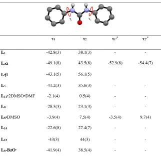
Considering the urea molecular unit, the comparison of the molecular conformation for
the ten crystal structures shows that in all the structures, urea NH groups are oriented
trans with respect to the carbonyl group, confirming the behaviour generally observed in crystal structures of urea derivatives. Furthermore, in most of them, both phenyl rings
are slightly tilted with respect the plane of the urea function (Table 2). The only
exception is represented by the two solvated forms, L8•DMSO and L11•2DMSO•DMF,
in which the phenyl rings are co-planar with the urea plane. According to previous
observations, the planar conformation of the two solvate forms is stabilised by
intra-molecular C-HO hydrogen bonds involving the urea C=O group and aromatic CHs of
the phenyl groups (HO distances lie in the range 2.20-2.28 Å, CO distances lie in
the range 2.836(3)-2.876(3) Å). However, weak intra-molecular C-HO hydrogen
bonds are also observed in most of the structures which adopt a tilted conformation.
Excluding L15 and the two polymorphs L2 and L2, which show intra-molecular
interactions only on the substituted ring the structures (see Table S3, Supporting
Information) of the free receptors show a set of intramolecular C-HO interactions with
Å). In the case of L5 this intramolecular interaction is also assisted by a further
intramolecular N-HO hydrogen bond involving one of the urea NHs and the nitro
group in position ortho (HO distance is 2.24(5) Å, NO distance is 2.935(4) Å). Interestingly, L6-BzO-, adopts a conformation with the phenyl rings tilted out with
respect the urea plane, showing only one C-HO intramolecular interaction involving
[image:10.595.83.406.324.639.2]the CH in the ortho position on the iodo-substituted ring and the C=O of the urea group (HO distance is 2.47 Å, CO distance is 2.920(4) Å).
Table 2. Torsion angles 1 and 2
1 2 1’* 2’*
L1 -42.8(3) 38.1(3) - -
L2 -49.1(8) 43.5(8) -52.9(8) -54.4(7)
L2 -43.1(5) 56.1(5)
L5 -41.2(3) 35.6(3) - -
L11•2DMSO•DMF -2.1(4) 0.5(4) - -
L8 -28.3(3) 23.1(3) - -
L8•DMSO -3.9(4) 7.5(4) -3.5(4) 9.7(4)
L14 -22.6(8) 27.4(7) - -
L15 -43(3) 44(3) - -
L6-BzO- -41.9(4) 38.5(4) - -
* For crystal structures with Z’=2 we use 1’ and2’ to indicate torsional angles for the
second symmetrically independent molecule.
Most of the structures show the classical 1-D chains connected by three-centre N-HO
L11•2DMSO•DMF adopt alternative supramolecular synthons. In these structures, the
presence of the guest molecule with a set of competing hydrogen bond acceptors
prevents the formation of the typical urea-urea N-HO tapes. Accordingly, we discuss
separately the three structures L8•DMSO, L11•2DMSO•DMF and L6-BzO- and start
our discussion focusing on the supramolecular features of free receptors.
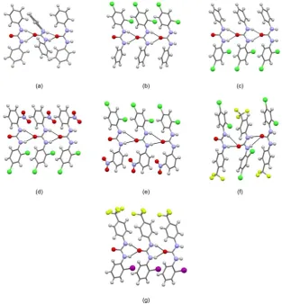
One-dimensional N-HO chains.
Structures of free receptors show 1-D urea chains, in most cases connected by the robust
bifurcated N-HO supramolecular synthon (HO distances are in the range 1.95-2.70
Å, NO distances are in the range 2.775(2)-3.406(6) Å). The shape of the 1-D chains is
very similar in all the structures, consisting of linear arrangements of molecules. The
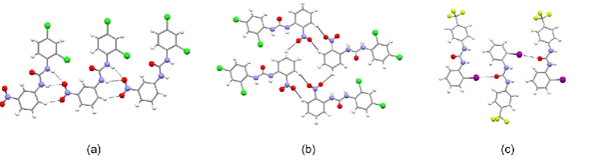
only exceptions are L1and L14 in which the chains adopt a zig-zag motif (Fig 1 a and f).
In the case of L1 the phenyl groups within the urea molecule are oriented approximately
perpendicular to each other with aromatic hydrogens pointing toward the centre of the
phenyl rings of adjacent urea units and forming T-shaped edge-to -face interactions8b
(C-HCentroid distances 2.99 (3) Å). In the case of L14, adjacent receptor molecules
are slightly tilted along the direction of propagation of the 1-D chain. As a consequence,
the N-HO hydrogen bond involves only one of the urea NH moieties (Fig 1 f). A
similar interaction is observed in L8 (Fig 1 e) but in this case the 1-D urea chain adopts
Fig 1. Ball and stick images of the one-dimensional urea chains for structures of free
receptors: (a) L1; (b) L2; (c) L2; (d) L5; (e) L8; (f) L14 and (g) L15. For structure L2
only one independent molecule is reported as representative of the shape of
one-dimensional urea chains. N-HO hydrogen bonds are indicated using black dashed
lines; atoms of iodine in purple, chlorine in dark green, fluorine in green/yellow,
nitrogen in blue, oxygen in red hydrogen in white and the carbon scaffold in grey.
Other interactions have been removed for clarity.
Contrary previous studies,7 while the substitution at the phenyl rings introduces
potential competing groups with respect to hydrogen bonding, no such competition is
observed in the free receptors reported herein. However, in the case of L8, the urea NHs
NO2 groups in position meta of adjacent 1-D chains (HO distance are 2.42 Å and 2.44
Å, NO distance are 3.142(2) Å and 3.145(2) Å). This particular supramolecular
synthon is not observed in the case of the substituted o-NO2 receptor L5, which, instead
forms centro-symmetric dimers with aromatic hydrogens of an adjacent urea unit via
C-HO interactions (Fig 2 b). In the case of L15, the urea C=O group is involved in a
second interaction (Fig 2 c) with the iodo substituents of adjacent chains [IO distance
is 3.50 (2) Å]. No such behaviour was observed in the crystal structure published by
Koshti et al.,5e which corresponds to our receptor L3, where the iodo- substituents only
[image:13.595.88.512.329.446.2]interacts via weak C-HI and II interactions with neighbouring molecules.
Fig 2. Further intermolecular interactions for L8 (a),L5 (b) and L15 (c).
The effect of varying the substituent groups, particularly the set of electron-withdrawing
groups chosen for the design of receptors L1-L15, seems to have no consistent effect on
the strength of the N-HO hydrogen bonds. Bond length analysis of the N-HO
intermolecular and weaker C-HO intra-molecular interaction (see Table S3 in
Supporting Information) reveals that for structures L1, L2, L2, L5 and L15 these are
very similar, with N-HO and C-HO distances in the range 1.952.24 Å and 2.44
-2.58 Å, respectively (NO distances in the range 2.775(2)-2.935(4) Å; CO distances
in the range 2.881(3)-2.958(8) Å). In the case of L8 and L14, when compared to L1, the
strength of the intra-molecular C-HO interactions, with HO distances decrease from
the range 2.44-2.53 Å for L1 to 2.30-2.40 Å for L8 and L14 (range of CO distances
decrease from 2.881(3)- 2.973(3) Å for L1 to 2.828(2)-2.920(7) Å for L8 and L14).
Solvates and receptor-anion structures.
Crystallisation of receptors L6, L8 and L11 in the presence of guest molecules such as
solvents or anions produced two solvates forms, L8•DMSO and L11•2DMSO•DMF,
corresponding to a DMSO and a DMSO/DMF solvate respectively and one benzoate
complex of the receptor L6, labelled as L6-BzO-.
As mentioned above, in the solvate compounds, the urea units adopt a conformation
with the phenyl rings approximately coplanar with the urea plane forming short
intramolecular C-HO interactions. This is a result of the increased acidity of the
aromatic hydrogens due to the presence of the electron-withdrawing groups. Previous
studies,5c, 5d, 6 have proposed that, in such case, the urea C=O is made a weaker
hydrogen bond acceptor and the urea NH groups preferentially interact with solvent
molecules instead, thus disrupting the 1-D urea-urea assemblies.
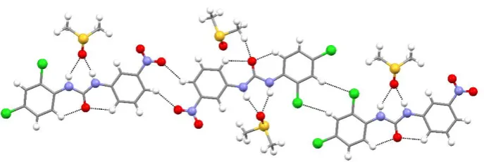
Structure L8•DMSO crystallises with two independent receptors in the asymmetric unit.
These interact with each other via C-HCl and C-HO interactions (HCl distances are 2.79 Å and 2.92 Å, CCl distances are 3.696(2) Å and 3.854(2) Å; HO distances
are 2.46 Å and 2.50 Å, CO distances are 3.244(3) Å and 3.317(3) Å) involving Cl and
NO2 groups in phenyl rings and the aromatic hydrogen in the para position to form a
1-D chain (Fig 3). Each independent receptor interacts with a molecule of 1-DMSO via
N-HO hydrogen bonds involving urea NHs (HO distances are in the range 1.87-2.25
Å, NO distances are in the range 2.737(3)- 3.037(3) Å). Solvent molecules also
DMSO (HO distances are 2.41 Å and 2.43 Å, CO distances are 3.203(3)Å and
3.383(3) Å). A more detailed description of the crystal packing is reported in
[image:15.595.92.436.183.298.2]Supporting Information.
Fig 3. Main intermolecular interaction for structure L8•DMSO.
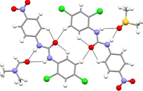
Structure L11•2DMSO•DMF formed a DMSO/DMF solvate with the two solvents
disordered to share the same molecular site in a 2:1 ratio, respectively. In this structure,
the receptor forms centro-symmetric dimers via C-HO interactions (HO distance is 2.51 Å, CO distance is 3.337(3) Å) involving the urea C=O group and the aromatic
hydrogen in a position meta to the di-chloro substituted phenyl ring (Fig 4). This dimer
exposes the urea NH groups which interact with neighbouring solvent molecules via
N-HO hydrogen bonds involving the S=O or C=O groups, depending on the solvent
present. The HO distances are in the range 1.95-2.48 Å (NO distances are in the
range 2.801(5)- 3.226(12)Å). Receptor molecules interact along the shortest axis of the
Fig 4. Main intermolecular interactions for structure L11•2DMSO•DMF. DMSO and
DMF have been separated for clarity.
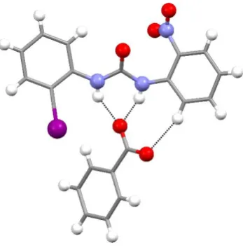
In the adduct L6-BzO- the urea NH groups are involved in the formation of strong
N-HO hydrogen bonds with the BzO- guest (HO distances are 1.92(4) Å and 2.10(4) Å,
NO distances are 2.717(3)Å and 2.878(3)Å). Interestingly, in this case the receptor
molecule has a non-planar conformation with the phenyl rings slightly tilted with
respect the urea plane. Accordingly, no intramolecular C-HO interactions are
observed between the urea C=O group and aromatic CHs in the ortho positions of the
phenyl rings. This can be explained considering that in order to have a planar
conformation stabilised by intramolecular C-HO interactions, the receptor must have
the substituted group in an ortho position and, both on the same side of the urea NHs. Such a case would present significant steric hindrance or electronic repulsion towards
the anionic guest. Accordingly, the best compromise seems to be a tilted conformation
with the I and NO2 group oriented mutually trans and the NO2 group on the opposite
side with respect the urea NHs. As a consequence of this conformation, in order to
interact with the receptor site, minimising the repulsion of the iodo substituent, the BzO
-specie is slightly shifted on the side of the nitro-phenyl ring, using one oxygen of the
hydrogen bond. The second oxygen forms a C-HO interaction with one aromatic CH
of the nitro-phenyl ring (HO distance is 2.46 Å, CO distance is 3.364(4) Å, see Fig
[image:17.595.212.386.161.340.2]5)
Fig 5. Receptor-anion interaction and conformation of receptor L6 in crystal structure of
L6-BzO-. Countercation has been omitted for clarity.
Solution studies
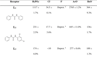
Anion binding affinity of receptors L1-L15 was evaluated by means of 1H-NMR
titrations in DMSO-d6/0.5% H2O towards a set of anions (F-, Cl-, H2PO4-, AcO-, and
BzO-, as their tetrabutylammonium salts). The experimental data were fitted according
to a 1:1 model and the stability constants (Table 3) were calculated using the
WINEQNMR programme.13
By means of a COSY (Correlation Spectroscopy) 2D-NMR experiment it was possible
to attribute the correct chemical shift value for each NH proton in the asymmetrical
receptors L2 and L3 (and therefore for all the other halogenated receptors): the NH
proton in close proximity of the phenyl moiety is downfield shifted with respect to the
NH protons near the 2,4-dichloro phenyl and the 2-iodophenyl fragments for L2 and L3,
Stability constants calculated following both NH proton signals were comparable so we
decided to follow the chemical shift of the NH proton signal in close proximity of the
non-halogenated phenyl ring.
The results observed for the triad L1, L2, L3 are in agreement with the degree of steric
hindrance increasing in the order L3>L2>L1. The presence of the chlorine or iodine
atom in an ortho position on the halogenated phenyl ring with respect to the urea function, for L2 and L3,respectively, partially obstructs the anion access to the
coordination site of the receptor. Several anion binding studies for receptor L1 are
reported in the literature, in particular recognition of carboxylates.12b, 14 The stability
constants obtained for the formation of the 1:1 adduct of L1 with acetate and benzoate at
[image:18.595.78.491.485.736.2]300 K are consistent with the values reported by Leito et al. at 298K (2138 M-1 and 661 M-1 for acetate and benzoate, respectively).
Table 3. Stability constants (Ka/M−1 ) of the 1:1 adducts of L1-L15 with F-, Cl-, H2PO4-,
AcO-, BzO-, as their tetrabutylammonium salts in DMSO-d6/0.5% H2O at 300 K. Receptor H2PO4- Cl- F- AcO- BzO
-L1 1117 ±
1.7%
34.5 ±
0.1%
Deprot. b 2765 ± 1.2% 364 ±
9.3%
L2 231 ±
2.5%
17.7 ±
3.6%
Deprot. b 445 ± 11.0% 136±
1.7%
L3 174 ±
6.0%
<10 Deprot. b 277 ± 0.6% 100 ±
L4 684 ±
5.9%
<10 Deprot. b 1283 ± 3.5% 314 ±
5.9%
L5 Deprot. <10 Deprot. b Deprot b 123.3 ±
5.9%
L6 a <10 Deprot. b 218.0 ± 3.9% 87.3 ±
11.0%
L7 a 57.8 ±
1.8%
Deprot. b 9620 ± 3.8% 3322 ±
1.8%
L8 a 35.2 ±
7.8%
Deprot. b 1883 ± 8% 567 ±
0.9%
L9 a 20.6 ±
2.3%
Deprot. b 1611 ± 4.3% 425 ±
2.8%
L10 a 68.6 ±
2.9%
Deprot. b 13467 ±
2.3%
3706±
1.0%
L11 a 36.8 ±
0.7%
Deprot. b 6833± 30% 780 ±
L12 a 26.2 ±
6.2%
Deprot. b 1470 ± 5.6% 681 ±
1.6%
L13 871.9 ±
32%
52.2 ±
3.3%
Deprot. b 2608 ± 17% 1980 ±
6.4%
L14 a 33 ± 4.8% Deprot. b 642 ± 4.8% 514 ±
11%
L15 a 14 ± 8.3% Deprot. b 583.5 ± 23% 301 ±
1.8%
a Significant downfield shift and broadening of the signals attributed to the urea
NHs suggesting strong interaction.
b Disappearance of the signals attributed to the urea NHs upon addition of 0.1
equivalent of fluoride, suggesting deprotonation.
The slight difference in values is probably due to the difference in the temperature at
which the experiments were conducted (300 K in our case, and 298 K for the data
reported in the literature).
In the triad L4-L6, the presence of the nitro group in an ortho position with respect to
the central urea function in addition to the presence of the chlorine and iodine atoms in
constants values compared to those of L1-L3. This result can be explained in terms of
both steric and electronic effects. In the triad L4-L6 the nitro group is in close proximity
to the urea function and it could obstruct the anion coordination. AcO- and H2PO4- cause
deprotonation of the receptor L5,presumably because of a combination of the elctron
withdrawing properties of the nitro group in ortho position that increase the acidity of the NH proton, and the steric hindrance that disfavour the anion binding favouring,
instead, the competitive deprotonation process in the case of basic anions.
In the series of receptors L7-L9 the stability constants increase with respect to the
previous triad L4-L6,probably due the meta positioning of the nitro group that allows a
more favourable interaction between the anions and the urea binding site. The
interaction of receptor L7 and anion guests was previously studiedby means of
UV-visible and 1H-NMR spectroscopy,12, 14 and the values of the stability constants reported
in Table 1 are in agreement with the literature.
The series of receptors L10-L12 shows the highest stability constants among all
the receptors bearing a nitro group. In particular, receptor L10,already known in the
literature,15 displays a good affinity for acetate as confirmed by the high value of the
stability constant (> 104 M-1). The reasons for the increasing anion coordinating ability
of these receptors could be ascribed to both steric and electronic factors. First, the nitro
group in the para position with respect to the active urea, should decrease the steric hindrance observed for the previous triads (L4-L9 ) allowing for easier access of the
anion in the pseudo-cavity of the receptors also for bigger anion like benzoate.
Moreover, an electron- withdrawingnitrogroup in the para position should influence in a positive way the coordination properties of the ligands, thereby increasing the acidity
The anion binding activity across this series is consistent with the trends previously
described for the other receptors. The stability constants decrease from L13 to L15
because of the varying steric hindrance of the halogen on the phenyl ring. By comparing
the stability constant of receptor L15 with that of receptor L3 (without substituents on the
non-halogenated phenyl ring) and receptor L12 (with a nitro group in place of the
tri-fluoromethyl unit), it is possible to define the increasing anion affinity in the order L3 <
L15 < L12..This evidence is in agreement with the lower acidity of the NH protons in the
unsubstituted receptor L3 compared to receptorsL15 and L12. Onthe other hand, between
receptors L15 and L12,the lower ability of receptor L15 to bind anions can be explained
by taking into account the electron- withdrawing nature of the CF3group with respect to
the NO2 group. The same behaviour can be found for the series L1-L10-L13 and L2-L11
Conclusions
In conclusion, we have described herein the synthesis and the anion binding properties
of fifteen N-N’-diphenylurea receptors substituted with electron-withdrawing groups
(namely nitro and trifluoromethyl) and halogens (chlorine and iodine). We were able to
obtain crystals suitable for single crystal X-ray diffraction for nine receptors (including
two polymorphs and two solvates) and the 1:1 adduct of L6 with benzoate. As expected,
the classic urea 1-D chains were observed in most of the structures. Only L6-BzO-,
L8•DMSO and L11•2DMSO•DMF adopted alternative supramolecular synthons
because of the presence of the anion guest or the solvents that prevents the formation of
the typical urea-urea N-HO tapes. Solution studies conducted by means of 1H-NMR
spectroscopic titrations allowed us to calculate the stability constant for the formation of
the 1:1 adducts with all receptors and a set of anions (F-, Cl-, H2PO4-, AcO-, BzO-). The
highest values of stability constants were obtained for the receptors L10-L12 bearing the
nitro group in the para position with respect to the urea moiety.
Acknowledgements
The authors thank the financial support from Fondazione Banco di Sardegna.
References
1. (a) Ashton, T. D.; Jolliffe, K. A.; Pfeffer, F. M., Luminescent probes for the
bioimaging of small anionic species in vitro and in vivo. Chemical Society Reviews
2015,44 (14), 4547-4595; (b) Busschaert, N.; Caltagirone, C.; Van Rossom, W.; Gale,
P. A., Applications of Supramolecular Anion Recognition. Chemical Reviews 2015,115
(15), 8038-8155; (c) Duke, R. M.; Veale, E. B.; Pfeffer, F. M.; Kruger, P. E.;
Gunnlaugsson, T., Colorimetric and fluorescent anion sensors: An overview of recent
developments in the use of 1,8-naphthalimide-based chemosensors. Chemical Society
Reviews 2010,39 (10), 3936-3953; (d) Gale, P. A.; Caltagirone, C., Fluorescent and colorimetric sensors for anionic species. Coordination Chemistry Reviews; (e) Gale, P. A.; Caltagirone, C., Anion sensing by small molecules and molecular ensembles.
Chemical Society Reviews 2015,44 (13), 4212-4227; (f) Ngo, H. T.; Liu, X.; Jolliffe, K.
A., Anion recognition and sensing with Zn(ii)-dipicolylamine complexes. Chemical
2. (a) Casula, A.; Bazzicalupi, C.; Bettoschi, A.; Cadoni, E.; Coles, S. J.; Horton, P. N.; Isaia, F.; Lippolis, V.; Mapp, L. K.; Marini, G. M.; Montis, R.; Scorciapino, M. A.; Caltagirone, C., Fluorescent asymmetric bis-ureas for pyrophosphate recognition in
pure water. Dalton Transactions 2016,45 (7), 3078-3085; (b) Amendola, V.;
Bonizzoni, M.; Esteban-Gómez, D.; Fabbrizzi, L.; Licchelli, M.; Sancenón, F.; Taglietti,
A., Some guidelines for the design of anion receptors. Coordination Chemistry Reviews
2006,250 (11-12), 1451-1470; (c) Antonisse, M. M. G.; Reinhoudt, D. N., Neutral
anion receptors: Design and application. Chemical Communications 1998, (4),
443-448; (d) Gale, P. A., Structural and molecular recognition studies with acyclic anion receptors. Accounts of Chemical Research 2006,39 (7), 465-475; (e) Hay, B. P.; Firman, T. K.; Moyer, B. A., Structural design criteria for anion hosts: Strategies for achieving anion shape recognition through the complementary placement of urea donor groups. Journal of the American Chemical Society 2005,127 (6), 1810-1819; (f) Li, A. F.; Wang, J. H.; Wang, F.; Jiang, Y. B., Anion complexation and sensing using
modified urea and thiourea-based receptors. Chemical Society Reviews 2010,39 (10), 3729-3745; (g) Nishizawa, S.; Bühlmann, P.; Iwao, M.; Umezawa, Y., Anion
recognition by urea and thiourea groups: Remarkably simple neutral receptors for
dihydrogenphosphate. Tetrahedron Letters 1995,36 (36), 6483-6486.
3. Casula, A.; Llopis-Lorente, A.; Garau, A.; Isaia, F.; Kubicki, M.; Lippolis, V.;
Sancenón, F.; Martínez-Máñez, R.; Owczarzak, A.; Santi, C.; Andrea Scorciapino, M.; Caltagirone, C., A new class of silica-supported chromo-fluorogenic chemosensors for
anion recognition based on a selenourea scaffold. Chemical Communications 2017,53
(26), 3729-3732.
4. Blažek Bregović, V.; Basarić, N.; Mlinarić-Majerski, K., Anion binding with
urea and thiourea derivatives. Coordination Chemistry Reviews 2015,295, 80-124.
5. (a) Capacci-Daniel, C. A.; Bertke, J. A.; Dehghan, S.; Hiremath-Darji, R.; Swift,
J. A., Concomitant polymorphs of 1,3-bis(3-fluorophenyl)urea. Acta Crystallographica
Section C 2016,72 (9), 692-696; (b) Custelcean, R., Crystal engineering with urea and
thiourea hydrogen-bonding groups. Chemical Communications 2008, (3), 295-307; (c)
Deshapande, S. V.; Meredith, C. C.; Pasternak, R. A., Crystallographic data on disubstituted symmetric ureas. Acta Crystallographica Section B 1968,24 (10), 1396-1397; (d) Etter, M. C., Hydrogen bonds as design elements in organic chemistry.
Journal of Physical Chemistry 1991,95 (12), 4601-4610; (e) Koshti, V. S.; Thorat, S. H.; Gote, R. P.; Chikkali, S. H.; Gonnade, R. G., The impact of modular substitution on crystal packing: the tale of two ureas. CrystEngComm 2016,18 (37), 7078-7094; (f) Taouss, C.; Thomas, L.; Jones, P. G., Packing principles for urea and thiourea solvates: structures of urea : morpholine (1 : 1), urea : 1,4-dioxane (1 : 1), thiourea : morpholine (4 : 3) and thiourea : 1,4-dioxane (4 : 1). CrystEngComm 2013,15 (34), 6829-6836.
6. (a) Etter, M. C.; Panunto, T. W., 1,3-Bis(m-nitrophenyl)urea: An exceptionally
good complexing agent for proton acceptors. Journal of the American Chemical Society
1988,110 (17), 5896-5897; (b) Etter, M. C.; Urbañczyk-Lipkowska, Z.; Zia-Ebrahimi,
M.; Panunto, T. W., Hydrogen bond directed cocrystallization and molecular
recognition properties of diarylureas. Journal of the American Chemical Society 1990, 112 (23), 8415-8426.
7. Reddy, L. S.; Chandran, S. K.; George, S.; Babu, N. J.; Nangia, A., Crystal
structures of N-aryl-N'-4-nitrophenyl ureas: Molecular conformation and weak
interactions direct the strong hydrogen bond synthon. Crystal Growth and Design 2007,
7 (12), 2675-2690.
Physical Chemistry Chemical Physics 2014,16 (22), 10943-10958; (b) Kirby, I. L.; Pitak, M. B.; Wenzel, M.; Wilson, C.; Sparkes, H. A.; Coles, S. J.; Gale, P. A.,
Systematic structural analysis of a series of anion receptor complexes. CrystEngComm
2013,15 (44), 9003-9010.
9. Desiraju, G. R.; Shing Ho, P.; Kloo, L.; Legon, A. C.; Marquardt, R.;
Metrangolo, P.; Politzer, P.; Resnati, G.; Rissanen, K., Definition of the halogen bond
(IUPAC recommendations 2013). Pure and Applied Chemistry 2013,85 (8),
1711-1713.
10. Chudzinski, M. G.; McClary, C. A.; Taylor, M. S., Anion receptors composed of
hydrogen- and halogen-bond donor groups: Modulating selectivity with combinations of
distinct noncovalent interactions. Journal of the American Chemical Society 2011,133
(27), 10559-10567.
11. Chutia, R.; Das, G., Hydrogen and halogen bonding in a concerted act of anion
recognition: F- induced atmospheric CO<inf>2</inf> uptake by an iodophenyl
functionalized simple urea receptor. Dalton Transactions 2014,43 (41), 15628-15637.
12. (a) Baggi, G.; Boiocchi, M.; Ciarrocchi, C.; Fabbrizzi, L., Enhancing the anion
affinity of urea-based receptors with a Ru(terpy) 22+ chromophore. Inorganic
Chemistry 2013,52 (9), 5273-5283; (b) Kadam, S. A.; Haav, K.; Toom, L.; Haljasorg, T.; Leito, I., NMR method for simultaneous host-guest binding constant measurement.
Journal of Organic Chemistry 2014,79 (6), 2501-2513.
13. Hynes, M. J., EQNMR: A computer program for the calculation of stability
constants from nuclear magnetic resonance chemical shift data. Journal of the Chemical
Society, Dalton Transactions 1993, (2), 311-312.
14. Kadam, S. A.; Martin, K.; Haav, K.; Toom, L.; Mayeux, C.; Pung, A.; Gale, P.
A.; Hiscock, J. R.; Brooks, S. J.; Kirby, I. L.; Busschaert, N.; Leito, I., Towards the
Discrimination of Carboxylates by Hydrogen-Bond Donor Anion Receptors. Chemistry
- A European Journal 2015,21 (13), 5145-5160.
15. Ghosh, A.; Jose, D. A.; Das, A.; Ganguly, B., A density functional study