processing by VCP/p97
Thesis by
Emily E. Blythe
In Partial Fulfillment of the Requirements for the
Degree of
Doctor of Philosophy
CALIFORNIA INSTITUTE OF TECHNOLOGY
Pasadena, California
2019
© 2019
Emily E. Blythe
ORCID: 0000-0001-6363-2644
ACKNOWLEDGEMENTS
First, I must thank my original scientific home, the Deshaies lab. Ray Deshaies
introduced me to the world of ubiquitin and challenged me to not shy away from
tackling the difficult questions in the field. I appreciate his encouragement and
mentorship, which have shaped the way I think about science. The Deshaies lab as a whole provided a stimulating environment, and I learned so much about biochemistry
and cell biology from my colleagues. I particularly want to thank Rati Verma for
sharing her extensive knowledge about Cdc48 to inform and improve my work; Jing
Li for working with me on the p97/proteasome substrate projects; Ruzbeh Mosadeghi
and David Sherman for their patience and wisdom when I was troubleshooting
assays; and Rob Oania, Heenam Park, and Daphne Shimoda for keeping the lab
running day-to-day.
My committee members—Pamela Bjorkman, David Chan, Shu-ou Shan, and
Alex Varshavsky—provided crucial project and career advice, and I thank them
for their support over the years. I particularly thank Shu-ou for her assistance in
designing fluorescence assays.
I appreciate those at Caltech who looked out for me after Ray’s departure.
Kai Zinn was my official advisor, and I am thankful for his effort to get up to speed
about my project. Rebecca Voorhees was my unofficial advisor, and I spent a year
with her and her lab. I greatly appreciate the logistical assistance, experimental help, and career advice I received from her.
I was so lucky to have a Berkeley adoptive lab that helped me bring this thesis
to completion. I am grateful to Andy Martin for taking me in and for his support
and mentorship during the end of my PhD. The Martin lab was very welcoming
and helped me to continue to grow as a biochemist. In particular, I must thank
“Team Cdc48”—Michal Olszewski, Cameron Williams, and Stephanie Gates—for
providing insightful discussion and fruitful collaboration.
Finally, I want to acknowledge my friends and family, especially my partner
ABSTRACT
Valosin-containing protein (VCP/p97) is an essential AAA+ ATPase that is critical
to numerous important cellular pathways, such as ER-associated degradation. p97
works in concert with a repertoire of adaptor proteins to extract ubiquitylated proteins
from membranes or complexes and, often, target them for degradation by the protea-some. The nature of the p97 system—dependent upon a complex network of
acces-sory proteins and targeted to substrates that are unstable and heterogeneous—makes
the mechanism of substrate processing challenging to study. Here, we developed
in vitro biochemical assays to reconstitute two important steps in the p97 pathway
for mechanistic study: adaptor binding and substrate processing. We showed that
p97-adaptor complexes are highly dynamic, recapitulating observations made in cell
lysate. Using a model p97 substrate, we demonstrated for the first time that p97
processes its substrates through unfolding, a fact long presumed but never
explic-itly proven. Finally, with these model systems in hand, we explored the effects of p97 mutations that cause the neurodegenerative disease multisystem proteinopathy
(MSP) on p97-adaptor-substrate complexes. MSP mutations cause faster substrate
unfolding, and we hypothesize that this increase is due to a higher affinity for the
requisite adaptors Ufd1-Npl4. Our biochemical data presents evidence for a gain of
PUBLISHED CONTENT AND CONTRIBUTIONS
(1) Blythe, E., Olson, K., Chau, V., and Deshaies, R., (2017). Ubiquitin-and ATP-dependent unfoldase activity of P97/VCP•NPLOC4•UFD1L is en-hanced by a mutation that causes multisystem proteinopathy.Proceedings of the National Academy of Sciences 114, E4380–E4388, DOI: 10.1073/pnas. 1706205114,
E.E.B. designed, performed, and interpreted all experiments related to p97 and contributed to the writing and editing of the manuscript.
(2) Xue, L., Blythe, E., Freiberger, E., Mamrosh, J., Hebert, A., Reitsma, J., Hess, S., Coon, J., and Deshaies, R., (2016). Valosin-containing protein (VCP)–Adaptor Interactions are Exceptionally Dynamic and Subject to Dif-ferential Modulation by a VCP Inhibitor.Molecular and Cellular Proteomics 15, 2970–2986, DOI: 10.1074/mcp.M116.061036,
TABLE OF CONTENTS
Acknowledgements . . . iii
Abstract . . . iv
Published Content and Contributions . . . v
Table of Contents . . . vi
List of Figures . . . vii
List of Tables . . . ix
Nomenclature . . . x
Chapter I: Introduction to the AAA+ ATPase VCP/p97 . . . 1
1.1 The ubiquitin-proteasome system (UPS) . . . 1
1.2 The p97 system . . . 2
1.3 p97 and neurodegenerative disease . . . 5
1.4 Remaining questions and research aims . . . 5
Chapter II: Probing p97-adaptor interactions by FRET . . . 10
2.1 Introduction . . . 10
2.2 Results . . . 11
2.3 Discussion . . . 16
2.4 Methods . . . 18
Chapter III: In vitro reconstitution of p97 unfoldase activity . . . 22
3.1 Introduction . . . 22
3.2 Results . . . 22
3.3 Discussion . . . 32
3.4 Methods . . . 35
Chapter IV: p97 MSP mutants are better unfoldases . . . 39
4.1 Introduction . . . 39
4.2 Results . . . 39
4.3 Discussion . . . 43
4.4 Methods . . . 46
LIST OF FIGURES
Number Page
1.1 The ubiquitin proteasome system. . . 2
1.2 p97/VCP function . . . 3
1.3 p97 structure and binding partners . . . 4
1.4 p97-adaptor network is dynamic in cell lysate . . . 7
2.1 p97-p47 FRET construct design . . . 12
2.2 p47TAMRAandCy5p97 show FRET . . . 12
2.3 Example p97-p47 FRET data . . . 13
2.4 p97-UN FRET construct design . . . 14
2.5 UTRITCN andCy5p97 show FRET . . . 14
2.6 Example p97-UN FRET data . . . 15
2.7 WT-A232E mixed hexamer purification and FRET . . . 17
3.1 Characterization of gp78RING-Ube2g2 chimera. . . 23
3.2 Substrate design and synthesis. . . 25
3.3 SDS-PAGE analysis of proteins used in this study. . . 26
3.4 p97 unfolds Ub-GFP in a UN-dependent manner. . . 27
3.5 p97 adaptors p47 and UBXD7 do not promote substrate processing. . 28
3.6 Unfolding by p97 is temperature dependent. . . 28
3.7 Unfolding reaction components are saturating. . . 28
3.8 Branched ubiquitin chains are better p97 substrates. . . 29
3.9 Two Ub-K48R can be added onto Ub-Ub-GFP. . . 29
3.10 ATPase activity of p97 is critical for and stimulated by substrate unfolding. . . 30
3.11 Substrate alone or in complex with other adaptors does not accelerate p97 ATPase. . . 31
3.12 UN recruits ubiquitylated substrate to p97. . . 32
4.1 MSP unfoldase assay design. . . 40
4.2 MSP mutants have moderately accelerated unfoldase rates. . . 41
4.3 Single-turnover assays are at saturating concentrations of p97 and UN. 41 4.4 Prep-to-prep variability in MSP mutant unfoldase rates. . . 41
4.6 Unfoldase rates correlate with the number of mutant subunits in a
LIST OF TABLES
Number Page
NOMENCLATURE
AAA+ ATPase. ATPases associated with diverse cellular activities.
ADP. Adenosine diphosphate.
ALS. Amyotrophic lateral sclerosis.
ATP. Adenosine triphosphate.
ATPγS. Adenosine 5-(γ-thio)triphosphate.
BSA. Bovine serum albumin.
Cryo-EM. Cryoelectron microscopy.
Cy5. Cyanine 5.
D1. First ATPase domain of p97.
D2. Second ATPase domain of p97.
DNA. Deoxyribonucleic acid.
DUB. Deubiquitinating enzyme.
E1. Ubiquitin-activating enzyme.
E2. Ubiquitin-conjugating enzyme.
E3. Ubiquitin ligase.
ERAD. Endoplasmic reticulum-associated degradation.
FRET. Fluorescence resonance energy transfer.
FTD. Frontotemporal dementia.
HEK293. Human embryonic kidney 293 cells.
IκB-α. Nuclear factor of kappa light polypeptide gene enhancer in B-cells inhibitor, alpha.
IBMPFD. Inclusion body myopathy with Paget’s disease of the bone and fron-totemporal dementia, also called MSP.
IP. Immunoprecipitation.
K11. Refers to lysine 11 on ubiquitin or describes chains of ubiquitin linked through lysine 11.
K29. Refers to lysine 29 on ubiquitin or describes chains of ubiquitin linked through lysine 29.
K48. Refers to lysine 48 on ubiquitin or describes chains of ubiquitin linked through lysine 48.
K6. Refers to lysine 6 on ubiquitin or describes chains of ubiquitin linked through lysine 6.
LDH. Lactate dehydrogenase.
MS. Mass spectrometry.
MSP. Multisystem proteinopathy, also called IBMPFD.
mTOR. Mammalian target of rapamycin.
N-domain. N-terminal domain of p97.
NADH. Nicotinamide adenine dinucleotide.
NBM. Npl4 binding motif.
NF-κB. Nuclear factor kappa-light-chain-enhancer of activated B cells.
Ni-NTA. Nickel-nitrilotriacetic acid agarose resin.
NMR. Nuclear magnetic resonance.
NPLOC4/Npl4. Nuclear protein localization protein 4, called NPLOC4 in human and Npl4 in yeast.
NSF. N-ethylmaleimide-sensitive factor.
PDB. Paget’s disease of the bone.
PEP. Phosphoenolpyruvate.
PK. Pyruvate kinase.
PQC. Protein quality control.
PTM. Post-translational modification.
SEC. Size exclusion chromatography.
SEP. Saccharomyces cerevisiaesuppressor of high-copy PP1 protein (shp1),Drosophila melanogaster eyes closed gene (eyc) and vertebrate p47 domain.
TAMRA. Tetramethylrhodamine.
TDP-34. Tar DNA binding protein-43.
TRITC. Tetramethylrhodamine-isothiocyanate.
Ub. Ubiquitin.
UBA. Ubiquitin-associated domain.
UBD. Ubiquitin-like domain.
UBX. Ubiquitin regulatory X domain.
UBXD1. Ubiquitin regulatory X domain-containing protein 1.
UBXD4. Ubiquitin regulatory X domain-containing protein 4.
UBXD9. Ubiquitin regulatory X domain-containing protein 9.
UFD. Ubiquitin fusion degradation.
UFD1L/Ufd1. Ubiquitin fusion degradation protein 1, called UFD1L in human and Ufd1 in yeast.
UN. Heterodimer of NPLOC4/Npl4 and UFD1L/Ufd1.
UPS. Ubiquitin proteasome system.
VAT. VCP-like ATPase fromThermoplasma acidophilum.
VCP. Valosin-containing protein, also called p97 or Cdc48 in yeast.
VIM. VCP-interacting motif.
VIMP. Valosin-containing protein-interacting membrane protein.
C h a p t e r 1
INTRODUCTION TO THE AAA+ ATPASE VCP/P97
1.1 The ubiquitin-proteasome system (UPS)
Cellular processes depend upon the spatiotemporal coordination of proteins;
therefore, maintenance of the proteome is critical for cell survival. Many
path-ways are involved in proteostasis, including protein synthesis and trafficking. The
ubiquitin-proteasome system (UPS) contributes to proteostasis by controlling
pro-tein degradation. In addition to its roles in programmed cellular pathways such as
signal transduction, the cell cycle, and stress response, the UPS is also responsible
for protein quality control (PQC), or the clearance of damaged or misfolded
pro-teins. The roles of the UPS and PQC underscore their importance in human health;
programmed protein degradation is particularly important during development, and PQC pathways are challenged during aging (1,2). Modulators of the UPS are being developed to treat a range of diseases, including viral infections, Alzheimer’s, cystic
fibrosis, type II diabetes, and cancer (1–3).
Figure 1.1 shows a summary scheme of the UPS. The 76-amino acid protein
ubiquitin (Ub)—nicknamed the "molecular kiss of death’—serves as a degradation
signal for proteins targeted to the UPS. Chains of ubiquitin are appended to proteins
through a ubiquitylation cascade involving three types of enzymes (4). First, the C-terminus of ubiquitin forms a thioester linkage with an active site cysteine in an E1 ubiquitin-activating enzyme, consuming ATP. Next, the ubiquitin is passed from
the E1 enzyme to a cysteine in an E2 ubiquitin-conjugating enzyme. This charged
E2 works in conjunction with an E3 ubiquitin ligase to catalyze the formation of an
isopeptide bond between the C-terminus of ubiquitin and the side chain of an internal
lysine in the target protein. The E2/E3 cycle is repeated to form chains of ubiquitin
by adding ubiquitin instead to an internal lysine in the ubiquitin already conjugated to
the target protein (5). Ubiquitin contains seven internal lysines and an N-terminus that can serve as acceptors for ubiquitin modification. Specific ubiquitin chain
target proteins into short peptides (9).
Figure 1.1: The ubiquitin proteasome system. First, the C-terminus of ubiquitin is ligated onto an active site cysteine of an E1 in an ATP-dependent manner, forming a thioester linkage. The ubiquitin is then transferred to an active site cysteine in an E2. The E2, along with an E3, transfers the ubiquitin to a lysine in the target protein, forming an isopeptide bond. The cycle repeats to build up chains of ubiquitin formed between the C-terminus of ubiquitin and an internal lysine in ubiquitin, often K48-linked. This polyubiquitin chain recruits the proteasome, which digests the target protein into peptides.
1.2 The p97 system
Sometimes proteins targeted to the UPS are in contexts that make them
in-accessible to the proteasome. For example, these proteins may be embedded in
a membrane or part of a large macromolecular assembly. In these cases, the cell
employs p97 to aid in degradation. p97, also called valosin-containing protein
(VCP) or Cdc48 in yeast, is a highly abundant AAA+ ATPase (ATPase associated
with various cellular activities). p97 acts as an unfoldase and segregase, extracting and unfolding proteins to facilitate their delivery to the proteasome. Numerous
cellular pathways rely on p97 activity, such as DNA repair and cell cycle control
(Figure 1.2) (10, 11), and the most well-studied of the p97-dependent pathways is endoplasmic reticulum-associated degradation (ERAD) (12). p97 also functions in some non-degradative pathways that require the action of an unfoldase or segregase,
such as membrane fusion (10). Due to its role in the UPS, p97 is a target for cancer therapeutics, and a number of inhibitors have been developed (3,13–20).
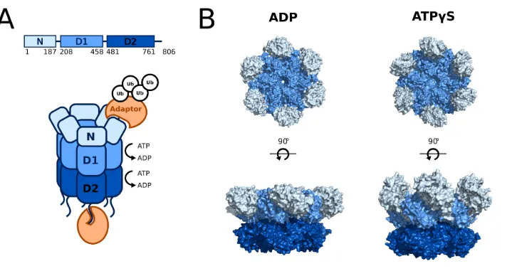
Structurally, p97 is a homohexamer, with its subunits forming a ring around a central pore. p97 comprises three domains: an N-terminal domain (N), two ATPase
domains (D1 and D2) stacked head-to-tail, and an unstructured C-terminal tail
(Fig-ure 1.3) (22–26). Hydrolysis of ATP by p97 generates its mechanical force from chemical energy. Each ATPase domain includes two key sequences: the Walker
A motif (GX4GK(T/S), where X is any amino acid), which is responsible for
Figure 1.2: p97/VCP function. Adapted from (21)
any hydrophobic amino acid), which is responsible for nucleotide hydrolysis (27). Both D1 and D2 are competent to hydrolyze ATP, but the relative contributions
of the two domains to processing substrates is controversial (28). D1 has a higher affinity for nucleotide while D2 has a higher hydrolysis rate, leading many to suggest
that D1 plays a structural role while D2 is the main generator of force (28–35). Nu-merous structural (22–26,31,36–43) and biochemical (28,33,44–47) studies have demonstrated the extensive communication among domains during the p97 ATPase
cycle. Our best understanding comes from a set of high resolution cryoelectron
microscopy (cryo-EM) structures of p97 in various nucleotide states (22) (Figure 1.3B). When both domains are in the ADP state, the N-domain is oriented in a
position planar to the D1 ring. Upon binding of ATPγS to D1, the N-domains flip
upward into a position coaxial with respect to the D1-D2 barrel. D2 shows rotation
with respect to the D1 upon binding of ATPγS to D2, which is in agreement with
biochemical studies that suggest a flexible di-glycine linker between D1 and D2 is critical for domain communication (45,46).
p97 does not function alone but instead relies upon its interactions with
Figure 1.3: p97 structure and binding partners. (A) Cartoon representation of a p97 homohexamer showing three folded domains (N, D1, D2) as well as an unstructured C-terminal tail. Adaptor proteins (orange), many of which recruit ubiquitin, bind to the N-domains and C-terminal tails. (B) Cryo-EM structures of p97 in two nucleotide states. Upon exchange of ADP for ATPγS in both D1 and D2, the N-domains move from a coplanar to a coaxial position with respect to the D1-D2 barrel, and D2 rotates downward. PDB #5FTK and #5FTN (22)
domains (49–51) or other specialized domains (33, 52), which recruit UPS sub-strates to p97. Other adaptors serve to process subsub-strates through post-translational
modifications (PTMs), such as addition of ubiquitin through E3 ligases, removal
of ubiquitin by deubiquitinating enzymes (DUBs), or removal of oligosaccharides
(53–57). Previous research has implicated some adaptors in specific p97 pathways (58–62); for example, p47 is critical for Golgi reassembly after mitosis (63, 64). However, the functions of many adaptors are still unclear (49,51). Further compli-cating our understanding of the p97-adaptor network is the fact that some adaptors
are able to co-bind p97 while other adaptors are mutually-exclusive binders (49,65, 66).
Most adaptors bind to the N-domains or C-terminal tails of the p97 hexamer
with various stoichiometries (27, 51, 66,67) (Figure 1.3A). Two p97 adaptors are notable exceptions and bind in unqiue ways. First, ubiquitin regulatory X
domain-containing protein 9 (UBXD9) disassembles p97 hexamers to bind p97 monomers
instead (68, 69). Second, ubiquitin regulatory X domain-containing protein 1 (UBXD1) binds both to the N-domain and the D1-D2 barrel of p97 hexamers (70, 71). A few common p97 binding motifs are employed by adaptors that bind to the N-domains. The ubiquitin regulatory X (UBX) domain, found in thirteen adaptors,
interacts with the hydrophobic groove of an N-domain through an R...FPR motif
N-domains using the consensus sequences RX5A2X2R and A2X2R (71,74), while the
SHP motif binds to a separate site on the N-domains using the sequence FXGXGX2H
(43,75).
Binding of adaptors alters the struture and activity of p97 hexamers.
Low-resolution cryo-EM structures of p97 in complex with various adaptors suggest that
the N-domains must be in an axial position with respect to D1-D2 for adaptors to
bind to the N-domains, biasing the conformations that p97 samples (36, 66, 67, 76–78). Adaptor binding to N-domains also propagates conformational changes to D2 (36, 76). Some adaptors, such as p47, modulate ATPase activity while others, such as UN, do not (47,79,80).
1.3 p97 and neurodegenerative disease
Mutations in p97 have been linked to neurodegenerative diseases such as
amyotrophic lateral sclerosis (ALS) and multisystem proteinopathy (MSP), also
called inclusion body myopathy with Paget’s disease of the bone and
frontotempo-ral dementia (IBMPFD) (81–83). MSP is a fatal disease of the muscle, bone and brain that develops in middle age (reviewed in (84)). Myopathic symptoms include atrophy and weakness of the skeletal, cardiac, and respiratory muscles due to
intra-cellular inclusions containing ubiquitin and other proteins such as Tar DNA binding
protein-43 (TDP-43). Paget’s disease of the bone (PDB) causes bone fragility and
deformities due to increased osteoclast activity. Neuronal loss in the frontal and temporal lobes of the brain due to inclusions containing ubiquitin, TDP-43, and tau
cause loss of reasoning, judgement, and social awareness in frontotemporal
demen-tia (FTD). Not all MSP patients display the same symptoms. Approximately 90%
of patients develop myopathy, approximately 40% of patients develop PDB, and
approximately 30% of patients develop FTD; only around 10% of patients develop
all three disorders (85). While over 50 unique missense mutations in p97 have been discovered to cause MSP (86), there are no strong correlations between genotype and phenotype due to the background genetic variability among individuals (85).
1.4 Remaining questions and research aims
While biochemical and structural studies have shed light on some of p97’s
p97-adaptor dynamics
It remains unclear how the vast p97 network is regulated. Little is known about
the dynamics of p97-adaptor association and whether other factors, such as substrates
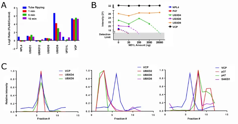
or PTMs, affect these processes (48). Previous work in the Deshaies lab began to answer these questions by examining p97 complexes from cells through a variety of
mass spectrometry (MS) experiments (Figure 1.4)(87). First, the stabilities of p97-adaptor complexes were probed by immunoprecipitation (IP)-MS. Cells expressing
p97 tagged at the genomic locus with the FLAG epitope were grown in isotopically
“light” media while cells with untagged p97 were grown in “heavy” media. Cell
lysates were incubated with anti-FLAG antibodies, and the captured proteins were
assessed by MS. If the complexes are stable, only light adaptors would be captured
by FLAG-p97, representing endogenous complexes. However, if the complexes are
dynamic, the bound adaptors would be a 1:1 mixture of heavy and light due to
exchange. As seen in Figure 1.4A, most adaptors display rapid exchange, with the
notable exception of UBXD9.
To further validate these dynamics, a competition IP-MS experiment was
performed. Cell lyates were spiked with increasing amounts of a truncated p97
(N-D1-L) which is competent to bind adaptors but is not recognized by the antibody
used for IP. If complexes are stable, the pull-down efficiency of adaptors should be
impervious to added p97-ND1L, but if complexes are unstable, the adaptors should
be chased by added p97-ND1L. With the exception of UBXD9 and UBXD4, all
adaptors were readily depleted in the IP by the addition of exogenous p97-ND1L,
further underscoring the dynamic nature of p97-adaptor interactions (Figure 1.4B). Additionally, size exclusion chromatography (SEC)-MS was used to assess
p97-adaptor complexes in cell lysate. In agreement with the other MS experiments,
only UBXD4 and UBXD9 were observed to co-elute with p97, suggesting no other
adaptors form stable complexes with p97 (Figure 1.4C).
A multitude of factors could influence the rapid dynamics of the p97-adaptor
network in the cell (48). The proteins themselves could have intrinsically dynamic interactions, and these interactions could be further modulated by PTMs, oligomeric
states, and conformational changes. Outside factors such as competition for binding sites or additional binding partners could also affect the network. In the previous
experiments in cell lysate, it is impossible to tease apart the effects of these different
influences; therefore, the first goal of this work is to probe the dynamics of the
Figure 1.4: p97-adaptor network is dynamic in cell lysate. (A) Exchange rate of p97 adaptors during IP-MS. Cells expressing FLAG-p97 and WT p97 were grown in light and heavy media, respectively. Lysates were incubated with anti-FLAG antibodies for various amounts of time, and the ratio of heavy to light adaptors recovered was determined by MS. All adaptors show rapid equilibration (as evidenced by a light:heavy ratio close to 1), except UBXD9. (B) A p97 fragment, ND1L, which can bind adaptors but not the antibody used for IP, was added in increasing amounts to an IP of cell lysates, and the recovery of adaptor proteins was plotted. While the p97-UBXD9 complex is insensitive to added p97-ND1L, all other adaptors have reduced recovery as rapid exchange allows p97-ND1L to compete with full length p97 for adaptor binding. (C) Fractionation profiles in a SEC-MS experiment for a variety of p97 adaptors. Only UBXD9 and UBXD4 co-elute with p97 after fractionation on a Superose 6 sizing column. All panels are modified from data previously published (87).
Substrate processing by p97
Due to its functions in cellular pathways and its homology to other AAA machines such as ClpA (88), the proteasome 19S regulatory particle (89, 90), the VCP-like ATPase from Thermoplasma acidophilum (VAT) (91, 92), and N-ethylmaleimide-sensitive factor (NSF) (93), most have assumed that p97 remodels its protein substrates through unfolding. However, this function has never been
explicitly demonstrated. Many AAA ATPases unfold their substrates by feeding
the polypeptide through their central pores (e.g. (94)). However, p97 does not contain the requisite hydrophobic pore residues in its D1 domain for this threading
mechanism, though other pore residues in D1 have been implicated in substrate
processing (34,95). Additionally, structural studies of p97 have revealed a narrow central pore that may not be able to accommodate a threaded polypeptide (22, 25). Many other mechanisms for unfolding have been proposed, such as unfolding exclusively in the D2 pore through an arginine "denaturation collar" or exclusively
through the N-domain movements without direct participation of the pore, similar
to that of NSF (93,95).
research into these questions. Native p97 substrates are complex, unstable,
hetero-geneous, and highly modified by ubiquitin, making their purification for biochemical studies challenging. Therefore, the presumed function of p97—protein unfolding—
has never been directly tested, and previous studies on p97 function have instead
relied on indirect assays, such as measurements of basal ATP hydrolysis rates and
reconstitutions with cellular components (e.g.(95)).
In addition to whether or not p97 is a bona fide unfoldase, many other
fun-damental questions about p97 mechanism remain unanswered. First, the minimal
components necessary to reconstitute substrate processing remains obscure. It is assumed that p97 requires adaptors to recruit substrates (48), but p97’s multitude of adaptors complicates our understanding. Though it is unclear what role each
adap-tor may play in substrate unfolding, a good candidate for p97’s general unfolding
partner is the heterodimer Ufd1/UFD1L and Npl4/NPLOC4 (UN), as this adaptor
has been implicated in numerous proteasome-dependent degradative pathways such
as ERAD (10,65,96,97). In particular, UN is a critical component of, and the only substrate adaptor linked to, the ubiquitin fusion degradation (UFD) pathway, a
p97-dependent degradation process (34, 98). ATP is also likely required for substrate processing, as ATPase activities in D1 and D2 are critical for substrate processing
in cells (34).
Second, it is unclear what the requirements are for the substrate itself in
order for it to be processed by p97. Unlike the proteasome, p97 does not require
a disordered region on the substrate to initiate processing (34, 99). Due to p97’s role in PQC, p97-dependent processes have been connected to numerous ubiquitin
linkage types, including K6, K11, K29 and K48 (98,100–102). In particular, Ufd1 binds K48-linked chains specifically, while Npl4 associates with poly-ubiquitin
chains in a linkage type-independent manner (33, 52, 77). Therefore, it is likely substrates require ubiquitylation, but the linkage types and chain topologies that
support proessing by p97 are undefined.
In order to address these critical gaps in our understanding of the basic
biochemical activity that underlies p97’s cellular functions, the second aim of this
work is to develop an in vitro reconstitution of p97 substrate processing using
Perturbations caused by p97 disease mutations
The mechanism by which mutations in p97 cause neurodegenerative
dis-eases remains highly controversial. Defects in numerous cellular pathways, such
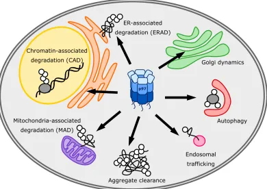
as autophagy, endosomal trafficking, lysosomal function, mitochondrial
homeosta-sis, ERAD, mTOR regulation, energy balance, aggregate clearance, and NF-κB
signaling (58,84,103–114), have all been reported in MSP mutants, yet the under-lying cellular perturbations that cause disease pathology remains a mystery. On a
more basic level, it even remains unclear whether the the molecular-level defect in
these dominantly-inherited MSP mutations causes a gain or loss of p97 function.
This question is particularly relevant for treatment, as many p97 inhibitors, such as
NMS-873 and CB-5083, have already been developed for cancer treatment (14,15, 115).
The majority of MSP mutations lie on the interface between the N and D1
domains (83), and in vitro studies have provided some clues as to the effects of these mutations on p97 structure and enzymology. Under basal conditions, MSP mutants
show an increase in ATPase activity in D2 (28, 47, 116–119). The functional significance of this activity is hotly debated. Some have speculated that this higher
activity is not a productive gain of function but instead represents an uncoupling
of ATP hydrolysis from mechanochemical transduction in the ring due to severed
communication between the regulatory N domain and the D1 ATPase domain (83, 120). A dominant-negative mechanism in which non-functional mutant protomers poison the activity of WT-MSP mixed hexamers could then explain the inheritance
pattern of MSP.
Structurally, mutants show an abnormal N-D1 conformation, with the N
do-mains occupying a more axial, ATP-like conformation with respect to D1 regardless
of nucleotide state due to a destabilization of ADP binding (118,119,121–124). IPs from cells suggest altered associations with adaptors in MSP mutants, with some
adaptors being more abundant and others less (116, 125). In particular, the loss of UBXD1 binding, which is linked to endosomal sorting, has been proposed as a
major cause of disease (58,126).
With no biochemical assay available to directly test the effects of these
muta-tions on p97’s basic function—protein unfolding—it has been impossible to discern
whether MSP mutants represent a true gain or loss of function. Therefore, the third
aim of this work is to use the tools described above to explore how MSP mutations
C h a p t e r 2
PROBING P97-ADAPTOR INTERACTIONS BY FRET
The p97-p47 results and p97-p47 methods were previously published in: Xue, L.,
Blythe, E.E., Freiberger, E.C., Mamrosh, J.L., Hebert, A.S., Reitsma, J.M., Hess,
S., Coon, J.J., and Deshaies, R.J. (2016) Valosin-containing protein (VCP)-adaptor
interactions are exceptionally dynamic and subject to different modulation by a
VCP inhibitor. Mol Cell Proteomics 15, 2970-2986. © the American Society for Biochemistry and Molecular Biology.
2.1 Introduction
The first goal of this work was to investigate the properties of p97-adaptor
interactions in order to gain mechanistic insight into the how the incredibly dynamic
p97-adaptor network is regulated. We chose two p97-adaptor pairs on which to
focus. First, we developed a fluorescence resonance energy transfer (FRET) assay to
quantify the interaction between p97 and p47. p47, also called NSFL1C, is involved
in Golgi reassmebly after mitosis by binding to syntaxin 5 (63, 64, 127). p47 is a homotrimer comprising three domains: a UBA domain that preferentially binds
mono-ubiquitin, a SEP domain that mediates trimerization, and a UXB domain (65, 76,128) (Figure 2.1A). One p47 trimer binds one p97 hexamer through interactions between both its UBX domain and a SHP motif located in SEP-UBX linker on
separate sites on the p97 N domain (63, 72, 76, 129). p47 was an appealing target not only because its role in membrane dynamics was well established but also
because a crystal structure of its UBX domain bound to p97-ND1 was published,
facilitating the design of FRET probes (72).
Second, we focused on developing a FRET binding assay for p97-UN. Due to its participation in a wide array of p97 processes as discussed in Chaper 1, UN
was an important target for this assay. Npl4 comprises a ubiquitin-like domain
(UBD), which binds to the p97 N domain, and a zinc finger (ZF) domain that binds
Using these designed FRET assays of p97-p47 and p97-UN, we demonstrate
that the rapid dynamics of p97-adaptor complexes observed in our previous cell-based MS experiments are due to the intrinsic properties of these proteins, suggesting
there is not a requirement for an exogenous factor to facilitate p97-adaptor complex
assembly or disassembly. However, these rapid dynamics suggest that some
stabi-lizing factors may exist to promote the formation of p97-adaptor complexes during
cellular processes. In agreement with structural work, the affinities of these
in-teractions are highly dependent on the nucleotide state of p97, underscoring the
importance of N-domain movement for adaptor binding. Additionally, we observed
enhanced binding of both p47 and UN in MSP mutants, suggesting adaptor balance
defects in mutant cells may underlie some MSP pathology.
2.2 Results
p97-p47
Using the crystal structure of p47 bound to the N-domain of p97 as a guide
(72) (Figure 2.1B), we first mutagenized the C-terminal amino acid (Thr370) of p47 to cysteine, and then reacted the purified recombinant protein with
maleimide-TAMRA to generate p47TAMRA. For p97, we used the ybbR tagging method (132) to attach a Cy5 tag at its N terminus (Cy5p97). Upon mixing p47TAMRAandCy5p97 and exciting with 540 nm light, we observed a significant reduction in TAMRA
fluorescence coupled to an increase in Cy5 emission (Figure 2.2). This FRET
signal was because of specific interaction of p47TAMRAandCy5p97, because it was competed by addition of excess unlabeled p97 (Figure 2.2).
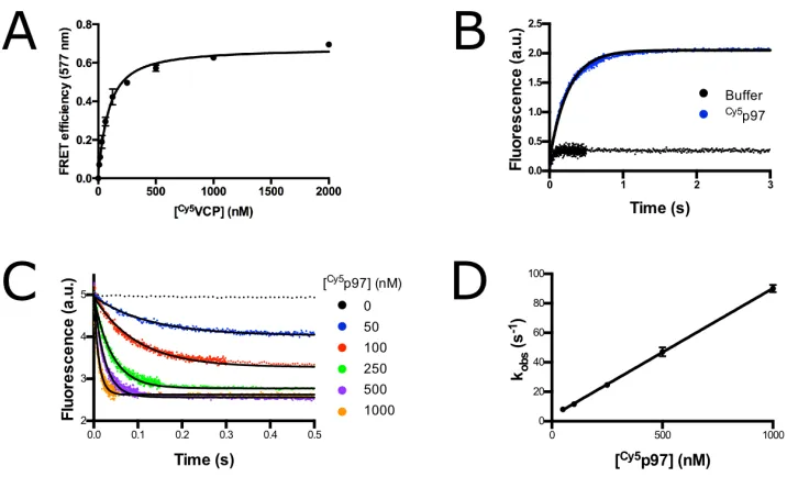
By titrating Cy5p97, we estimated a KD of 65 nM for interaction of the two
proteins in the absence of nucleotide (Figure 2.3A and Table 2.1). This affinity
is 10-fold tighter than what was reported from isothermal titration calorimetry (ITC) studies (50, 76), but is close to the affinity measured in a pair of surface plasmon resonance (SPR) studies (20–31 nM) (133, 134). However, there are significant problems with measuring p47-p97 interactions by SPR because of the
oligomeric nature of both proteins. In addition, none of the studies cited above
addressed the crucial issue of the dynamics of p47-p97 interaction. To investigate
binding dynamics, we measured the konfor complex formation and koff for complex
dissociation in the absence of nucleotide (“apo”) or in the presence of ADP or
ATPγS with and without VCP inhibitors. Examples of koff and kon measurements
Figure 2.1: p97-p47 FRET construct design. (A) Cartoon diagram of p97 (blue) and p47 (orange) showing domain architecture. (B) Crystal structure of p47 UBX domain bound to p97-ND1, with the positions of FRET dyes labeled. A mutant p47 (T370C) was labeled with TAMRA donor dye via maleimide chemistry. p97 was labeled with an N-terminal ybbR tag to which a Cy5 acceptor dye was enzymatically conjugated. PDB#1S3S
2.3D. As shown in Table 2.1, kon values were essentially invariant, ranging from
8–11x107M-1sec-1regardless of nucleotide state. Meanwhile, koff showed slightly
more variation, ranging from 2.5 s-1 in the presence of ATP and NMS-873 to 9.5 s-1 in the presence of ADP. Consistent with the lack of an effect of NMS-873 on co-IP of p47 with p97 from lysates (87), addition of NMS-873 in the presence of ATP had less than a twofold effect on koff (Table 2.1). These results confirm that
purified p47TAMRA exhibited extremely dynamic association with purified Cy5p97 in accordance with the behavior of these proteins in HEK293 cell extracts, and that
their association was relatively insensitive to modulation by NMS-873.
Figure 2.2: p47TAMRA and Cy5p97 show FRET. Fluorescence emission spectra of 16.7 nM p47TAMRAtrimer, 83.3 nMCy5p97 hex-amer, and a mixture excited at 540 nm shows a 20% loss of TAMRA donor fluorescence in the presence ofCy5p97. Loss of fluorescence is prevented by preincubation of p47TAMRA with 833 nM unlabeled p97 hexamer.
p97-UN
In order to further investigate the dynamics of p97-adaptor complexes, we
developed a FRET assay to probe the p97-UN complex. As shown in Figure 2.4,
Figure 2.3: Example p97-p47 FRET data. (A) Equilibrium titration of 50 nM p47TAMRAtrimer withCy5p97 hexamer in the absence of nucleotide. Fit to a quadratic binding equation yields a KDof 65±7 nM. Error bars
represent S.D., with n=3. (B) Change in donor fluorescence of 50 nM p47TAMRAtrimer preincubated with
300 nMCy5p97 hexamer upon addition of 3µM unlabeled p97 hexamer in the absence of nucleotide. Curve was fit to a single exponential to give koffof 4.04 s-1. (C) Change in donor fluorescence of 50 nM p47TAMRA
trimer upon addition ofCy5p97 hexamer at various concentrations in the absence of nucleotide. Curves were fit to a single exponential. (D) Exponential fits measured in panel (C) plotted against the concentration ofCy5p97 hexamer. Linear slope gives konof 8.68x10⁷±0.09x10⁷M-1s-1. Error bars represent S.D., with n=5.
p97 p97-R155H
Buffer
KD (nM)
(eq)
kon(x107 M-1s-1) koff(s
-1) KD(nM) (koff/kon)
KD (nM)
(eq)
kon(x107 M-1s-1) koff(s
-1) KD(nM) (koff/kon)
Apo 65±7 8.68± 0.09
4.04±
0.09 47 49±6 9.2±0.1
2.496±
0.005 27
ATPγS n.d. 10.5± 0.1
3.30±
0.02 31 n.d.
3.89± 0.08
2.04±
0.03 52
ADP n.d. 8.30±
0.07
9.50±
0.08 114 n.d. 7.9±0.1
3.44±
0.06 44
Apo +
CB-5083 n.d. 8.0±0.1
4.80±
0.03 60 n.d.
4.07± 0.06 2.06± 0.04 51 ATP + NMS-873 n.d. 11.01± 0.01 2.50±
0.03 23 n.d.
7.03± 0.05
1.98±
0.01 28
Table 2.1: Kinetic and equilibrium binding constants for p47TAMRA-Cy5p97.
the N-terminus of Npl4 with a donor TRITC fluorophore via sortase (135) was designed to complement theCy5p97 used in the previously described p47-p97 FRET assay. Incubation of UTRITCN withCy5p97 lead to a decrease in TRITC fluorescence, indicative of FRET (Figure 2.5). Importantly, the loss of fluorescence can be rescued
by the addition of unlabeled UN, providing evidence that this FRET assay is specific
[image:25.612.124.493.393.555.2]Figure 2.4: p97-UN FRET construct design. (A) Cartoon diagram of p97 (blue) and the UN heterodimer (orange and yellow, respectively) showing domain architecture. Adapted from (75). (B) Structure of Npl4 UBD domain bound to p97 N domain, with the positions of FRET dyes labeled. The N-terminus of Npl4 was labeled with a TRITC dye via sortase labeling, while the N-terminus of p97 was labeled with Cy5 via a ybbR tag as described above for p97-p47 FRET. Composite of PDB #2PJH and #5FTN
With this assay in hand, we were able to measure the equilibrium and kinetic
constants for p97-UN in various nucleotide states (Figure 2.6, Table 2.2). The
equilibrium dissociation constant is 2-fold less in ATP versus ADP and has a further
reduction in ATPγS. These KD measurements are close to that measured by SPR
(100-400µM) (133) but 2-10-fold tighter than that measured by ITC in the absence of nucleotide (1.7 µM) (50). SPR studies also observed an increase in UN affinity when p97 was in an ATP state (133). Due to the low affinity in ADP, we were able to measure on- and off-rates for the ATP and ATPγS states only (Table 2.2).
The two conditions have similar rates, with ATPγS showing a slightly faster on-rate
and slightly slower off-rate. Compared to p47, UN has a nearly 80-fold slower
on-rate and 10-fold slower off-rate, suggesting that while p97-adaptor dynamics are
generally rapid, variation among adaptors is present.
Figure 2.5: UTRITCN and Cy5p97 show FRET. Fluorescence emission spectra of 50 nM UTRITCN heterodimer shows a 20% loss of fluorescence in the presence of 250 nM
Cy5p97 hexamer. Loss of fluorescence is
[image:26.612.128.492.560.663.2]Figure 2.6: Example p97-UN FRET data. (A) Equilibrium KD titration of 50 nM UTRITCN withCy5p97 hexamer in the presence of ATPγS. Data were fit to a quadratic binding equation. Error bars represent S.D., with n=3. (B) Change in donor fluorescence of 50 nM UTRITCN preincubated with 250 nMCy5p97 upon
addition of 2.5µM unlabeled UN in the in the presence of ATPγS. Curve was fit to a single exponential to give koff. (C) Change in donor fluorescence of 50 nM UTRITCN upon addition of 50 nMCy5p97-A232E in the
presence of ATPγS. Curve was fit to a single exponential to give kobs. (D) Exponential fit like those measured
in panel (C) plotted against the concentration ofCy5p97-A232E. Linear slope gives kon. Error bars represent
S.D., with n=5.
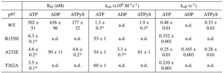
KD(nM) kon(x106M-1s-1) koff(s-1)
p97 ATP ADP ATPγS ATP ADP ATPγS ATP ADP ATPγS
WT 302±
51 636± 96 177± 32 1.3± 0.3* n.d. 1.9± 0.3* 0.48± 0.01 n.d. 0.33± 0.01
R155H 6.3±
0.1* n.d. n.d. 53±1 n.d. n.d.
0.332±
0.001 n.d. n.d.
A232E 4.6±
0.2* 50±11 4.6±
0.2* 54±1
3.3±
0.7* 61±1
0.25± 0.01 0.165± 0.003 0.28± 0.01
T262A 3.5±
0.1* n.d. n.d. 60±1 n.d. n.d.
0.210±
0.001 n.d. n.d.
Table 2.2: Kinetic and equilibrium binding constants for UTRITCN-Cy5p97. Error represents S.D., with n≥3. *Calculated from other values.
Effects of disease mutants on adaptor binding
Because pull-downs from cells showed differential adaptor binding in MSP mutants (125), we were interested in investigating how binding of p47 and UN changes in MSP mutants. For p47, we measured binding constants for the MSP
mutant R155H in the presence of various nucleotides and inhibitors (Table 2.1). The
largest difference we observed among all parameters was the off-rate in ADP. While
WT p97 has a nearly 3-fold faster off-rate in ADP versus ATPγS, R155H retains a
similar off-rate regardless of nucleotide state. This observation is in agreement with
[image:27.612.117.496.374.498.2]well as two other mutants, A232E and T262A (Table 2.2). We characterized UN
binding of A232E most fully and, as with WT p97, we observed a slower on-rate and therefore a decrease in affinity in the ADP state compared to ATP or ATPγS.
Strikingly, A232E has a 10-60-fold higher UN affinity compared to WT regardless
of nucleotide state, and this increase is the result of a significantly faster on-rate.
R155H and T262A have very similar affinities to A232E in the presence of ATP,
suggesting that this phenomenon is a common feature of MSP mutations.
Patients with MSP have one mutated allele and one WT allele, so in cells we
expect mixed WT-MSP hexamers to exist. In order to probe the adaptor affinity in mixed hexamers, we devised a purification scheme to prepare mixed hexamers from
E. coli. WT and His-tagged A232E were co-expressed, and hexamers with different WT:A232E ratios were broadly pooled into three fractions using a stepwise elution
from a nickel affinity column (Ni-NTA) (Figure 2.7A). The UN affinity of ’Mixed
2’ was indirectly measured by a FRET competition assay (Figure 2.7B). Briefly,
UTRITCN and Cy5A232E were incubated with unlabeled p97 hexamers, and the inhibition of FRET was measured. The UN binding affinity Kiwas then calculated
from a curve fit of IC50using the known concentrations and KDfor A232E-UN. As
seen in Figure 2.7B, the UN affinities for pure WT and A232E hexamers agree fairly
well with those measured directly (Table 2.2), and the affinity for the mixed hexamers falls in between that of the pure hexamers. No protomer exchange occurred during
the time scale of the competition FRET experiment (Figure 2.7C). Therefore, the
UN affinity of p97 titrates with the number of mutant subunits.
2.3 Discussion
The p97-p47 and p97-UN complexes are sensitive to the nucleotide state of
p97, and that these adaptors have high affinity for p97 in an ATP-bound state is in
agreement with published structural information. The N-domains of p97 adopt an
“up” conformation with respect to the ATPase rings in ATP whereas they adopt
a more “down” conformation in ADP (22). Cryo-EM structures of p97-p47 and p97-UN show the adaptors perched on top of p97, suggesting that the N-domains need to be in an “up” or ATP-bound conformation to bind adaptor (67,76,78).
Both complexes have altered affinities in MSP mutants, which have altered
N-domain conformations. p47 binding becomes insensitive to nucleotide state
with mutant p97, and the same phenomenon has been reported for two other p97
Figure 2.7: WT-A232E mixed hexamer purification and FRET (A) Mixed hexamer purification scheme. WT and A232E-His were co-expressed inE. coli, and three fractions of mixed hexamers with increasing amounts of A232E protomers were crudely purified by stepwise elution from a Ni-NTA column. (B) Competition FRET assay of 25 nM UTRITCN-Cy5A232E by unlabeled p97 in the presence of ATP. Kiwas calculated from a curve
fit of IC50using the known KD for A232E. Error bars represent S.D., with n=3. (C) Protomer exchange is slow.Cy5p97-His was incubated with anti-FLAG resin in the presence or absence of p97-FLAG for ten minutes. Coomassie stain and fluorescence scan of an SDS-PAGE gel show no evidence ofCy5p97-His binding to the resin despite robust binding of p97-FLAG.
(124,137). UN behaves differently, showing increased affinity for mutant p97 under all nucleotide conditions. Structural studies have consistently shown than MSP mutants favor N-domains in an up conformation even in ADP (118,121,123,138), which would explain why adaptors are able to maintain high affinity interactions
in ADP with mutant p97. Nuclear magnetic resonance (NMR) studies measuring
the dynamics of N domain movements suggest that WT and mutant p97 have
similar up/down equilibrium in ATPγS (121). We still observe a large difference in UN affinity under those conditions, which would suggest instead that differences
in up/down dynamics remain regardless of nucleotide state. Alternatively, MSP
mutations could be altering p97 structure beyond its normal up/down movement that
further promotes UN binding. NMR studies with mixed WT and mutant hexamers demonstrated cooperativity in N domain orientation among adjacent protomers,
which implies that adaptor affinity in mixed hexamers is not linearly related to
the number of mutant protomers (123). Our biochemical data is consistent with differential adaptor binding to WT versus mutant p97 in IPs from cell lysate, though
the rapid exchange of these factors suggest these interactions were formed in lysate
adaptors such as UBXD1 conversely lose binding to mutant p97, preferring instead
to bind to the ADP-like down conformation (58, 121, 123). This question will be addressed more by functional assays in Chapter 4.
The rapid binding kinetics we observe with the p97-p47 and p97-UN
com-plexes are in agreement with the rapid adaptor exchange we observed in cell lystate
by MS, suggesting that some network regulation may come from rapid p97
sam-pling of adaptors. However, the fast off-rates of adaptors raises the question of how
p97-adaptor complexes can properly function in the cell. The ATPase rate of p97 is
between 0.75-5.2 ATP per second per hexamer (28,32,95,118), yet the off-rates of adaptors we measured ranged from 0.2-2 per second. In the slowest case, p97-UN
would hydrolyze 3 to 25 ATP per hexamer during the lifetime of the complex, and
in the extreme case of p47, at most 1 to 2 ATP would be hydrolyzed since p47
also retards p97 ATPase activity (47,79). Given these rates, it is unclear how p97 can engage substrate and sustain unfoldase activity without other mechanisms that
modulate the lifetimes of these complexes. p97 and its adaptors are decorated with
PTMs (48, 139); for example, methylation of p97 at K315 has been proposed to affect its ATPase activity (140). Covalent modifications or interactions with other proteins, such as substrates, may serve to stabilize p97-adaptor complexes.
Con-sistent with this hypothesis is the observation that p47 forms an extremely stable complex with p97 when purified from rat liver yet did not coelute with p97 in our
SEC-MS experiments (63).
2.4 Methods
p97-p47 FRET reagents
Full-length p97 was amplified by PCR from human p97 pET15T (28) and ligated into pET24b using NdeI/SalI to produce a noncleavable C-terminal His-tagged construct. For FRET studies, p97 coding sequences were amplified by
PCR and ligated into a modified pET28a vector to produce a construct with a
noncleavable C-terminal His-tag and an N-terminal ybbR tag with a short linker
(MDSLEFIASKLAGGGS). The construct for full-length p47 with a noncleavable
N-terminal His-tag (80) was obtained through Addgene (#21268), and site-directed mutagenesis was used to make a p47-Thr370Cys mutation. Proteins were expressed
and purified as described previously (28), with the exception that p47 was expressed in TOP10 cells for 3 h at 37 °C. For FRET, p47-Thr370Cys was incubated with
described (132). Thirty micromolar ybbR-VCP was incubated for at least 3 h at room temperature with 60µM Cy5-CoA conjugate and 12µM Sfp in 50 mM HEPES pH 7.4, 10 mM MgCl2prior to gel filtration. All proteins were purified on a Superose
6 gel filtration column.
UN FRET reagents
An N-terminal sortase recognition motif was added to the N-terminus of Npl4
(50) to form MGGG-Npl4. To prevent labeling of His-Ufd1 (125), site directed mu-tagenesis was used to remove residue G2. This modified UN was expressed and
purified as previously described (141) with one modification. Prior to gel filtration, 30 µM UN was labeled using 500 µM peptide (TRITCWSHPQFEKLPETGG, Gen-Script) and 5 µM sortase (135) in labeling buffer (25 mM Hepes pH 7.4, 150 mM NaCl, 10 mM CaCl2, 1 mM DTT) at 25 °C for 2 hours. The reaction was diluted in
Strep-tag buffer (100 mM Tris pH 8.0, 150 mM NaCl, 1 mM EDTA) and incubated
with 2 mL of Strep-Tactin resin (IBA Lifesciences) for 10 minutes at 4 °C with
agitation. Resin was washed with 5 column volumes of Strep-tag buffer and eluted
with Strep-tag buffer plus 50 mM biotin.
Mixed hexamer preparation
A plasmid for co-expression of p97 and p97-A232E-His was constructed
via Gibson cloning to insert sequences from p97-His and p97-A232E-His into a
pCOLA-Duet backbone. Protein was expressed in Rosetta DE3 overnight at 16 °C
overnight with 0.4 mM IPTG. Cell pellet was resuspended in nickel binding buffer
(50 mM Tris pH 7.4, 500 mM KCl, 5 mM MgCl2, 20 mM imidazole, 5% glycerol,
2 mM β-mercaptoethanol) with protease inhibitors and sonicated. Clarified lysate was bound to a HisTrapHP (GE Healthcare) and eluted in a stepwise gradient of 12%
(wash), 20% (’Mixed 1’), 35% (’Mixed 2’), and 55% (’Mixed 3’) nickel binding
buffer with 300 mM imidazole. Proteins were purified on a Superose 6 column (GE
Healthcare) in storage buffer (20 mM Hepes pH 7.4, 250 mM KCl, 1 mM MgCl2,
5% glycerol, 0.5 mM TCEP), concentrated, and flash frozen.
p97-p47 FRET measurements
All FRET measurements were carried out in 20 mM Hepes, pH 7.4, 100 mM
KCl, 3 mM MgCl2, 1 mM TCEP, and 1 mg/ml ovalbumin (Sigma). Nucleotides
were optionally present at 2 mM, and inhibitors were optionally present at 15 µM.
excitation at 540 nm and emission scan 555–750 nm. Stopped-flow experiments
were carried out on a Kintek SF-300X instrument with excitation at 540 nm and a 580/20 emission filter. Data were analyzed using Prism 6 (GraphPad).
p97-UN FRET measurements
Assays were carried out at room temperature in assay buffer (20 mM Hepes
pH 7.4, 150 mM KCl, 20 mM MgCl2, 1 mM TCEP, 1 mg/mL bovine serum
al-bumin (BSA)) supplemented with 2mM ADP, 2 mM ATPγS, or ATP regeneration
system (5 mM ATP, 30 mM creatine phosphate, 50µg/mL creatine phosphokinase).
Equilibrium binding assays were measured on a fluorometer (Photon Technology
In-ternational, Inc) with 50 nM UTRIRCN. Data were fit to a parabolic binding equation. Kinetic constants were measured using an AutoSF-120 stopped flow fluorimeter
(KinTek) with an excitation wavelength of 540 nm and an emission filter of 576 ± 31 nm. For measurements of kon, the final concentrations were 50 nM UTRITCN
and 50-400 nM Cy5p97. Data were fit to single exponential decay equation to fit kobs, and linear regression of kobs versus [Cy5p97] were used to calculate kon. For
measurements of koff, 100 nM UTRITCN and 500 nMCy5p97 were incubated before
mixing with 5 µM UN (final dilution 2X). Data were fit to a single exponential
association equation to extract koff. For competition FRET experiments, final
con-centrations were 25 nM UTRITCN, 25 nM Cy5p97-A232E, and various values of unlabeled p97 protein. Fluorescence intensity was measured on a Synergy Neo2
plate reader (BioTek) using a 540 nm excitation filter and 590 nm emission filter.
FRET efficiencies were calculated from a control UTRITCN sample, and percent inhibition was calculated relative to a UTRITCN-Cy5p97-A232E sample. Inhibition curves were fit to an IC50 equation, and Kiwas calculated using the known KD of
UTRITCN-Cy5p97-A232E and protein concentrations.
Protomer exchange assay
A plasmid encoding p97-FLAG for bacterial expression was made through the
addition of a FLAG tag to a plasmid encoding untagged p97 in a pET24b backbone
(RDB#3376). Protein was expressed in Rosetta DE3 as previously described (141). Harvested cells were resuspended in lysis buffer (50 mM Tris pH 7.4, 150 mM KCl, 5
mM MgCl2, 5% glycerol) with protease inhibitors (Roche) and sonicated. Clarified
lysate was incubated with 1 mL anti-FLAG resin (Sigma) at 4 °C for 1 hour. Resin was washed with 3 column volumes of wash buffer (50 mM Tris pH 7.4, 400 mM
followed by one wash with elution buffer without FLAG peptide (25 mM Hepes pH
7.4, 150 mM KCl, 2 mM MgCl2). Protein was eluted with 2 mL of elution buffer
containing 0.2 mg/mL 3XFLAG peptide for ten minutes with rotation. Elution was
repeated, and resin was drained with a final 2 mL of elution buffer. After elution,
concentrated protein was purified by gel filtration (Superose 6) in storage buffer (20
mM Hepes pH 7.4, 250 mM KCl, 1 mM MgCl2, 5% glycerol, 0.5 mM TCEP) before
flash freezing.
A solution of 250 nM ofCy5p97-His with or without 250 nM p97-FLAG was incubated for ten minutes at 25 °C with 25 µL anti-FLAG resin (Sigma) in 100
µL exchange buffer (50 mM Tris pH 7.4, 150 mM NaCl, 10 mM MgCl2, 0.01%
Triton X-100, ATP regeneration system). Resin was washed three times with 500
µL of exchange buffer without ATP regeneration system, and resin was boiled in SDS-PAGE sample buffer to elute. Samples were run on a 7% SDS-PAGE gel and
C h a p t e r 3
IN VITRO RECONSTITUTION OF P97 UNFOLDASE ACTIVITY
The entirety of this chapter was originally published in: Blythe, E.E., Olson, K.C.,
Chau, V., Deshaies, R.J. (2017) Ubiquitin- and ATP-dependent unfoldase activity
of p97/VCP • NPLOC4 • UFD1L is enhanced by a mutation that causes multisystem
proteinopathy. Proc. Natl. Acad. Sci. USA114, E4380-E4388.
3.1 Introduction
The common mechanism that underlies all of p97’s cellular jobs is presumed to
be the extraction and unfolding of ubiquitylated proteins (21,142). The nature of the majority of known p97 substrates—unstable, scarce, modified by ubiquitin, and not
readily divorced from their contexts—presents challenges for studying the enzymatic
activity of p97 in a systematic manner. A major barrier to progress has been the
absence of a simple, rapid, quantitative assay using defined components, that can
be employed to dissect in detail the mechanism of action of p97. To address this
obstacle, we have developed a soluble, monomeric p97 substrate. Our substrate is
based on a non-cleavable ubiquitin fusion protein, UbG76VGFP, which is targeted for proteolysis by the Ub fusion degradation (UFD) pathway (143). Normally, ubiquitin fusions are co-translationally cleaved by a deubiquitinating enzyme to remove the
ubiquitin (144). However, if the C-terminal glycine is mutated, processing is blocked and the fusion is rapidly degraded. Previous studies have demonstrated that the
degradation of these non-cleavable Ub fusion proteins, including UbG76VGFP, is dependent upon p97•UN in human,Drosophila, and yeast cells (34,98,143, 145). We show that p97 can unfold UbG76VGFP modified with a K48-linked polyUb chain and that this reaction is dependent upon the nature of the Ub chain, UN, and p97
ATPase activity in D2. Our system provides the first direct demonstration of a p97
unfoldase activity that depends on predicted physiological requirements and will be
an invaluable tool for further study of p97 mechanism.
3.2 Results
Substrate and Assay Design.
(34) and is a well-behaved protein whose folding state can be easily monitored by fluorescence. As p97 substrates are often polyubiquitylated, and p97•UN binds polyubiquitin (33), we reasoned that UbG76VGFP would need to be polyubiquitylated to be recognized. To efficiently ubiquitylate it, we developed a chimera of the RING
domain from the E3 ubiquitin ligase gp78 and the E2 enzyme Ube2g2. Prior studies
have shown that these enzymes catalyze formation of K48-linked ubiquitin chains
(33). Notably, these two enzymes function upstream of p97 in ERAD, attesting to the physiological relevance of the use of these enzymes to generate a p97 substrate for
our assay (146,147). Compared with Ube2g2 alone or unfused Ube2g2 with added gp78RING (Figure 3.1A), the gp78RING-Ube2g2 chimera produced unanchored
polyubiquitin chains with very high efficiency (Figure 3.1B).
Figure 3.1: Characterization of gp78RING-Ube2g2 chimera. Time-courses comparing the rate of ubiquitin polymerization by 0.1 µM Ube2g2 alone or in the presence of 20 µM gp78RING (A), or when Ube2g2 is fused di-rectly to gp78RING (B; assayed at 0.1µM). The chimera is>6-fold faster than the unfused proteins.
Using this tool, we used various strategies to produce three types of potential
p97 substrates. To simplify notation, linearly fused proteins are shown by dashes,
and the length of the K48-linked ubiquitin chain attached to a particular ubiquitin is shown with a superscript preceding the initiator ubiquitin. First, we aimed
to create a substrate with a short ubiquitin chain of defined length. Although
the minimal requirement for recognition of ubiquitylated proteins by p97 is not
known, the minimum ubiquitin chain length for recognition by the proteasome
is four (8). Therefore, we enzymatically ligated Ub3, in which the ubiquitins
were joined via K48 linkages and the distal ubiquitin carried a K48R mutation,
base substrate containing two or more linearly fused ubiquitins (Ub-Ub-GFP or
Ub-Ub-Ub-GFP; Figure 3.2C). Heterogeneous substrates were fractionated by size-exclusion chromatography to enrich for different chain lengths (Figure 3.2D). One
concern we had was the potential for GFP to refold after being processed by p97,
leaving the assay without an observable endpoint. To address this, we added an
ATPase mutant of the chaperonin GroEL. The GroEL D87K “trap” retains the
ability to bind unfolded proteins but can no longer release those proteins (150). This dead-end complex sequesters unfolded GFP, preventing it from refolding, and has
been used previously to provide assay endpoints for other unfolding machines (88).
GFP is unfolded by p97 in an Ub- and UN-dependent manner
To explore the unfolding potential of p97, we first compared a set of
Ub-GFP substrates bearing K48-linked ubiquitin chains of varying lengths (Figure 3.4A). When mixed with p97, UN, and GroEL (Figure 3.2), Ub-GFP and Ub3 GFP showed no appreciable loss of GFP fluorescence (Figure 3.4B). However,
Ub-GFP with both “medium” (>4 Ub,UbMUb-GFP) and “long” (>12 Ub,UbLUb-GFP) ubiquitin chains showed a modest decrease in fluorescence over time ( 30%, Figure
3.4). Whereas UbLUb-GFP did show improved unfolding relative to UbMUb-GFP, the difference was quite small ( 5% signal loss).
We examined further the requirements for p97-dependent unfolding using
UbLUb-GFP, since this substrate gave the largest signal. When incubated with only
p97 or p97 and GroEL, no unfolding was observed (Figure 3.4C). A similar result
was obtained when UN was replaced with p47 or UBXD7, p97 adaptors involved in
Golgi reassembly after mitosis (63) and regulation of cullin-RING Ub ligases (49, 60, 151), respectively, despite both of these adaptors binding to p97 and substrate (Figure 3.5). Therefore, UN is required for p97-catalyzed unfolding, and it cannot
be replaced by other adaptors. Additionally, GroEL was essential to provide an
endpoint for the assay. Fluorescence loss was amplified by the addition of GroEL
(Figure 3.4C), indicating GFP was able to refold to some degree after processing
by p97•UN. However, GroEL did not unfold substrate on its own (Figure 3.4C), and immunoprecipitation of GroEL showed that p97•UN was required for GroEL
interaction with substrate (Figure 3.4D). Unfolding of substrate by p97 was also
highly temperature dependent, with the rate and extent of unfolding increasing
between 22-42 °C (Figure 3.6).
Figure 3.2: Substrate design and synthesis. (A) Preassembled, K48-linked Ub3 chains containing a K48R
mutation on the distal ubiquitin were ligated onto a noncleavable linear His6-Ub-GFP fusion protein to produce
pureUb3Ub-GFP. (B) E1, E2, ubiquitin, and ATP were added to His6-Ub-GFP to elongate K48-linked ubiquitin
chains of varying lengths on the ubiquitin fused to GFP. These resulting substrates were purified from free ubiquitin chains via Ni-NTA resin and crudely fractionated according to chain length via size-exclusion chro-matography to produce pools of “long-”, “medium-”, and “short”-chain substrates (UbLUb-GFP,UbMUb-GFP, andUbSUb-GFP, respectively). (C) To produce branched chains, ubiquitin chains of varying length were enzy-matically elongated on a di- or triubiquitin linear fusion protein, Ub-Ub-GFP, or Ub-Ub-Ub-GFP, similar to B. (D) Size-exclusion chromatogram and corresponding SDS/PAGE gel for the purification of substrate described in B.
typically lost in our unfolding assays even though all components of the system
were at or very near saturation (Figure 3.7), suggesting that there was another
factor influencing substrate competence that remained to be discovered. Three Ub
binding sites with different chain linkage preferences are available on p97•UN (33). Therefore, we tested whether substrates carrying branched Ub chains would be more
effectively unfolded, because a branch would enable two separate ubiquitin chains to
be elaborated from a single attachment point (in this case, Met1 of GFP). As a proxy
Figure 3.3: SDS-PAGE analysis of proteins used in this study.
additional ubiquitins in tandem and used these proteins as substrates for subsequent
enzymatic polyubiquitylation (Figure 3.8A). Interestingly, a substrate in which
K48-linked polyubiquitin chains were polymerized on Ub-Ub-GFP (UbMUb-UbMUb-GFP) showed significant improvement in unfolding by p97 as compared to UbMUb-GFP (Figure 3.8B) despite the latter having a greater amount of ubiquitin conjugation as
judged by mobility upon SDS-PAGE (Figure 3.8A, lanes 1 and 2). Neither further
extension of the chains to formUbLUb-UbLUb-GFP nor use of a triubiquitin fusion significantly increased the rate or extent of unfolding (Figure 3.8B). Although we
cannot directly visualize ubiquitin chains branching from each ubiquitin in the Ub-Ub-GFP fusion protein, reactions run with Ub-K48R indicate that both Ub moieties
were efficiently conjugated with ubiquitin under our reaction conditions (Figure 3.9).
Incidentally, this same reaction confirms the linkage specificity of our Ube2g2–gp78
E2–E3 chimera. Taken together, our data suggest that the physical arrangement of
the Ub chains is important for unfolding by p97•UN, with the enzyme preferring
substrates with at least one branch point that enables nucleation of more than one
chain of K48-linked ubiquitins.
Substrate unfolding is dependent upon ATP hydrolysis and stimulates p97
ATPase activity
Next we examined the energy-dependence of p97-catalyzed unfolding. UbL
Ub-UbLUb-GFP was not unfolded by p97 in the absence of nucleotide, and ADP or the
Figure 3.4: p97 unfolds Ub-GFP in a UN-dependent manner.(A) SDS/PAGE analysis of GFP substrates with different ubiquitin chain structures stained with Coomassie Brilliant Blue. (B) Upon addition of ATP, 75 nM p97, 150 nM UN, and 250 nM GroEL, 25 nM Ub-GFP, andUb3-GFP did not appreciably lose fluorescence over time. However, Ub-GFP with “medium” or “long” K48-linked chains (UbMUb-GFP andUbLUb-GFP) exhibited 26% and 32% loss of signal after 15 min, respectively. Representative traces shown (n≥3). (C) Fluorescence ofUbLUb-GFP did not change over time with the addition of p97, GroEL, or p97 plus GroEL. Upon addition of p97 plus UN, a small decrease in signal was observed, and this decrease was augmented with the addition of GroEL. Representative traces shown (n≥2). (D)UbLUb-GFP coimmunoprecipitated with GroEL only in the presence of p97 and UN.
Two p97 ATPase inhibitors, the allosteric inhibitor NMS-873 (14) and the D2-specific, ATP-competitive inhibitor CB-5083 (15), also prevented ATP-dependent substrate processing (Figure 3.10A). p97 with a D1 domain Walker B motif mutation (p97-E305Q) that blocks nucleotide hydrolysis but not binding exhibited only mild
defects in substrate unfolding. By contrast, the same mutation in D2 (p97-E578Q)
completely abolished unfoldase activity (Figure 3.10B; Table 3.1). Together, these
data demonstrate that ATP hydrolysis in D2 powers the unfolding of substrate by
p97•UN.
Some adaptors modulate p97 ATPase activity (79), so we examined the effects of substrate processing on the hydrolysis of ATP by p97 and p97•UN. Addition of
long unanchored K48-linked ubiquitin chains (UbLUb), Ub-GFP, Ub3Ub-GFP or
Figure 3.5: p97 adaptors p47 and UBXD7 do not promote substrate processing. (A) In the presence of p97 and GroEL, UN pro-moted a decrease in fluorescence ofUbL
Ub-UbLUb-GFP, indicating substrate unfolding.
However, p47 and UBXD7 were not able to replace UN. Representative traces shown, n
≥2. (B) Both p47 and UBXD7 bindUbL
Ub-UbLUb-GFP and mediate its interaction with
p97, as shown by co-immunoprecipitation with purified proteins. Reactions contained 100 nM p97, 200 nM adaptor, and 200 nM substrate and were immunoprecipitated with antibodies to either GFP or p97, as indicated.
Figure 3.6: Unfolding by p97 is tempera-ture dependent. Unfolding reactions using
UbLUb-UbLUb-GFP were carried out at 22
°C, 37 °C, and 42 °C. Higher temperatures in-creased both the rate and extent of the unfold-ing reaction. Representative traces shown, n
≥2.
Figure 3.7: Unfolding reaction components are saturating. (A) When the tion of GroEL was halved or the concentra-tion of UN was doubled, minimal changes in the rate of unfolding ofUbLUb-UbL Ub-GFP were observed. Representative traces shown, n = 2. (B) When the concentration of p97 was doubled, virtually no change in the rate of unfolding ofUbLUb-UbLUb-GFP was observed. Representative traces shown, n = 2.
UN did not significantly affect the ATPase activity of p97, the further addition of