METHODOLOGIC PERSPECTIVES
Multivariate Classification of Blood Oxygen Level–Dependent
fMRI Data with Diagnostic Intention: A Clinical Perspective
B. Sundermann, D. Herr, W. Schwindt, and B. Pfleiderer
ABSTRACT
SUMMARY: There has been a recent upsurge of reports about applications of pattern-recognition techniques from the field of machine learning to functional MR imaging data as a diagnostic tool for systemic brain disease or psychiatric disorders. Entities studied include depression, schizophrenia, attention deficit hyperactivity disorder, and neurodegenerative disorders like Alzheimer dementia. We review these recent studies which— despite the optimism from some articles—predominantly constitute explorative efforts at the proof-of-concept level. There is some evidence that, in particular, support vector machines seem to be promising. However, the field is still far from real clinical application, and much work has to be done regarding data preprocessing, model optimization, and validation. Reporting standards are proposed to facilitate future meta-analyses or systematic reviews.
ABBREVIATIONS:ADHD⫽attention deficit hyperactivity disorder; CV⫽cross-validation; LDA⫽linear discriminant analysis; MVPA⫽multivariate pattern analysis; SVM⫽support vector machine
F
unctional MR imaging based on blood oxygen level– depen-dent signal changes that are measured by using fast T2* -sensi-tive echo-planar imaging techniques provides an indirect mea-sure of neural activity in the brain. It has an enormous impact on basic research in the field of cognitive neurosciences1and has been applied in numerous group studies with the aim of clarifying disease mechanisms in psychiatric and neurologic disorders, some of which do not exhibit obvious structural alterations (eg, Zhang and Raichle2and Chen et al3). However, the applicability of fMRI to single subjects in clinical settings has been limited to a few indications, mainly in the context of surgery planning.4,5Although there has been a substantial effort to identify neuro-imaging biomarkers for psychiatric disorders6,7(eg, schizophre-nia,8depression,9and neurodegenerative disorders like Alzhei-mer dementia10with the goal of including biomarkers in official diagnostic criteria,11), to date capturing functional aspects in di-agnostic imaging is almost limited to tracer studies in certain kinds of neurodegeneration.6,12,13In clinical practice, neurora-diologic MR imaging examinations are broadly confined to the
exclusion of gross structural abnormalities, but normally, actual disease mechanisms are not used as further information in a ma-jority of these individuals. Voxel-based morphometry, DTI, and fMRI have been proposed as potential MR imaging biomarkers that might help overcome this shortcoming in the future.7,8
A prime drawback of fMRI is the rather high inter- and intra-individual variability of measures in conventional analyses, even in healthy individuals,14-16that foils many such attempts. Con-ventional fMRI methods mainly comprise univariate activation or cofluctuation (functional-connectivity) analyses based on av-eraged signals in a few regions of interest or mass-univariate anal-yses across the whole brain,1which come along with high require-ments to control for multiple comparisons.17
Overview of Machine-Learning-Based Classification Techniques for fMRI
In the case of intertrial variability in individual subjects, the prob-lem of differentiating single trials has been overcome in recent years by the rise of multivariate supervised learning methods de-rived from the fields of machine learning and pattern recognition. Such methods, often termed multivariate or multivoxel pattern analyses (MVPAs), are increasingly adopted in psychologically motivated fMRI studies. The concept of such analyses is that at first an algorithm is used to derive a decision rule (classifier) on the basis of a set of labeled training data (eg, comprisingⱖ2 class-es; eg, different stimuli categories or tasks). This rule is applied to classifying an independent set of test data as belonging to one of these classes in a second step. A general overview of this approach
Received May 31, 2013; accepted after revision June 19.
From the Department of Clinical Radiology (B.S., W.S., B.P.), University Hospital Mu¨nster, Mu¨nster, Germany; and Department of Psychiatry and Psychotherapy (D.H.), University of Cologne, Cologne, Germany.
Please address correspondence to Benedikt Sundermann, MD, Department of Clinical Radiology, University Hospital Mu¨nster, Albert-Schweitzer-Campus 1, Geba¨ude A1, 48149 Mu¨nster, Germany; e-mail: [email protected]
Indicates article with supplemental on-line tables
is shown in Fig 1. In contrast to conventional analyses, these tech-niques are based on patterns of brain activation or connections not on individual regions or voxels.18-20Recently this concept has been extended to classifying individual subjects with a diagnostic purpose (for earlier, methodologically oriented reviews see Klop-pel et al21and Orru` et al22). This article gives a comprehensive overview of MVPA applications to fMRI from a more clinical, particularly neuroradiologic, point of view.
Although there are a large number of supervised machine-learning techniques that can, in principle, be applied in this con-text,232 groups of methodologies are of particular importance: support vector machines (SVMs) and linear discriminant analy-ses (LDAs). In SVMs, the classification problem is operationalized as defining a hyperplane that best distinguishes groups of subjects. The classifier is trained by using a kernel by maximizing the mar-gin of separation between 2 groups on the basis of the examples closest to the separating hyperplane.22-24In a typical LDA variant, all data points are projected to a 1D space with the aim of maxi-mizing intergroup separation and minimaxi-mizing intraclass varia-tion.22,23LDA and support vector machines are very heterogeneous groups, depending on the actual operationalization or the kernel used. Certain kinds of SVMs are mathematically very similar to cer-tain types of LDAs, while there can be important differences between different support vector machine formulations and parameter sets.23 The distinction made is, therefore, somewhat artificial.
fMRI datasets usually comprise several thousand nonindepen-dent voxels. Yet the number of subjects is usually limited to doz-ens. This difference poses a certain problem for MVPA because most methods cannot deal with a high dimensionality of the data compared with the number of samples. There is a high risk of overfitting. This means that the classifier is perfectly trained to
separate the samples used for training but has a poor ability to generalize to the successful classification of new data. This issue can be dealt with by the selection of classification methods that are less sensi-tive to a high dimensionality, such as SVMs. In contrast, LDA is usually very sensitive to this. Still, a strict dimension-ality reduction is necessary: Primary data are preprocessed to concatenate redun-dant information by feature extraction, and features that are decisive are identi-fied before actual classifier training by feature selection or weighting. Filter ap-proaches, partially by using conventional univariate statistics or wrapper-based ap-proaches, are commonly applied for fea-ture selection.19
An issue that has to be overcome in diagnostic classification is interindividual structural variability regarding the mor-phology of the cerebral sulci and gyri as well as their relation to histologically and functionally relevant brain areas.25 With-in-subject MVPA analyses often rely on fine-grained patterns on a single-voxel level.18,19In contrast, most diagnostic MVPA studies reviewed here focus on another spatial scale: larger functionally coherent brain areas.
A specific feature present in the design of most MVPA-based fMRI studies is that datasets are often small and that classification performance is assessed through cross-validation (CV). Here, fea-ture selection and classifier training are repeated several times. Each time a different range of datasets, often exactly one in the case of leave-one-out CV, is excluded and used as a test set.19
Recent Diagnostic fMRI Approaches Based on MVPA
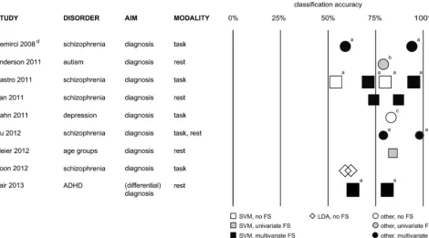
There has recently been a remarkable upsurge of scientific articles from the interdisciplinary functional neuroimaging community reporting successful applications of MVPA on fMRI data to vari-ous diagnostic problems, especially in the past 3 years. This con-stitutes a paradigm shift from comparative univariate to discrim-inative multivariate analyses of fMRI data. An exhaustive overview of these previous studies by using either task-based26-63 or task-free55,64-97fMRI is given in On-line Tables 1 and 2. An overview of particularly reliable studies with above-average statis-tical power is presented in Fig 2.
Although they are promising at first glance, there is a high degree of methodologic heterogeneity of classification algorithms and data-preprocessing steps in these studies. Some of the re-ported results seem to be mostly add-ons to studies whose de-signs were primarily aimed at clarifying disease mechanisms or were focused on computational aspects, not primarily done with the aim of developing a diagnostic tool. Until now, no single effort in this field has provided sufficient large-scale validation and sys-tematic optimization of methodologic choices leading to an ap-plication in a real medical diagnostic setting. Due to this feature extraction
(preprocessing, dimensionality reduction) fMRI training data (multiple subjects)
analogous features from
new clinical fMRI data diagnostic decision or
feature weighting/selection (multivariate wrapper)
+ classifier training
feature weighting/selection
(univariate filter)
classifier training
classifier (decision rule)
[image:2.594.58.368.47.313.2]heterogeneity and because strategies to assess the statistical significance of diagnostic accuracy vary considerably between studies, we did not perform a formal meta-analytical compar-ison of these reports.
Data Acquisition and Preprocessing. By now a majority of
re-ported approaches are based on conventional task-based fMRI.26-63This means that patients have to perform a specific, mainly neuropsychological task in the MR imaging scanner. Sta-tistical models are designed to evaluate the amount of variance in the acquired EPI data caused by this task modification of brain activity. This corresponds to “brain activation” in conventional fMRI studies.1An advantage of this approach is a rather straightfor-ward functional interpretability of such data. Yet in addition to mainly psychologically motivated studies in young healthy partici-pants, patients’ adherence to task instructions constitutes an impor-tant source of variability in real clinical settings and may even inter-fere with diagnostic decision-making.
Recently, a significant number of studies55,64-97have been based on task-free fMRI acquisitions, so-called resting-state fMRI, which focuses on the functional connectivity of distant brain regions in terms of signal cofluctuations and therefore on the integrity of large-scale brain networks.98,99A potential benefit of this method is that typical networks seem to be robustly iden-tifiable in individual subjects. However, reports focusing on the reliability of typical resting-state fMRI measures highlight the problem that these are highly dependent on potentially con-founding factors such as wakefulness or autonomic arousal.100,101 Although most resting-state fMRI findings in basic neuroscience are based on short acquisitions of approximately 5 minutes,
which seem to be sufficient for network detection,102there is re-cent evidence that retest reliability can be significantly improved by longer acquisitions.103Only a minority of resting-state func-tional connectivity– based MVPA approaches have used acquisi-tions lasting at least 7 minutes.67,72,79,84,87,91
As a common analysis step on a single subject level, feature-extraction methods are used to extract meaningful information from and simultaneously reduce the high dimensionality of the raw EPI time-series data. Prevailing approaches based on prior knowledge are activation modeling, based on general linear models for task-based acquisitions,1and seed-to-voxel or region-of-interest to region-of-interest correlation analyses for task-free acquisitions. In addition, recently more complex graph-theoretic approaches have been derived from the ROI– based methods. Another way of analyz-ing task-based and task-free studies relies on data-driven approaches such as independent component analyses.98,104
Recent further developments in diagnostic MVPA are not solely based on one of these methods. For example, Du et al55 combined both task- and task-free fMRI in schizophrenia in a small study. Additionally, combinations of fMRI measures with volumetric data,41,48-50,63,76,78-81,86,89DTI,46,49,92as well as ge-netics42and behavioral data,40,41,50,76have been used as features in MVPA analyses. However, results reported so far do not allow veri-fied statements about the benefit of such multimodal acquisitions.
Feature Selection, Classifier Training, and Assessment of
Classifi-cation Accuracy. Figure 2 and On-line Tables 1 and 2 contain
[image:3.594.60.531.48.309.2]based on conventional univariate analyses or whether it was also guided by multivariate information of distributed network patterns.
Apparently there are 2 groups of actual classification methods that have been applied successfully repeatedly: variants of LDA26,53,59,61,65-67,70,73,79,81,82,94 and support vector ma-chines.35,36,38,40-49,51,52,54,58,60,62,67-71,74-78,80,81,83-85,88-90,92,93,95-97 Al-though a certain number of articles report on conceptually different machine-learning techniques27-34,37,39,41,45,48,50,53-57,62,64,67,70,72,86-88,91 (eg, neural networks31,39,64,67,70 and decision tree– based ap-proaches41,50,81,86,87,91), each has only been applied occasionally, making reliable conclusions about their specific benefits and drawbacks in this context practically impossible.
With a few exceptions,72,75-78,80,81,86,88,89,95the small sample sizes in most studies did not allow testing the classification accu-racy in datasets completely independent of those used for classi-fier training. As stated above, a trick makes approximative assess-ments of classification accuracy of a set of trained classifiers possible: Most studies use CV to show the generalizability of strongly overlapping classifiers to new test data. This means that in most reports only the diagnostic ability of 1 particular set of dependent classifiers23is proved. There is usually no formal test that allows conclusions regarding the ability of whole MVPA ap-proaches (acquisition⫹feature extraction⫹feature selection⫹ classifier training) to construct successful diagnostic tools in a particular clinical setting because CV is only used to assess classi-fication of new data but not reliable classifier training indepen-dent from particular subjects. Additionally, setting up CV loops that do not strictly keep the test set and training set apart is a known source of error, leading to overoptimistic estimates of di-agnostic accuracy. There is still some uncertainty regarding the most appropriate test of significance to be applied in the CV setting.19
Only a small subset of reports contains systematic compari-sons and optimizations of larger sets of classification models used.49,68,70,75-78,80,81,86,88,89,95
Potential Clinical Applications and Integration in Diagnostic Workflows
To this point, most studies report applications to distinguish healthy controls and patients with a specific disease. These are a necessary step in developing and accessing diagnostic tools, but is it currently really clinically desirable to strive for such a tool?
In the context of practically illness-defining brain alterations, as in certain kinds of neurodegeneration, MVPA-fMRI methods might compete with radioactive tracer studies in the future. Re-garding psychiatric diseases, it seems, for example, desirable to identify patients with a high risk of disease recurrence or progres-sion. Especially in the case of major depressive disorders, there are a number of patients who do not respond to standard pharmaco-logic treatment; this outcome hints at potentially underlying di-vergent biologic mechanisms. Prediction of treatment response to a certain group of drugs seems to be a valuable objective as well.9 To date, some MVPA-fMRI studies have already attempted to classify subjects regarding prognostically relevant sub-groups.36,38,40,46,49,50,52,53,56,58 Another important but overlap-ping clinical question may be how to distinguish patients with
neurobiologically different disease entities but with a similar initial clinical presentation such as unipolar and bipolar depression. Such differential diagnostic aspects have been addressed in a few recent MVPA-fMRI studies as well.32,39,51,54,57,71,73,75,78,80,81,88,89,94,95
In this context, specific features of most psychiatric diseases should be taken into account when discussing the results of these analyses: The etiology and progression of disease are complex and only partly attributable to biologic causes. The biopsychosocial model of pathogenesis includes major influences of social and life event–related factors105,106that do not necessarily lead to corre-lates that are approachable by biologic measures such as fMRI.107 Furthermore, many diagnostically relevant symptoms are, by def-inition, subjective (eg, depressed mood).108The burden of suffer-ing is often decisive in terms of indications for treatment.109 Therefore, fMRI-MVPA– based measures should not be expected to become the criterion standard in diagnostics and replace in-depth history-taking. The accuracies of the studies reviewed here support this theoretic argument. Still, imaging-based multivariate tools might be able to provide clinically useful additional infor-mation: When important information (eg, regarding prognosis) is, by definition, not deducible from the course of disease, these tools might provide the clinician with crucial hints,7unraveling the “biologic share” of disease.
In nearly all fMRI-MVPA studies, there was a significant amount of misclassifications (On-line Tables 1 and 2). Partially, they may be attributable to inherent noise in the data and remain-ing methodologic weaknesses in data analysis. However, misclas-sification might also be based on biologically and medically mean-ingful information like the effects of medication110and age.69,85 Sex effects are a much-debated issue in fMRI as well.111,112 Fur-ther investigation of misclassified subjects might even pose a start-ing point to identify biologically different disease subgroups. Sup-posedly, a practical problem is that the referring physician and the radiologist cannot easily grasp what leads to a single diagnostic decision by fMRI-MVPA. In comparison with other types of di-agnostic imaging, it is therefore not directly possible to appreciate the extent of potentially biasing features in a specific subject. Only 5 recent studies have tried to overcome this issue by introducing individual confidence measures.39,48,54,56,57
As seen in Fig 2 and On-line Table 1, MVPA-fMRI has already been applied to a larger number of psychiatric disease entities. For depression,35,36,38,45,47,48,54,57,68,84 schizo-phrenia,26,32-34,37,39,42-44,55,58,61,62,64,66,70,74,90,91,96 and Alz-heimer dementia,26,27,41,50,53,65,71,73,79,82,94 there is now a larger body of independent work.
summarized in the Table. Before MVPA-fMRI could be applied in real clinical settings, potential interscanner variability113should also be taken into account.
CONCLUSIONS
Approximately 70 studies at the proof-of-principle level that use MVPA of fMRI data with a diagnostic intention have been re-ported. However, there is wide range of different methodologic decisions, from data-acquisition strategies through preprocessing and feature selection to actual diagnostic classification algorithms and parameter settings and, therefore, a high flexibility in study design. Results reported as yet are mainly based on small sets of subjects. Therefore, one has to be cautious in drawing reliable conclusions on the basis of this literature. Published results may just represent the tip of the iceberg, with a lot more unsuccessful unpublished attempts to apply this methodology. Therefore, there might be an important publication bias, and published re-sults regarding the statistical significance of successful diagnostic classification should be interpreted in the light of a potential need to correct for multiple comparisons.114Nevertheless, it can be regarded meanwhile as an independently replicated finding that building on task-based and resting-state fMRI as well support vector machines as LDA approaches has the potential to differen-tiate patients from healthy subjects in psychiatric disorders with most repeated findings in dementia, schizophrenia, and depression.
In contrast, there is apparently more uncertainty regarding optimal strategies for data preprocessing and feature selection, advisable steps to allow the classification algorithm to work de-spite a very high dimensionality and noise level of the original data. Many of these methods are derived from conventional fMRI analysis methods. Hardly any effort seems to have been made to systematically compare and evaluate the influence of these differ-ent approaches and parameter-setting selections on diagnostic accuracy.
In conclusion, here is some evidence that MVPA-fMRI is promising for overcoming long-known reliability issues in fMRI and providing clinically important prognostic and differential di-agnostic information in psychiatric disorders beyond pure exclu-sion of gross structural alterations. Despite the optimism coming from the recent discussion in the interdisciplinary functional
neu-roimaging community, this method is still rather new, and work has to be done to validate methodologic choices and identify those specific clinical settings that really allow a beneficial application. Moreover, a conceivable integration of MVPA-based fMRI into clinical workflow will depend critically on tackling diagnostic problems with a real clinical benefit and effects on therapeutic decision-making.
Disclosures: Wolfram Schwindt—UNRELATED: payment for manuscript preparation:
Ro¨Fo(Executive Editor).
REFERENCES
1. Huettel SA, Song AW, McCarthy G.Functional Magnetic Resonance Imaging.Sunderland, Massachusetts: Sinauer Associates; 2009 2. Zhang D, Raichle ME.Disease and the brain’s dark energy.Nat Rev
Neurol2010;6:15–28
3. Chen CH, Suckling J, Lennox BR, et al.A quantitative meta-analysis of fMRI studies in bipolar disorder.Bipolar Disord2011;13:1–15 4. Bartsch AJ, Homola G, Biller A, et al.Diagnostic functional MRI:
illustrated clinical applications and decision-making.J Magn Reson Imaging2006;23:921–32
5. Pillai JJ.The evolution of clinical functional imaging during the past 2 decades and its current impact on neurosurgical planning. AJNR Am J Neuroradiol2010;31:219 –25
6. Gruber O.Neuroimaging markers: their role for differential diag-nosis and therapeutic decisions in personalized psychiatry[in Ger-man].Nervenarzt2011;82:1404, 406, 408
7. Linden DE.The challenges and promise of neuroimaging in psychi-atry.Neuron2012;73:8 –22
8. Oertel-Kno¨chel V, Bittner RA, Knochel C, et al.Discovery and devel-opment of integrative biological markers for schizophrenia.Prog Neurobiol2011;95:686 –702
9. Mo¨ssner R, Mikova O, Koutsilieri E, et al.Consensus paper of the WFSBP task force on biological markers: biological markers in de-pression.World J Biol Psychiatry2007;8:141–74
10. Hampel H, Lista S, Khachaturian ZS.Development of biomarkers to chart all Alzheimer’s disease stages: the royal road to cutting the therapeutic gordian knot.Alzheimers Dement2012;8:312–36 11. Hyman SE.Can neuroscience be integrated into the DSM-V?Nat
Rev Neurosci2007;8:725–32
12. de Souza LC, Lehericy S, Dubois B, et al.Neuroimaging in demen-tias.Curr Opin Psychiatry2012;25:473–79
13. Jack CR, Jr.Alzheimer disease: new concepts on its neurobiology and the clinical role imaging will play.Radiology2012;263:344 – 61 14. Mueller S, Wang D, Fox MD, et al.Individual variability in func-Proposed reporting standards for future studies applying multivariate classification techniques to fMRI data with a diagnostic intention in addition to general reporting standards in fMRI research
Proposed Reporting Standards Desirable Aspects of Study Design
Report of overall classification accuracies/generalization rate (in addition to sensitivity and specificity if applicable)
Sufficiently large, equally sized groups (25 subjects per group desirable, but depending on specific methodologies used and power analyses)
Rigorous statistical tests Sufficiently long scan duration for resting state fMRI (at least 5
minutes, evaluation of a potential benefit of longer acquisitions desirable) Report of all tested models (not only optimized/best models) Systematic model optimization
Clearly identify inferential tests and explorative analyses Well-chosen and well-defined clinical problem Clearly identify feature extraction and feature selection/weighting
strategies
Multisite/multiscanner studies
Denominate origin of classification algorithms if an independent toolbox is used for pipeline building
Evaluation of functionally/anatomically interpretable classifiers and methods with individual reliability measures
Good clinical characterization of the sample including demographic data (psychometric data if applicable)
tional connectivity architecture of the human brain. Neuron
2013;77:586 –95
15. Raemaekers M, du Plessis S, Ramsey NF, et al.Test-retest variability underlying fMRI measurements.Neuroimage2012;60:717–27 16. Shehzad Z, Kelly AM, Reiss PT, et al.The resting brain:
uncon-strained yet reliable.Cereb Cortex2009;19:2209 –29
17. Nichols TE.Multiple testing corrections, nonparametric methods, and random field theory.Neuroimage2012;62:811–15
18. Norman KA, Polyn SM, Detre GJ, et al.Beyond mind-reading: multi-voxel pattern analysis of fMRI data. Trends Cogn Sci
2006;10:424 –30
19. Pereira F, Mitchell T, Botvinick M.Machine learning classifiers and fMRI: a tutorial overview.Neuroimage2009;45:S199 –209 20. Haynes JD, Rees G.Decoding mental states from brain activity in
humans.Nat Rev Neurosci2006;7:523–34
21. Klo¨ppel S, Abdulkadir A, Jack CR Jr, et al.Diagnostic neuroimaging across diseases.Neuroimage2012;61:457– 63
22. Orru` G, Pettersson-Yeo W, Marquand AF, et al.Using support vector machine to identify imaging biomarkers of neurological and psychiatric disease: a critical review.Neurosci Biobehav Rev
2012;36:1140 –52
23. Alpaydin E. Introduction to Machine Learning. Cambridge, Massachusetts: MIT Press; 2010
24. Vapnik VN.The Nature of Statistical Learning Theory.2nd ed. New York: Springer-Verlag; 2000
25. Derrfuss J, Brass M, von Cramon DY, et al.Neural activations at the junction of the inferior frontal sulcus and the inferior precentral sulcus: interindividual variability, reliability, and association with sulcal morphology.Hum Brain Mapp2009;30:299 –311
26. Ford J, Farid H, Makedon F, et al.Patient classification of fMRI activation maps.In: Ellis R, Peters T, eds.Medical Image Computing and Computer-Assisted Intervention - MICCAI 2003, Lecture Notes in Computer Science, Vol. 2879. Berlin, Germany: Springer-Verlag; 2003:58
27. Kontos D, Megalooikonomou V, Pokrajac D, et al.Extraction of dis-criminative functional MRI activation patterns and an application to Alzheimer’s disease.In: Barillot C, Haynor DR, Hellier P, eds.
Medical Image Computing and ComputerAssisted Intervention -MICCAI 2004, Lecture Notes in Computer Science, Vol. 3217.Berlin, Germany: Springer-Verlag. 2004;727–35
28. Bogorodzki P, Rogowska J, Yurgelun-Todd DA.Structural group classification technique based on regional fMRI BOLD responses. IEEE Trans Med Imaging2005;24:389 –98
29. Zhang L, Samaras D, Tomasi D, et al.Exploiting temporal informa-tion in funcinforma-tional magnetic resonance imaging brain data.Med Im-age Comput Comput Assist Interv2005;8:679 – 87
30. Shinkareva SV, Ombao HC, Sutton BP, et al.Classification of func-tional brain images with a spatio-temporal dissimilarity map. Neu-roimage2006;33:63–71
31. Chen R, Herskovits EH.Clinical diagnosis based on bayesian classi-fication of functional magnetic-resonance data.Neuroinformatics
2007;5:178 – 88
32. Calhoun VD, Maciejewski PK, Pearlson GD, et al.Temporal lobe and “default” hemodynamic brain modes discriminate between schizo-phrenia and bipolar disorder.Hum Brain Mapp2008;29:1265–75 33. Demirci O, Clark VP, Calhoun VD.A projection pursuit algorithm
to classify individuals using fMRI data: application to schizophre-nia.Neuroimage2008;39:1774 – 82
34. Demirci O, Clark VP, Magnotta VA, et al.A review of challenges in the use of fMRI for disease classification/characterization and a projection pursuit application from multi-site fMRI schizophrenia study.Brain Imaging Behav2008;2:147–226
35. Fu CH, Moura˜o-Miranda J, Costafreda SG, et al.Pattern classifica-tion of sad facial processing: toward the development of neurobio-logical markers in depression.Biol Psychiatry2008;63:656 – 62 36. Marquand AF, Moura˜o-Miranda J, Brammer MJ, et al.
Neuroanat-omy of verbal working memory as a diagnostic biomarker for de-pression.Neuroreport2008;19:1507–11
37. Michael AM, Calhoun VD, Andreasen NC, et al.A method to classify schizophrenia using inter-task spatial correlations of functional brain images.Conf Proc IEEE Eng Med Biol Soc2008;2008:5510 –13 38. Costafreda SG, Khanna A, Moura˜o-Miranda J, et al.Neural
corre-lates of sad faces predict clinical remission to cognitive behavioural therapy in depression.Neuroreport2009;20:637– 41
39. Arribas JI, Calhoun VD, Adali T.Automatic bayesian classification of healthy controls, bipolar disorder, and schizophrenia using in-trinsic connectivity maps from fMRI data.IEEE Trans Biomed Eng
2010;57:2850 – 60
40. Saur D, Ronneberger O, Kummerer D, et al.Early functional mag-netic resonance imaging activations predict language outcome af-ter stroke.Brain2010;133:1252– 64
41. Tripoliti EE, Fotiadis DI, Argyropoulou M, et al.A six stage approach for the diagnosis of the Alzheimer’s disease based on fMRI data. J Biomed Inform2010;43:307–20
42. Yang H, Liu J, Sui J, et al.A hybrid machine learning method for fusing fMRI and genetic data: combining both improves classifica-tion of schizophrenia.Front Hum Neurosci2010;4:192
43. Castro E, Martinez-Ramon M, Pearlson G, et al.Characterization of groups using composite kernels and multi-source fMRI analysis data: application to schizophrenia.Neuroimage2011;58:526 –36 44. Costafreda SG, Fu CH, Picchioni M, et al.Pattern of neural
re-sponses to verbal fluency shows diagnostic specificity for schizo-phrenia and bipolar disorder.BMC Psychiatry2011;11:18 45. Hahn T, Marquand AF, Ehlis AC, et al.Integrating neurobiological
markers of depression.Arch Gen Psychiatry2011;68:361– 68 46. Hoeft F, McCandliss BD, Black JM, et al.Neural systems predicting
long-term outcome in dyslexia. Proc Natl Acad Sci U S A
2011;108:361– 66
47. Moura˜o-Miranda J, Hardoon DR, Hahn T, et al.Patient classifica-tion as an outlier detecclassifica-tion problem: an applicaclassifica-tion of the one-class support vector machine.Neuroimage2011;58:793– 804
48. Nouretdinov I, Costafreda SG, Gammerman A, et al.Machine learn-ing classification with confidence: application of transductive con-formal predictors to MRI-based diagnostic and prognostic markers in depression.Neuroimage2011;56:809 –13
49. Rizk-Jackson A, Stoffers D, Sheldon S, et al.Evaluating imaging bio-markers for neurodegeneration in pre-symptomatic Huntington’s disease using machine learning techniques. Neuroimage
2011;56:788 –96
50. Tripoliti EE, Fotiadis DI, Argyropoulou M.A supervised method to assist the diagnosis and monitor progression of Alzheimer’s disease using data from an fMRI experiment.Artif Intell Med2011;53:35– 45 51. Weygandt M, Schaefer A, Schienle A, et al.Diagnosing different binge-eating disorders based on reward-related brain activation patterns.Hum Brain Mapp2012;33:2135– 46
52. Abdulkadir A, Ronneberger O, Wolf RC, et al.Functional and struc-tural MRI biomarkers to detect pre-clinical neurodegeneration. Curr Alzheimer Res2013;10:125–34
53. Andersen AH, Rayens WS, Liu Y, et al.Partial least squares for dis-crimination in fMRI data.Magn Reson Imaging2012;30:446 –52 54. Grotegerd D, Suslow T, Bauer J, et al.Discriminating unipolar and
bipolar depression by means of fMRI and pattern classification: a pilot study.Eur Arch Psychiatry Clin Neurosci2013;63:119 –31 55. Du W, Calhoun VD, Li H, et al.High classification accuracy for
schizophrenia with rest and task fMRI data.Front Hum Neurosci
2012;6:145
56. Moura˜o-Miranda J, Oliveira L, Ladouceur CD, et al.Pattern recog-nition and functional neuroimaging help to discriminate healthy adolescents at risk for mood disorders from low risk adolescents. PLoS One2012;7:e29482
57. Moura˜o-Miranda J, Almeida JR, Hassel S, et al.Pattern recognition analyses of brain activation elicited by happy and neutral faces in unipolar and bipolar depression.Bipolar Disord2012;14:451– 60 58. Nejad AB, Madsen KH, Ebdrup BH, et al.Neural markers of negative
of antipsychotic-naive schizophrenia patients.Int J Neuropsychop-harmacol2013;16:1195–204
59. Ramezani M, Abolmaesumi P, Marble K, et al.Classification of indi-viduals based on sparse representation of brain cognitive patterns: a functional MRI study. Conf Proc IEEE Eng Med Biol Soc
2012;2012:2688 –91
60. Weygandt M, Blecker CR, Schafer A, et al.fMRI pattern recognition in obsessive-compulsive disorder.Neuroimage2012;60:1186 –93 61. Yoon JH, Nguyen DV, McVay LM, et al.Automated classification of
fMRI during cognitive control identifies more severely disorga-nized subjects with schizophrenia.Schizophr Res2012;135:28 –33 62. Rish I, Cecchi G, Thyreau B, et al.Schizophrenia as a network
disease: disruption of emergent brain function in patients with au-ditory hallucinations.PLoS One2013;8:e50625
63. Kim J, Lee JH.Integration of structural and functional magnetic resonance imaging improves mild cognitive impairment detection. Magn Reson Imaging2013;31:718 –32
64. Jafri MJ, Calhoun VD.Functional classification of schizophrenia using feed forward neural networks.Conf Proc IEEE Eng Med Biol Soc2006;(suppl):6631–34
65. Wang K, Jiang T, Liang M, et al.Discriminative analysis of early Alzheimer’s disease based on two intrinsically anti-correlated net-works with resting-state fMRI.Med Image Comput Comput Assist Interv2006;9:340 – 47
66. Shi F, Liu Y, Jiang T, et al.Regional homogeneity and anatomical parcellation for fMRI image classification: application to schizo-phrenia and normal controls.Med Image Comput Comput Assist In-terv2007;10(pt 2):136 – 43
67. Zhu CZ, Zang YF, Cao QJ, et al.Fisher discriminative analysis of resting-state brain function for attention-deficit/hyperactivity dis-order.Neuroimage2008;40:110 –20
68. Craddock RC, Holtzheimer PE 3rd, Hu XP, et al.Disease state pre-diction from resting state functional connectivity.Magn Reson Med
2009;62:1619 –28
69. Supekar K, Musen M, Menon V.Development of large-scale func-tional brain networks in children.PLoS Biol2009;7:e1000157 70. Shen H, Wang L, Liu Y, et al.Discriminative analysis of resting-state
functional connectivity patterns of schizophrenia using low dimen-sional embedding of fMRI.Neuroimage2010;49:3110 –21
71. Zhou J, Greicius MD, Gennatas ED, et al.Divergent network con-nectivity changes in behavioural variant frontotemporal dementia and Alzheimer’s disease.Brain2010;133:1352– 67
72. Anderson JS, Nielsen JA, Froehlich AL, et al.Functional connectivity magnetic resonance imaging classification of autism. Brain
2011;134:3742–54
73. Chen G, Ward BD, Xie C, et al.Classification of Alzheimer disease, mild cognitive impairment, and normal cognitive status with large-scale network analysis based on resting-state functional MR imag-ing.Radiology2011;259:213–21
74. Fan Y, Liu Y, Wu H, et al.Discriminant analysis of functional con-nectivity patterns on Grassmann manifold. Neuroimage
2011;56:2058 – 67
75. Brown MR, Sidhu GS, Greiner R, et al. ADHD-200 global competition: Diagnosing ADHD using personal characteristic data can outperform resting state fMRI measurements.Front Syst Neu-rosci2012;6:69
76. Bohland JW, Saperstein S, Pereira F, et al.Network, anatomical, and non-imaging measures for the prediction of ADHD diagnosis in individual subjects.Front Syst Neurosci2012;6:78
77. Cheng W, Ji X, Zhang J, et al.Individual classification of ADHD patients by integrating multiscale neuroimaging markers and ad-vanced pattern recognition techniques. Front Syst Neurosci
2012;6:58
78. Colby JB, Rudie JD, Brown JA, et al.Insights into multimodal imag-ing classification of ADHD.Front Syst Neurosci2012;6:59 79. Dai Z, Yan C, Wang Z, et al.Discriminative analysis of early
Alzhei-mer’s disease using multi-modal imaging and multi-level charac-terization with multi-classifier (M3).Neuroimage2012;59:2187–95
80. Dai D, Wang J, Hua J, et al. Classification of ADHD children through multimodal magnetic resonance imaging.Front Syst Neu-rosci2012;6:63
81. Eloyan A, Muschelli J, Nebel MB, et al.Automated diagnoses of at-tention deficit hyperactive disorder using magnetic resonance im-aging.Front Syst Neurosci2012;6:61
82. Koch W, Teipel S, Mueller S, et al.Diagnostic power of default mode network resting state fMRI in the detection of Alzheimer’s disease. Neurobiol Aging2012;33:466 –78
83. Long D, Wang J, Xuan M, et al.Automatic classification of early Parkinson’s disease with multi-modal MR imaging. PLoS One
2012;7:e47714
84. Ma Q, Zeng LL, Shen H, et al.Altered cerebellar-cerebral resting-state functional connectivity reliably identifies major depressive disorder.Brain Res2013;1495:86 –94
85. Meier TB, Desphande AS, Vergun S, et al.Support vector machine classification and characterization of age-related reorganization of functional brain networks.Neuroimage2012;60:601–13
86. Olivetti E, Greiner S, Avesani P.ADHD diagnosis from multiple data sources with batch effects.Front Syst Neurosci2012;6:70
87. Richiardi J, Gschwind M, Simioni S, et al.Classifying minimally dis-abled multiple sclerosis patients from resting state functional con-nectivity.Neuroimage2012;62:2021–33
88. Sato JR, Hoexter MQ, Fujita A, et al.Evaluation of pattern recogni-tion and feature extracrecogni-tion methods in ADHD predicrecogni-tion.Front Syst Neurosci2012;6:68
89. Sidhu GS, Asgarian N, Greiner R, et al.Kernel principal component analysis for dimensionality reduction in fMRI-based diagnosis of ADHD.Front Syst Neurosci2012;6:74
90. Tang Y, Wang L, Cao F, et al.Identify schizophrenia using resting-state functional connectivity: an exploratory research and analysis. Biomed Eng Online2012;11:50
91. Venkataraman A, Whitford TJ, Westin CF, et al.Whole brain resting state functional connectivity abnormalities in schizophrenia. Schizophr Res2012;139:7–12
92. Wee CY, Yap PT, Zhang D, et al.Identification of MCI individuals using structural and functional connectivity networks.Neuroimage
2012;59:2045–56
93. Zeng LL, Shen H, Liu L, et al.Identifying major depression using whole-brain functional connectivity: a multivariate pattern analy-sis.Brain2012;135:1498 –507
94. Zhang Z, Liu Y, Jiang T, et al.Altered spontaneous activity in Alz-heimer’s disease and mild cognitive impairment revealed by re-gional homogeneity.Neuroimage2012;59:1429 – 40
95. Fair DA, Nigg JT, Iyer S, et al.Distinct neural signatures detected for ADHD subtypes after controlling for micro-movements in resting state functional connectivity MRI data. Front Syst Neurosci
2012;6:80
96. Yu Y, Shen H, Zhang H, et al.Functional connectivity-based signa-tures of schizophrenia revealed by multiclass pattern analysis of resting-state fMRI from schizophrenic patients and their healthy siblings.Biomed Eng Online2013;12:10
97. Zhang D, Liu B, Chen J, et al.Determination of vascular dementia brain in distinct frequency bands with whole brain functional con-nectivity patterns.PLoS One2013;8:e54512
98. van den Heuvel MP, Hulshoff Pol HE.Exploring the brain network: a review on resting-state fMRI functional connectivity.Eur Neuro-psychopharmacol2010;20:519 –34
99. Lee MH, Smyser CD, Shimony JS.Resting-state fMRI: a review of methods and clinical applications.AJNR Am J Neuroradiol2013;34: 1866 –72
100. Fan J, Xu P, Van Dam NT, et al.Spontaneous brain activity relates to autonomic arousal.J Neurosci2012;32:11176 – 86
func-tional connectivity as a tool for human connectomics: theory, properties, and optimization.J Neurophysiol2010;103:297–321 103. Anderson JS, Ferguson MA, Lopez-Larson M, et al.Reproducibility
of single-subject functional connectivity measurements.AJNR Am J Neuroradiol2011;32:548 –55
104. Margulies DS, Bottger J, Long X, et al.Resting developments: a review of fMRI post-processing methodologies for spontaneous brain activity.MAGMA2010;23:289 –307
105. Adler RH.Engel’s biopsychosocial model is still relevant today. J Psychosom Res2009;67:607–11
106. Engel GL.The need for a new medical model: a challenge for bio-medicine.Science1977;196:129 –36
107. Smith RC, Fortin AH, Dwamena F, et al.An evidence-based pa-tient-centered method makes the biopsychosocial model scien-tific.Patient Educ Couns2013;91:265–70
108. Fuchs T.Subjectivity and intersubjectivity in psychiatric diagno-sis.Psychopathology2010;43:268 –74
109. Bu¨chi S, Sensky T, Sharpe L, et al.Graphic representation of illness: a novel method of measuring patients’ perceptions of the impact of illness.Psychother Psychosom1998;67:222–25
110. Marquand AF, De Simoni S, O’Daly OG, et al.Pattern classification of working memory networks reveals differential effects of meth-ylphenidate, atomoxetine, and placebo in healthy volunteers. Neuropsychopharmacology2011;36:1237– 47
111. Biswal BB, Mennes M, Zuo XN, et al.Toward discovery science of human brain function. Proc Natl Acad Sci U S A
2010;107:4734 –39
112. Gong G, He Y, Evans AC.Brain connectivity: gender makes a dif-ference.Neuroscientist2011;17:575–91
113. Friedman L, Glover GH, Krenz D, et al.Reducing inter-scanner variability of activation in a multicenter fMRI study: role of smoothness equalization.Neuroimage2006;32:1656 – 68 114. Ioannidis JP.Why most published research findings are false.PLoS